- 5 -
Inverted Papilloma
Kyung-Rae Kim, M.D.
INTRODUCTION
Inverted papilloma is a relatively rare disease of the nasal cavity and paranasal sinus that constitutes 0.5- 4% of all nasal tumors.
1-3)Even though it is a benign tumor that originates from the ectoderm and inverts into the matrix, it carries characteristics such as local, aggressive growth, multicentricity, high recurrence rate, association with carcinoma and malignant transfor- mation and therefore requires surgical removal and elaborate postoperative observation.
4)Advanced im- aging such as computed tomography (CT) and mag- netic resonance imaging (MRI) have enhanced the ability of the surgeon to diagnose, localize and resect the tumor with greater precision than ever before.
Patients with inverted papilloma are typically men who present with unilateral nasal obstruction. Its highest incidence was in the fourth and sixth decades of life.
1-4)In the study by Kim et al.,(2001) there were no patients under the age 30 and there were no ch- ildren noticed at all.
5)Although the cause of inverted papilloma is unknown, a viral etiology has been su- spected. However, despite the fact that children are susceptible to viral diseases and still there were no incidences of inverted papilloma in children, it can be speculated that there might be a different etiology other than a viral one.
Inverted papilloma was first described by Ward in
1854 and after one year, Billroth reported 2 cases under the title of “villiform cancer”.
6)7)Rinngetz, in 1938, drew attention to the microscopic characteristic of a cylindrical or transitional cell type lesion reversing into the matrix.
8)Due to this characteristic growing pattern, there are over 20 different synonyms used, e.g.
Ewing Papilloma, cylindrical cell papilloma, papillary sinusitis, transitional cell papilloma, nasal epithelial papilloma, papillary respiratory epithelial carcinoma.
Further confusion was added by the term “Schnei- derian” after Victor Conrad Schneider described an epithelial tumor deriving from ectoderm. The term “in- verted” was coined by Lampertico et al., in 1963.
9)ETIOLOGY
The etiology of inverted papilloma is not known.
Allergy, chronic inflammation, environmental toxins and viruses have been suggested as etiological factors.
Allergy does not seem to be a causative factor, as a positive history of allergy is not found to be common.
Allergic polyposis is characteristically bilateral situ- ated, whereas inverted papilloma usually is of unila- teral appearance. Moreover, inverted papilloma does not respond to therapy directed against allergy. Hyams found no evidence to relate inverted papilloma to local chronic inflammation or allergy.
10)Phillips et al. and Majumdar and Beck found no correlation between occupational history and inverted papilloma in their study.
11)12)The most popular theory describes a human papilloma virus to be involved in the development of the tumor. However, electron microscopical and im- munohistochemical studies have failed to reveal virus particles or virus antigens.
13)14)However, in 1987, Res- pler et al., were able to demonstrate the presence of human papillomavirus 11 in inverted papilloma spe- cimens using in situ hybridization teachniques.
13)DNA Department of Otorhinolaryngology, College of Medicine, Han-
yang University, Seoul, Korea
Address correspondence and reprint requests to Kyung-Rae Kim, M.D., Department of Otorhinolaryngology, Hanyang University Hospital, 17 Haengdang-Dong, Sungdong-Ku, Seoul 133-792, Korea
Tel:82-2-2290-8580, Fax:82-2-2293-3335 E-mail:[email protected]
Accepted for publication on July 21, 2001
sequences of HPV 11 and HPV 16 were isolated in inverted papilloma in 19% (5/26) of patients studied by Furuta, et al., and types 6b and 11 were isolated in inverted papilloma in 76% (16/21) of the cases exa- mined by Weber et al.,
14)15)Even more intriguing is the possible association of HPV with inverted papilloma- associated malignancies. Using differenet molecular biological techniques, Furuta, et al., Syrjanen, et al.
and Brandwein, et al., each reported HPV 16 in in- verted pailloma associated with squamous cell carci- noma.
14)16)17)PATHOLOGY
On gross examination, inverted papillomas have lobulated configuration with a pink-to-gray velvety surface which appears far less translucent than an all- ergic nasal polyp. Although the precise site of origin is often difficult to determine owing to the advanced stage of the disease, the lateral nasal wall predomi- nates in all large reported series.
1-3)The most characteristic microscopic feature is a hypercellular thickening of the surface epithelium with downward cryptiform invaginations into the supporing stroma.
8)The basement membrane appears intact and of normal thickness.
10)The neoplastic epithelium is either a squamous, transitional or atypical type. In most instances epithelial maturation appears uniform with minimal nuclear pleomorphism or atypical mi- totic activity. The stroma is occasionally inflamed, evidencing a migration of chronic inflammatory cells but lacking the edematous, myxoid quality of allergic polyps as well as the eosinophilia.
18)RADIOLOGY
The radiologic appearance of inverted papilloma is often distinctive but not pathognomonic. Plain ra- diographs can be entirely negative or more often the appearances are non-specific with opacification of one maxillary antrum indistinguishable from inflammatory sinus disease.
19)The characteristic feature on CT is that of a mass continuous from the middle meatus into the adjacent maxillary antrum through a widened maxillary ostium.
The maxillary antrum is most commonly affected al-
though the ethmoids, the frontal and occasionally the sphenoid sinus may be involved with, or without, antral involvement. Tumors with an atypical location are much more difficult to diagnose, but the presence of internal hyperdensity or irregular sclerosis and defor- mation of the sinus walls may point to the diagnosis.
CT does have the limitation that the boundaries of the tumor are difficult or impossible to distinguish from adjacent soft tissue density due to retained secretion in adjacent sinuses where drainage is obstructed by the tumor.
The main advantage of MRI over CT is in defining the extent of inverted papilloma rather than making the diagnosis.
20)This is important for the extent of the disease which primarily determines the choice of sur- gical approach. T2-weighted MRI is very accurate in distinguishing papilloma (intermediate signal) from adjacent inflammatory change (very high signal).
However, there are no distinctive signal intensity or enhancement characteristics to help differentiate inv- erted papilloma from various sinus malignancies. In common with CT, MRI is unable to differentiate foci of coexistent squamous cell carcinoma from inverted papilloma.
21)Frank destruction of sinus walls due to malignant change is less easy to recognize than by CT, but invasion of tumor into the orbit and nasopharynx is optimally shown by unenhanced T1, or enhanced T1 weighted sequences with fat suppression. Disorder that need to be differentiated are antrochoanal polyp, ma- lignant sinus tumor, chronic rhinosinusitis and fungal disease.
SYMPTOMS AND SIGNS
The most common presenting symptom, by far, was
unilateral nasal obstruction. Rhinorrhea, facial pain or
pressure, epistaxis, frontal headache and anosmia con-
stituted the majority of the remaining complaints.
1-3)11)18)Less frequent symptoms included foul taste, otalgia,
numbness, neck mass, or a mass presenting at the nasal
vestibule. Rarely, there were patients who were asym-
ptomatic and the mass was discovered incidentally on
CT scans done for unrelated symptoms. Numerous
patients had undergone prior nasal sinus surgeries from
polypectomy and intranasal ethmoidectomy to Cald-
well-Luc operation.
RECURRENCE
It was Ringertz, in 1938, who first suggested that incomplete resection of tumor due to connective tissue stromal invasion was the key factor in the recurrence of inverted papilloma.
8)Others have stressed a mul- ticentric origin of the tumor to account for its tendency to recur.
10)22)23)Multifocality is difficult to assess clini- cally and even histopathologically, although reports as high as 12% and 30% have been reported by Wissler, et al. and Morris, respectively.
23)24)The significant in- cidence of recurrences of inverted papilloma follow- ing radical surgery that exenterates a large area sug- gests multifocal or unrecognized residual disease. The sites of tumor recurrence correlates primarily with the original tumor location. However, areas such as the sphenoid sinus, cribriform plate, and frontal sinus were involved at proportionately higher rates when comp- ared with their low frequency of initial involvement. In these patients, it is conceivable that tumor implantation may have occurred at the time of the initial surgery, or unrecognized microscopic disease was present.
2)The presence of areas of metaplasia seen at tumor margins has led some authors to speculate that failure to excise these areas may result in recurrence.
25)Others have suggested that inverted papillomas exhibiting cellular atypia are associated with a higher rate of re- currence.
9)26)However, most investigators have failed to correlate cellular atypia with recurrence.
11)27-29)When the tumor does recur, it may not become cli- nically evident for several years. Recurrence usually occurs within the first 2 years following surgery.
24)30)31)According to Lawson, a 2-year and even a 4-year- disease-free interval does not necessarily imply cure.
2)MANAGEMENT
Surgery is the agreed primary therapeutic modality of choice and a range of surgical approaches has been described. It is clear that limited surgery such as con- ventional polypectomy is not surprisingly associated with recurrence rates of up to 75%.
18)This has led to a greater reliance on more concerted attempts to com- pletely remove the papilloma via external approaches such as the lateral rhinotomy or mid-facial degloving approach accompanied by commensurately lower re-
currence rates of between zero and 29%.
2)3)26)31)32)Phillips stated that transnasal operations are associated with a high recurrence rate and should be avoided.
Myers proposed that complete removal of the ipsila- teral sinus mucosa along with medial maxillectomy must be carried out.
11)32)However, even before nasal endoscopy was introduced, the possibilities of con- servative surgery have been presented by numerous authors.
In 1991, Kamel reported 4 cases where endoscopic surgery has been executed.
33)After a 23 month follow- up, recurrence did not occur in all 4 cases. Waitz and Wigand stated that the recurrence rate following endo- scopic interventions was 17%, as compared to 19%
after extranasal operations.
31)In 1993, Stankiewicz reported 5 recurrence cases after medial maxillectomy, that were treated by endoscopic surgery.
34)Domes- tically, Lee (1996) reported 1 out of 11, Kwon (1997) reported 2 out of 16, Park (1998) reported 6 out of 41 recurrence cases. Dhong (1998) and Jang (1998) re- ported no recurrence cases at all.
35-39)In patients where the tumor originates from the la- teral wall of the maxillary sinus and the frontal sinus, endoscopic surgery is contraindicated. With the radi- ologic advances such as CT and MRI, the ability of the surgeon to localize unilateral nasal and paranasal sinus disease and determine preoperatively whether a tumor is present has been greatly enhanced. Also, based on the fact that the main reason of recurrence is incom- plete excision, with the use of an angled telescope, better visualization and margin control has made surgery of recess areas in the lateral nasal wall, frontal recess, supraorbital cell, cribriform plate, sphenoid sinus and maxillary sinus that were hard to reach with simple intranasal surgery much more convenient. A high recurrence rate does not accelerate the transfor- mation to malignant tumors. Also, a regular follow up using endoscopy makes early detection of recurrence much more simple that can lead to easier removal in cases of recurrence. In endoscopic surgery, structures of the nasal cavity and nasal mucosa are preserved and the function of the nasal cavity can also be maintained.
Inverted papilloma related to multifocal disease, ma- lignant transformation, invasion of the frontal sinus and lateral maxillary wall require an external approach using an en bloc medial maxillectomy.
The endoscopic surgery of inverted papilloma does
not differ greatly from the endoscopic surgery of chr- onic sinusitis or polyps. It must be exerted to find the origin of the tumor and remove it with En bloc and to keep a safe margin, frozen biopsies of different areas must be sent during surgery and surrounding sinuses must be affirmed. However, removal of all surroun- ding sinus mucosa is unnecessary. To confirm invasion of the maxillary sinus during follow-up, the middle meatal antrostomy must be made as big as possible. In cases of maxillary origin or wide invasion, Caldwell approach followed by Denker operation is executed.
This is necessary not only for complete removal but also for confirmation of recurrence in the maxillary sinus during follow-up. In cases of invasion inside the maxillary sinus, there is a possibility for the tumor to remain in the recess area which must be checked with an angled telescope. For complete removal of the tumor, the bony tissue surrounding the invaded area is either drilled with a diamond burr or the lamina papyracea is removed. Malignant tumors may accompany at times, therefore, after tissue removal, malignancy must be affirmed by postoperative biopsy.
Radiation therapy may be of some assistance in ca- ses of inverted papilloma that is related to squamous carcinoma.
1)32)Snyder, Perzin, Trible and Lekagul stated that radiation therpy has no effect in the trea- tment of pure inverted papilloma.
40)41)Mayberry re- ported 4 out of 14 tumors underwent malignant trans- formation. Snyder and Perzin reported 2 such cases and Suh, et al., as well as Woodson, et al., presented 1 case each.
25)27)40)There have been reports, however, that have suggested that radiation therapy can effect- tively control locally advanced inverted papilloma, particularly in cases of incomplete resection or mul- tiple recurrences. Hug, et al., treated 7 patients with inverted papilloma with radiation therapy:only 1 re- quired further surgery and all remained disease-free for several years.
43)In 3 of these cases, however, the gross tumor had been removed. Radiation was recomm- ended solely on the basis of previous recurrences.
ASSOCIATION WITH CARCINOMA
Squamous cell carcinoma that has occurred in the inverted papilloma is divided into synchronous and metachronous. Synchronous tumor is when carcinoma and inverted papilloma exist in the same anatomic
region without evidence that the papilloma gave rise to the cancer. Metachronous tumor is where carcinoma develops in the same anatomic area where a previously benign disease was resected.
44)Domestically, the frequency of inverted papilloma was 10.3% (3/29), 10.5% (2/19) and 9.7% (3/31) ac- cording to Min, et al., Ahn, et al. and Kwon, et al., res- pectively (Table 1).
35)45)46)Symptoms such as unilateral nasal obstruction, nasal cavity mass, epistaxis, diplopia etc. may appear, how- ever, according to Lesperance and Esclamado, the distinction between benign and malignancy based on symptoms is not possible.
47)Kamel stated that despite complete removal of the tumor, if there is fast rec- urrence, invasion to surrounding tissues, frequent nose bleeding, facial pain suggest bone or nerve invasion, malignancy must be considered. However, Hyams stated that there is no correlation between the number of recurrence and malignant transformation.
10)33)Benninger stated that malignant transformation may be expected if it has been diagnosed in the fifth and seventh decade of life, when epistaxis is greater than nasal obstruction or when symptoms manifest within 6 months.
48)The exact etiology of malignant transfor- mation is unknown, however, Ahn, et al., stated that there is a correlation between the area of metaplasia and malignant transformation. Nielsen, et al., was not able to find any specific characteristic of malignant
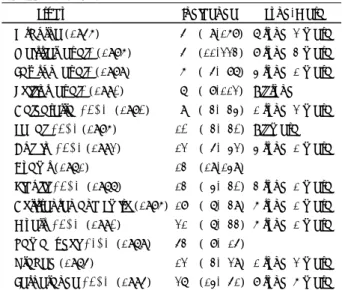
Table 1. Incidence of Squamous Cell Carcinoma (SCC) in In- verted Papilloma
Study Incidence Sync/Meta
Osborne (1956) 5% ( 9/168) 7 sync, 2 meta Weissler, et al., (1986) 5% (11/223) 8 sync, 3 meta Lawson, et al., (1989) 6% ( 5/ 87) 4 sync, 1 meta Phillips, et al., (1990) 7% ( 8/112) All sync Calcaterra, et al., (1980) 9% ( 3/ 34) 1 sync, 2 meta Segal, et al.,(1986) 10% ( 3/ 30) All meta Dolgin, et al.,(1992) 12% ( 5/ 42) 4 sync, 1 meta Hyams (1971) 13% (19/149)
Ridolfi, et al., (1977) 13% ( 4/ 30) 3 sync, 1 meta Christensen and Smith (1986) 18% ( 7/ 39) 6 sync, 1 meta Myers, et al., (1990) 21% ( 7/ 33) 6 sync, 1 meta Yamaguchi, el al., (1979) 53% ( 8/ 15)
Vrabec (1975) 12% ( 3/ 29) 1 sync, 2 meta Lesperance et al., (1995) 27% (14/ 51) 8 sync, 6 meta Modified from Lawson et al.,(1989)
Unable to determine synchronicity vs metachronicity from re- ference
Sync=synchronous;meta=metachronous
transformation after a histopathologic study based on 42 inverted papilloma cases.
46)49)After Furuta, et al.
reported that human papillomavirus participates in the development of inverted papilloma, the correlation of malignant transformation and human papillomavirus has brought much attention.
14)Kashima described 24 cases of squamous cell carcinoma irrelevant with in- verted papilloma, and was able to detect human pa- pillomavirus type 18 in 1 case. Furuta, et al., described 7 cases of squamous cell carcinoma that arose from inverted papilloma and among these cases they were able to find 2 cases of human papillomavirus type 16 and therefore stated that type 16 is related with ma- lignant transformation. However, due to a detection rate less than 30%, a significant verification was not established.
14)50)Beck, et al., isolated human papillo- mavirus type 6 and 11 in 12 out of 22 cases of inverted papilloma and among 17 cases of malignant trans- formation accompanied with metaplasia, type 16 was detected in 4 cases and type 18 in 1 case. Human pa- pillomavirus was classified as type 6/11 as the low risk group, and type 16/18 as the high risk group. A sta- tistical correlation was described between the high risk group and the severity of the lesion.
44)On the other hand, according to Byun, et al., the low risk group type 6 and 11 were detected in 81.8% in an inverted pa- pilloma group irrelevant with malignant transforma- tion and 71.5% was detected in the carcinomatous area of tissues accompanied with squamous cell carcinoma.
Unfortunately, there was no significant difference among the two groups and it was concluded that the clinical course cannot be determined only by the subtype but a different risk factor may be involved.
51)Lesperance and Esclamado stated that the modified Kadish staging system, that classifies inverted pa- pilloma accompanied by malignant transformation according to the degree of severity, may be useful in estimating the prognosis.
47)REFERENCES
1) Lawson W. Ho BT, Shaari CM, Biller HF. Inverted papilloma: A report of 112 cases. Laryngoscope 1995;105:282-8.
2) Vrabec DP. The inverted schneiderian papilloma: A 25-year study 1994;104:582-605.
3) Kerschner JE, Futran ND, Chaney V. Inverted papilloma associated with squamous cell carcinoma and adenocarcinoma: case report and review of the literature. Am J Otolaryngol 1996;17:257-9.
4) Lawson W, Benger JL. Som P, Bernard PJ, Biller HF. Inverted pa-
illoma: An analysis of 87 cases. Laryngoscope 1992;102:917-22.
5) Kim JH, Hong JP, Choi EC, Yoon JH. Treatment outcomes of pri- ary and recurred inverting papilloma: An analysis of 96 cases.
Korean J Otolaryngol 2001;44:731-5.
6) Ward N. A mirror of the practice of the medicine and surgery in the hospital of London: London Hospital. Lancet 1854;2:480-2.
7) Billroth T. Ueber dem Bau des Schleimpolyp Berlin Reimer;1855. p.11.
8) Ringertz N. Pathology of malignant tumors arising in the nasal and paranasal cavities and maxilla. Acta Otolaryngol Suppl (Stockh) 1938;27:31-42.
9) Lampertico P, Russel WO, MacComb WS. Squamous papilloma of the upper respiratory epithelium. Arch Pathol Lab Med, 1963;7:
293-302.
10) Hyams VJ. Papillomas of the nasal cavity and paranasal sinuses. A clinicopathological study of 315 cases. Ann Oto Rhinol Laryngol 1971;80:192-206
11) Phillips PP, Gustafson RO, Facer GW. The clinical behavior of in- verted papilloma of the nose and paranasal sinuses: Report of 112 cases ad review of the literature. Laryngoscope 1990;100:463-9.
12) Majumdar B, Beck S. Inverted papilloma of the nose: Some aspects of aetiology. J Laryngol Otol 1984;98:467-70.
13) Respler DS, Jahn A, Pater A. Isolation and characterization of pa- pillomavirus DNA from nasal inverting (schneiderian) papillomas.
Ann Oto Rhinol Laryngol 1987;96:170-3.
14) Furuta Y, Shinohara T, Sano, K. Molecular pathologic study of hu- man papillomavirus infection in Inverted papilloma and squamous cell carcinoma of the nasal cavities and paranasal sinuses. Laryn- goscope 1991;101:79-85.
15) Weber RS, Shillitoe EJ, Robbins KT. Prevalence of human papillo- mavirus in inverted nasal papilloma. Arch 0tolaryngol Head Neck Surg 1988;114:23-6.
16) Syrjanen S, Happonen RP, Virolainen E. Detection of human pa- pillomavirus (HPV) structural antigens and DNA types in inverted papillomas and squamous cell Carcinomas of the nasal and paran- asal sinuses. Acta Otolaryngol Suppl (Stockh) 1987;104:334-41.
17) Brandwein M, Steinberg B, Thung S. Human papillomavirus 6/11 and 16/18 in schneiderian inverted papilloma. In situ hybridization with human papillomavirus RNA probes. Cancer 1989;63:1708-13.
18) Calcaterra TC, Thompson JW, Paglia DE. Inverting papillomas of the nose and paranasal sinuses. Laryngoscope 1980;90:53-60.
19) Savy L, Lloyd G, Lund VJ, Howard D. Optimum imaging for in- verted papilloma. J Laryngol Otol 2000;114(11):891-3.
20) Lund VJ. Optimum management of inverted papilloma. J Laryngol Otol 2000;114:194-7.
21) Roobottom CA, Jewell FM, Kabala J. Primary and recurrent inver- ting papilloma: appearance with magnetic resonance imaging. Clin Radiol 1995;50:472-5.
22) 0sborn DA. Transitional cell growths of the upper respiratory tract.
J Laryngol Otol 1956;70:574-88.
23) Norris HJ. Papillary lesions of the nasal cavity and paranasal sin- uses. Part I: Exo-phytic (squamous) papillomas. A study of 28 cases.
Laryngoscope 1962;72:1784-97.
24) Weissler MC, Montgomery WW, Turner PA. Inverted papilloma. Ann Otol Rhinol Laryngol 1986;95:215-21.
25) Woodson J, Robbins K, Michaels L. Inverted papilloma. Arch Ot- olaryngol 1985;111:806-11.
26) Dolgin SR, Zaveri VD, Casiano RR. Different options for treatment of inverting papilloma of the nose and paranasal sinuses: A report of 41 cases. Laryngoscope 1992;102:231-6.
27) Suh KW, Facer GW, Devine KD. Inverting papilloma of the nose and paranasal sinuses. Laryngoscope 1977;87:35-46.
28) Smith 0, Gullane PJ. Inverting papilloma of the nose: analysis of 48 cases. J Otolaryngol 1987;16:154-6.
29) Ridolfi RL, Lieberman PH, Erlandson RA. Schneiderian papillo-
mas: A clinicopathologic study of 30 cases. Am J Surg Pathol 1977;
1:43-53.
30) Segal K, Atar E, Mor C. Inverting papilloma of the nose and para- nasal sinuses. Laryngoscope 1986;96:394-8.
31) Waitz G, Wigand ME. Results of endoscopic sinus surgery for the treatment of inverted papillomas. Laryngoscope 1992;102:917-22.
32) Myers EN, Fernau JL, Johnson, JT. Management of inverted pa- pilloma. Laryngoscope 1990;100:481-90.
33) Kamel R. Conservation endoscopic surgery in inverted papilloma.
preliminary report. Arch Otolaryngol Head Neck Surg l992;ll8:
649-53.
34) Stankiewicz JA, Girgis SJ. Endoscopic surgical treatment of nasal and paranasal sinus inverted papilloma. Otolaryngol Head Neck Surg 1993;109:988-95.
35) Lee CH, Lee SJ, Suh SJ, Kang JK. Treatment of inverted papilloma:
Role fo conservative surgery using nasal endoscope. Korean J Otolaryngol l996;39:1151-60.
36) Kwon SH, Park BA, Yun HW, Hong KH, Kim YK, Yoon YJ, Min YG. Treatment of inverted papilloma: A comparison of extranasal operation and conservative surgery using nasal endoscope. Korean J Otolaryngol l997;40(8):1097-102.
37) Park JH, Lee SD, Kwon YW, Park HJ, Lee YB. Outcomes of en- doscopic surgical treatment in 4l cases nasal and sinus inverted papilloma. Korean J Otolaryngol 1998;41(8):1025-8.
38) Jang TY, Song SY, Jung DH, Yun YS, Kim JH. Endoscopic surgery for inverted papilloma. Korean J Otolaryngol l998;4l(3):333-7.
39) Dhong HJ, Kim HY, Chung SK. Endoscopic management of benign tumors of the nose and paranasal sinuses. Korean J Otolaryngol l998;41(7):896-900.
40) Snyder RM and Perzin KH. Papillomatosis of the nasal cavity and paranasal sinuses(inverted papilloma, squamous papilloma). A clin- icopathologic study. Cancer 1972;30:668-90.
41) Trible WM and Lekagul S. Inverting papilloma of the nose and pa-
ranasal sinuses: report of 30 cases. Laryngoscope 1971;81:663-8.
42) Mayberry TE, Define KD and Harrison EG. The problem of mali- gnant transformation in a nasal papilloma. report of a case. Arch Otolaryngol 1965;82:296-300.
43) Hug EB, Wang CC, Montgomery WW. Management of inverted pa- pilloma of the nasal and paranasal sinuses: Importance of radiation therapy. Int J Radiat Oncol Biol Phys 1993;26:67-72.
44) Beck JC, McClatchey KD, Lesperance MM, Esclamado RM, Carey TE, Bradford CR. Human papillomavirus types important in pro- gression of inverted papilloma. Otolaryngol Head and Neck Surg 1995;113:558-63.
45) Min YG, Hong SH, Kim HS, Rhee CS. A clinical study on inverted papilloma of the nose and paranasal sinuses. Korean J Otolaryngol 1991;34(5):962-7.
46) Ahn HY, Yeo SG, Seok SR, Hong NP, Cho JS, Cha CI. Inverted pa- pilloma and malignant transformation. Korean J Otolaryngol 1994;
37(2):304-15.
47) Lesperance MM, Esclamado RM. Squamous cell carcinoma arising in inverted papilloma. Laryngoscope 1995;105:178-83.
48) Benninger MS. Roberts JK, Sebek BA, Levine HL, Tucker HM, Lavertu P. Inverted papilloma and associated with squamous cell carcinoma. Otolaryngol Head and Neck Surg 1990;103:457-61.
49) Nielsen PL, Buchwald C, Nielsen LH, Tos M. Inverted papilloma of the nasal cavity: Pathological aspects in a follow-up study.
Laryngoscope 1991;101:1094-101.
50) Kashima HK, Kessis T, Hurban KH, Wu TC, Zinreich SJ, Shah KV.
Human papillomavirus in sinonasal papillomas and squamous cell carcinoma. Laryngoscope 1992;102:973-6.
51) Byun JY, Cho JS, Hong IH, Kim WS, Lee DY, Cha CI. A study on the expression of p53 and the detection of human papillomavirus in the sinonasal inverted papilloma associated with carcinoma.
Korean J Otolaryngol 1998;41(2):188-94.