- 70 -
A FGF23-Positive Maxillary Sinus Tumor Associated with Oncogenic Osteomalacia
Na Hyun Kim, MD, Ji Hoon Kim, MD, Jin Ho Kwon, MD and Kyung-Su Kim, MD
Department of Otorhinolaryngology, Gangnam Severance Hospital, Yonsei University College of Medicine, Seoul, Korea
ABSTRACT
Oncogenic osteomalacia is a rare acquired paraneoplastic syndrome caused by excessive renal phosphate wast- ing that causes hypophosphatemia and osteomalacia. Fibroblast growth factor 23 (FGF23) has been identified as the causative factor of oncogenic osteomalacia and is also known as its diagnostic marker in immunohistochem- istry studies. Recently, the authors of the present study experienced a female patient with a prolonged history of osteomalacia; a rare maxillary sinus tumor was confirmed by anti-FGF23 immunohistochemical staining as the causative tumor. The present case is the first reported case in Korea to have been diagnosed as a head and neck tumor associated with oncogenic osteomalacia according to anti-FGF23 immunohistochemical staining.
KEY WORDS : Osteomalacia · Hypophosphatemia · Fibroblast Growth Factor 23 · Maxillary Sinus Neoplasms.
INTRODUCTION
Oncogenic osteomalacia or tumor-induced osteomala- cia is a rare acquired paraneoplastic syndrome character- ized by hypophosphatemia due to renal phosphate wast- ing. Its clinical features include adult-onset bone pain, atrophy of proximal muscles, gait disturbance, and mul- tiple bone fractures. Due to the slow-growing nature and absence of localized symptoms of the associated tumor, it takes an average of five years from the time of clini- cal presentation to diagnosis of the disease.1) Recently, fi- broblast growth factor 23 (FGF23) has been identified as the causative factor of oncogenic osteomalacia in several studies.2) 3) FGF23, which belongs to the FGF subfamily, plays a major role in phosphate homeostasis, and its over- expression seems to cause hypophosphatemic conditions.
In this case, we report a female patient with prolonged history of osteomalacia with a rare maxillary sinus tumor that was confirmed as the causative tumor by FGF23 im- munohistochemical staining. The present case is the first reported case in Korea to have been diagnosed by FGF23 immunohistochemical staining as a head & neck tumor
associated with oncogenic osteomalacia.
CASE
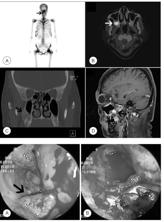
A 58-year-old female patient was referred to the De- partment of Otorhinolaryngology because of a bony lesion at the right maxilla in bone scan (Fig. 1A) and a mass lesion on the posterior wall of the right maxillary sinus in positron-emission tomography computerized tomog- raphy (PET-CT) (Fig. 1B). She suffered from multiple pathologic fractures on both legs and osteomalacia with high serum alkaline phosphate, severe hypophosphatemia, and slightly low 1,25-dihydroxy vitamin D3. She had no nasal symptoms or trismus. Clinical examination with a nasal endoscope revealed no remarkable findings. Para- nasal sinus CT & magnetic resonance imaging (MRI) de- tected a mass lesion on the postero-inferior wall of the right maxillary sinus, which correlated with her PET-CT and bone scan findings. The 1.5x1.5x2 cm-sized mass was expanding to the right pterygopalatine fossa, which was well-enhanced on a T1-weighted gadolinium-enhanced view (Fig. 1C, D).
Biopsy of tumor via a sublabial approach was per- formed. The anterior wall of the right maxillary sinus was punctured, and the mass was observed on the postero-in- ferior portion. It bled easily and was friable. Curettage and piecemeal resection was performed (Fig. 2). Histologic ex- amination revealed proliferation of spindle cells with dark and uniform nuclei, and proliferation of the capillary ves-
J Rhinol 19(1), 2012 www.ksrhino.or.kr
Address correspondence and reprint requests to Kyung-Su Kim, MD, De- partment of Otorhinolaryngology, Gangnam Severance Hospital, Yonsei University College of Medicine, 211 Eonju-ro, Gangnam-gu, 135-720 Seoul, South Korea
Tel: +82-2-2019-3460, Fax: +82-2-3463-4750 E-mail: [email protected]
Received for publication on November 9, 2011 Accepted for publicatoin on February18, 2012
Kim et al : Osteomalacia & Maxillary Tumor / 71
Fig. 1. A. Whole body bone scan showing multiple pathologic fracture foci.
Notice the bony lesion on the right maxilla (arrow). B.
PET-CT illustrating focal FDG uptake on the posterior wall of the right maxillary sinus (arrow). C. PNS CT showing a mass lesion on the poste- ro-inferior wall of the right maxillary sinus (arrow). D.
Sagittal MRI (T1 gadolinium- enhanced view) illustrat- ing well-enhanced lesion on the right maxillary sinus expanding to the pterygo- palatine fossa.
Fig. 2. A. Intraoperative pho- to showing the mass on the posteroinferior portion of the right maxillary sinus (ar- row). B. The same portion of the right maxillary sinus is observed after curettage &
piecemeal resection.
A C
B
B D
sels (Fig. 3A). Immunohistochemical staining for CD34, CD31, HHV-8, desmin, SMA, and S-100 was performed.
Tumor cells were positive for CD34, CD31, and negative for HHV-8, desmin, SMA, and S-100, which supported a diagnosis of spindle cell hemangioma. Immunohis- tochemical staining for FGF23 using monoclonal antibody (Enzo Life Sciences International, Inc., Farmingdale, NY) was also performed, and the cytoplasm of tumor cells was clearly positive, which confirmed the pathologic diagnosis of oncogenic osteomalacia (Fig. 3B).
The serum phosphate level slightly increased at 2 months post-operation. We warned the patient about the possibility of remnant tumors, and recommended revision
of the surgery (e.g. maxillectomy including pterygopala- tine fossa), but she refused angiography and surgery, and failed to follow-up.
DISCUSSION
Oncogenic osteomalacia is a rare acquired paraneo- plastic syndrome caused by excessive renal phosphate wasting. Renal phosphate wasting is an important mech- anism for developing osteomalacia. Less than 100 cases of oncogenic osteomalacia have been reported world- wide. Laboratory findings show characteristically pro- found hypophosphatemia and hyperphosphaturia with A
72 / J Rhinol 19(1), 2012
reduced or inappropriately normal 1, 25-dihydroxyvi- tamin D. Serum alkaline phosphate levels are elevated, along with normal serum calcium and PTH level. In this case, the patient showed low preoperative serum phos- phate and low 1, 25-dihydroxyvitamin D with elevated alkaline phosphatase.4) 5)
Oncogenic osteomalacia in the maxillary sinus is very limited. Only eight cases of oncogenic osteomala- cia involving the maxillary sinus have been reported in the published literature in English, including the present case.6-9) Patients were aged 35-75 with a mean age of 46.
Oncogenic osteomalacia is known to affect people in their 30s without preponderance of either gender.10) Among the maxillary sinus origin oncogenic osteomalacia patients, two were male and six were female. Because of the slow- growing nature of the associated tumor, the average time taken from clinical presentation to identification of the tumor was five years. The associated tumor was hard to identify in the current case because it was small and slow- growing. There was no clinical presentation involving the maxilla-facial area of the patient, hence head & neck im- aging was not performed until its appearance on the PET- CT. It was located at the posterior wall of right maxillary sinus expanding to the pterygopalatine fossa. This case suggests the need for a thorough head & neck examination of patients with suspected oncogenic osteomalacia.
Recently, FGF23 has been identified as the causative factor of oncogenic osteomalacia and X-linked hypo- phosphatemia in several studies.2) 3) In the present case, we found that the tumor was positive for FGF23, which repre- sents that it was associated with oncogenic osteomalacia.
The present case is the first reported case in Korea that was diagnosed by FGF23 immunohistochemical staining as oncogenic osteomalacia associated with a head & neck tumor. Measurement of serum FGF23 levels can be also useful in the diagnosis and identification of the tumor as- sociated with oncogenic osteomalacia.11)
To treat the tumor, we first performed piecemeal resec-
tion and biopsy because the mass was well-enhanced on MRI. We also planned a maxillectomy after pathologic confirmation and angiography. However, the patient re- fused further evaluation and treatment. Piecemeal remov- al of tumor caused the serum phosphate level to be elevat- ed, but the serum phosphate level was not restored to its normal level. The possible mechanism of elevated serum phosphate can be explained as follows; hyperphosphatu- ria decreases as serum FGF 23 level is decreased by the removal of tumor, which in turn causes increase of serum phosphate level. Thus, a radical operation was necessary for complete cure of the disease.
저자역할(Author Contributions)
김경수는 본 연구에서 모든 자료에 접근할 수 있으며 자료의 완전성과 자료 분석의 정확성에 책임을 지고 있습니다. 연구 기획 : 김경수. 자 료 해석 및 분석 : 김나현, 김지훈, 권진호. 논문초안 : 김나현. 논문수 정 : 김경수, 권진호, 김나현. 연구 총괄 : 김경수.
REFERENCES
1) Ruppe MD, Jan de Beur SM. Disorders of phosphate homeostasis.
In: Rosen CJ, editor. Primer on the metabolic diseases and disor- ders of mineral metabolism. 7th ed. Washington DC: American Society of Bone and Mineral Research; 2008. p.317-25.
2) Shimada T, Mizutani S, Muto T, Yoneya T, Hino R, Takeda S, et al. Cloning and characterization of FGF23 as a causative fac- tor of tumor-induced osteomalacia. Proc Natl Acad Sci USA.
2001;98:6500-5.
3) Jonsson KB, Zahradnik R, Larsson T, White KE, Sugimoto T, Imanishi Y, et al. Fibroblast growth factor 23 in oncogenic os- teomalacia and X-linked hypophosphatemia. N Eng J Med.
2003;348:1656-63.
4) Ahn J-M, Kim H-J, Cha C-M, Kim J, Yim S-G, Kim HJ. Onco- genic osteomalacia: induced by tumor, cured by surgery. Oral Surg Oral Med Oral Pathol Oral Radiol Endod. 2007;103:636-41.
5) Lewiecki EM, Urig EJ, Williams RC. Tumor-induced osteomala- cia: lessons learned. Arthritis Rheum. 2008;58:773-7.
6) Mori Y, Ogasawara T, Motoi T, Shimizu Y, Chikazu D, Tamura K, et al. Tumor-induced osteomalacia associated with a maxillofacial tumor producing fibroblast growth factor 23: report of a case and review of the literature. Oral Surg Oral Med Oral Pathol Oral Ra- Fig. 3. A. Microscopic image of the lesion showing cellular spindle cell proliferation with capillary vessel proliferation (Hematoxylin-Eosin stain- ing, original magnification x 100). B. Brownish stained cytoplasms, positively stained with FGF23, are noted (original magnification x 100).
A B
Kim et al : Osteomalacia & Maxillary Tumor / 73 diol Endod. 2010;109:57-63.
7) Savage CR, Zimmer LA. Oncogenic osteomalacia from pterygo- paltine fossa mass. J Laryngol Otol. 2009;123:1052-4.
8) Pedrazzoli M, Colletti G, Ferrari M, Rossetti G, Moneghini L, Auteli- tano L. Mesenchymal phosphaturic neoplasm in the maxillary sinus:
a case report. Int J Oral Maxillofac Surg. 2010;39:1027-32.
9) Koriyama N, Nishimoto K, Kodama T, Nakazaki M, Kurono Y, Yoshida H, et al. Oncogenic osteomalacia in a case with a maxil-
lary sinus mesenchymal tumor. Am J Med Sci. 2006;332:142-7.
10) Battoo A, Salih S, Unnikrishnan AG, Jojo A, Bahadur S, Lyer S, et al. Oncogenic osteomalacia from nasal cavity giant cell tumor.
Head Neck. 2010 Dec 9. [Epub ahead of print]
11) Takeuchi Y, Suzuki H, Ogura S, Imai R, Yamazaki Y, Yamashita T, et al. Venous sampling for fibroblast growth factor-23 confirms preoperative diagnosis of tumor-induced osteomalacia. J Clin En- docrinol Metab. 2004;89:3979-82.