- 87 -
흰쥐 비부비강의 횡단면의 구조 및 그 조직학적 특성
경상대학교 의과대학 이비인후과학교실,
1경상대학교 건강과학연구원
2성병기1·전시영1,2·김진평1,2·안성기1,2·박정제1,2·정재호1,2
Cross-Sectional Structure and its Histological Features of the Nasal Cavity and Paranasal Sinuses in the Rat
Byung Gi Sung, MD
1, Sea Yuong Jeon, MD
1,2, Jin Pyeong Kim, MD
1,2, Seong Ki Ahn, MD
1,2, Jung Je Park, MD
1,2and Jae Ho Jeong, MD
1,21Department of Otolaryngology, College of Medicine, Gyeongsang National University and
2Gyeongsang Institute of Health Sciences, Jinju, Korea
ABSTRACT
Background and Objectives:An understanding of the cross-sectional structures and histological features of experimental animals is necessary for conducting the research of rhinosinusitis in experimental animals. The aim of this study is to determine the most suitable cross-sectional level of rhinosinusitis in a rat model. Materials and Methods:The study examined the his- tological features of the mucosal epithelium, gland, lymphoid tissue, and vomeronasal organs using 4 week-old male rats weighing 60-70 g as test subjects. Whole mounted sinus-nose complexes were divided into four levels of areas which were sectioned according to the coronal plane and each section was stained with hematoxylin-eosin and observed under light microscopy.
Results:Level I revealed the nasal turbinate, maxilloturbinale, and nasolacrimal duct. Level II revealed the nasal turbinate, maxilloturbinale, vomeronasal organ, and nasolacrimal duct. Level III revealed the ectoturbinale I, II, endoturbinale II, III, maxillary sinus, Steno’s gland, maxillary sinus gland, and nasal associated lymphoid tissue (NALT). Level IV revealed the ectoturbinale II, endoturbinale III, IV, pharyx respiratorius, and NALT. The lining epithelia were squamous, respiratory and olfactory. However, the squamous epithelium was not observed in level III and IV. Conclusion:The Level III appears to be the most suitable for the rhinosinusitis animal rat model since we can observe the respiratory epithelium lined sinonasal airspace including the maxillary sinus and NALT.
KEY WORDS:Rats·Nose·Histology.
서 론
실험동물을 통한 비부비동염의 발생기전 및 병태 생리학 적 변화를 연구하는데 있어, 실험동물의 비강과 부비동 및 주변구조에 대한 해부학적 지식은 필수적이다. 실험 동물 중 흰쥐의 해부학적 구조에 대한 지식은 여러 문헌보고에서 기 술 되어 왔다. Hebel
1)이 흰쥐의 비강구조와 주변구조에 대하 여 해부학적 명칭과 횡단면의 구조를 전체적으로 기술하였 고 Bojsen-Moller
2)는 분비선의 해부학적 지도를 기술하여
보고 하였다. 하지만, 그들의 연구들은 흰쥐에 대한 일반적 인 해부학적 지식일 뿐 특정한 실험, 즉 비부비동염의 연구 에 있어서 필요한 해부학적 및 조직학적인 특징을 기술하지 는 못했다. 따라서 비부동염과 같은 특정 실험을 수행함에 있어 일반적인 해부학적인 지식이외에도 실험에 필요한 조 직학적인 특징과 어느 부위를 관찰하는 것이 실험에 유용한 지 아는 것이 필요하다. 실험동물에는 여러 가지가 있을 수 있으나, 최근에 흰쥐를 이용한 실험적 비부비동염 모델이 개 발되어 보고되고 있다.
3)4)흰쥐는 의학적 연구에 광범위하게 사용되며 신뢰도가 높은 동물이다. 또한 생쥐에 비하여 크기 가 커 실험적 조작이 용이하다.
저자들은 흰쥐 비부비강의 축상면 연속절편을 얻어 hema- toxylin-eosin(H & E) 염색을 한 후, 절단부위에 따른 횡단면 구조를 관찰하고, 그 조직학적인 특성으로, 점막상피, 분비선,
심사완료일:2006년 6월 2일교신저자:전시영, 660-702 경남 진주시 칠암동 90번지 경상대학교 의과대학 이비인후과학교실, 경상대학교 건강과학연구원 전화:(055) 750-8174·전송:(055) 759-0613
E-mail:[email protected]
림프조직 및 서비기관(vomeronasal organ)의 분포를 밝히고 자 한다. 또한 실험적 비부비동염 모델에 있어 연속절편 중 어 느 부위가 비부동염을 관찰하는데 가장 적합한지도 살펴보았다.
재료 및 방법
실험동물로는 체중 60~70 g의 외견상 건강하고, 비공이 깨끗한 4주령의 Sprague-Dawley계의 6마리의 수컷 흰쥐 를 사용하였으며, 실험기간 중에는 경상대학교병원 동물 사 육장에서 동일한 조건 하에서 사육하였다.
흰쥐의 정상 해부학적 구조를 관찰하기 위해 코주둥이에 서 비인강까지 4등분하여 각각 Level Ⅰ에서 Level Ⅳ로 명 명하였다(Fig. 1). 각 level의 경계는 level Ⅰ은 코 주둥이에 서 절치 바로 뒤까지, level Ⅱ는 절치 바로 뒤에서 절치 유 두까지, level Ⅲ는 절치 유두에서 제 1 대구치 앞까지, level
Ⅳ는 제 1 대구치 앞에서 구개주름까지이다.
Ketamine hydrochloride(Ketalar
®, 75 mg/kg), xylazine hydroch loride(Rampun
®, 10 mg/kg)의 혼합용액을 흰쥐 의 복강내 주사하여 마취하였다. 마취가 된 흰쥐는 기도확
보를 위해 머리를 약 30도 정도 낮게 한 다음, 관류고정은 먼저 200 ml 생리식염수를 관류시키고 이어서 2% parafor- maldehyde 첨가, 0.1 M 인산완충액의 고정액 200 ml 관류 고정을 하였다. 관류고정이 끝난 후 흰쥐의 머리를 적출하여 눈, 피부, 근육, 하악골과 혀를 저배율의 현미경에서 제거한 후 동일 고정액에 2시간 동안 4℃에 담궈 후고정을 하였다.
동일 고정액에 16시간 동안 더 침습고정 한 후 0.1 M 인산 완충용액에 5분간씩 3회 세척한 후에 5% nitric acid 용액 에 3일간 담궈 탈회를 실시하였다. 탈회가 되어 조직이 연하 게 되면 흰쥐의 머리부위를 Level Ⅰ에서 level Ⅳ 부위를 자 른 후 적출하여 흐르는 물에 약 16~18시간 동안 세척을 시 킨 후 탈수과정을 거쳐, 그 다음날에 파라핀에 조직을 고정
Table 1. The characteristics of the each cross-sectional level
Level I Level II Level III Level IV
Gross anatomy Nasal turbinate Nasal turbinate Ectoturbinale I,II Ectoturbinale II
Maxilloturbinale Maxilloturbinale Endoturbinale II,III Endoturbinale III,IV Nasolacrimal duct Vomeronasal organ Maxilary sinus Pharynx resphratorius,
Nasolacrimal duct Maxillary gland NALT Steno’s gland
NALT
Epithelium SE SE PSCC PSCC
PSCC PSCC OE OE
OE OE
SE:squamous epithelium, PSCC:pseudostratified ciliated columnar epithelium, OE:olfactory epithelium, NALT:nasal associated lymphoid tissue
Level IV III II I
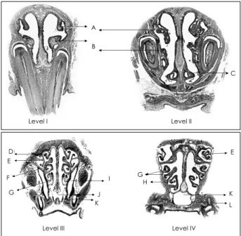
Fig. 1. Four levels of area for coronal section. Level I:from the snout to just posterior to the incisor, Leve II:from just posterior to the incisor to the papilla incisiva, Level III:from the papilla incisiva to just anterior to the first molar, Level IV:from just anterior to the first molar to the rugae palati.
Level I
A B
C
Level II
D E F G
I J K
G
E
H
K L
Level III Level IV
Fig. 2. Cross-sectional anatomy of each level. A:nasal turbinate.
B:maxilloturbinale. C:vomeronasal organ. D:ectoturbinale I. E:ectoturbinale II. F:endoturbinale II. G:endoturbinale III. H:
endoturbinale IV. I:maxillary sinus. J:nasopharyx. K:Pharynx respiratorius. L:nasal associated lympoid tissue.
시켰다. 파라핀에 조직을 고정시킨 후 4~5 μm 두께로 연속 절편을 제작하였고 탈파라핀화 과정을 거친 후 형태학적 관 찰을 위해 H & E 염색을 시행하였다.
광학현미경하에서 각 level의 해부학적 구조, 점막상피의 종류와 분포, 분비선의 종류와 분포, 림프조직과 서비기관 을 관찰하였다.
결 과
각 level에서 관찰되는 해부학적 구조 및 조직학적 특징은, level Ⅰ에서는 비비갑개(nasal turbinate), 상악비갑개(maxillo- turbinale), 누관, 상피는 편평상피, 호흡상피, 후각상피를 볼수 있었다. Level Ⅱ에서는 비비갑개, 상악비갑개, 서비기관, 편평
상피, 호흡상피, 후각상피를 볼 수 있었다. Level Ⅲ는 ectotur- binale Ⅰ, Ⅱ, endoturbinale Ⅱ, Ⅲ, 상악동, nasal-associa- ted lymphoid tissue(NALT), 상악동선(maxillary sinus gland), 외측비선(Steno ’ s gland), 호흡상피, 후각 상피를 볼 수 있었다.
Level Ⅳ는 ectoturbinale Ⅱ, endoturbinale Ⅲ, Ⅳ, 호흡인두 (pharynx respiratorius), NALT, 호흡상피, 그리고 후각상피 를 볼 수 있었다(Table 1) (Fig. 2).
Level Ⅰ에서의 상피분포는 편평상피, 호흡상피, 후각상피 모두 관찰되었다(Fig. 3, 4). 코바닥만 편평상피로 되어 있 었고, 코 천정은 후각상피로 되어 있었으며 나머지는 모두 호흡상피로 되어 있었다. level Ⅱ에서는 서비기관 주위와 코 바닥은 편평상피, 코 천정은 후각상피로 되어 있었고 나머지 는 호흡상피로 되어 있었다. level Ⅲ에서는 편평상피는 관찰
Level I Level II Level III Level IV
Fig. 5. The distribution of the lining epithelia in each level (blue line:stratified squamous epithelium, red line:peudostratified columnar epithelium, green line:olfactory epithelium).
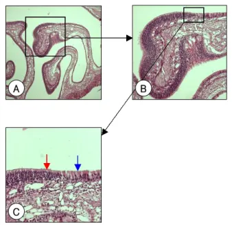
Fig. 3. Light micrographs of the lining epithelium. These show pseudostratified columnar epithelium (blue arrow) and olfac- tory epithelium (red arrow) on the endoturbinale III in the level III (original magnification;A:×40, B:×100, C:×200).
A B
C
A B
C
Fig. 4. Light micrographs of the lining epithelium. These show columnar epithelium (blue arrow) and squamous epithelium (red arrow) in the floor of the level I (original magnification;
A:×40, B:×100, C:×200).
할 수 없었다. 상악동과 코 바닥, 비인두(nasopharynx)로서 중격 열림(septal opening) 부위, endoturbinale Ⅲ의 일부는 호흡상피로 되어 있었고, 나머지는 모두 후각상피였다. level
Ⅳ에서는 호흡인두는 호흡상피로 되어 있었고 endoturbinale
Ⅳ의 일부만이 호흡상피로 되어 있었으며 나머지는 모두 후 각상피로 되어 있었다. 즉 level Ⅰ, Ⅱ에서는 편평상피가 코 바닥에 존재하였으나 Ⅲ와 Ⅳ에서는 존재하지 않았다. 호흡 상피는 level Ⅰ에서 level Ⅳ로 갈수록 점점 줄어들었고, 후각 상피는 level Ⅰ에서 level Ⅳ로 갈수록 점점 늘어났다(Fig. 5).
분비선은 상악동 주위에서 분비선의 군집을 확인 할 수 있 었는데, 상악동선과 외측비선이었다. 특히 level Ⅲ에서 상악 동이 가장 크게 보였고 주위 분비선도 가장 잘 확인 할 수 있 었다(Fig. 6). Level Ⅲ와 level Ⅳ의 아래 부분에서 NALT를 확인할 수 있었고, 특히 level Ⅲ에서 더 잘 확인 할 수 있었다 (Fig. 7). 서비기관은 level Ⅱ에서 확인 할 수 있었다(Fig. 8).
고 찰
이전부터 실험동물에 대한 해부학적 구조에 대한 연구는 지속되어 왔다. Hebel
1)이 흰쥐의 비강구조와 주변구조에 대 하여 해부학적 명칭과 횡단면의 구조를 전체적으로 기술하 였고 Bojsen-Moller
2)는 분비선의 해부학적 지도를 기술하 여 보고 하였다. 하지만 그들의 연구들은 동물에 대한 일반적 인 지식일 뿐 특정한 실험, 즉 비부비동염의 연구에 있어서 필요한 해부학적 및 조직학적인 특징을 기술하지는 못했다.
본 연구에서는 그들이 기술해 놓은 해부조직학적인 지식의 토대위에 실험적 비부비동염 모델에 필요한 해부학 및 조직 학적인 특징들을 자세히 관찰하고 그들의 연구와 비교하였다.
또한 실험적 비부비동염 모델에 있어 연속절편 중 어느 부위 가 비부비동염을 관찰하는데 가장 적합한지도 살펴보았다.
문헌 보고에 의하면, 흰쥐의 코의 상피는 세 가지 종류가 관찰된다.
1)2)5)편평상피, 호흡상피, 후각상피가 그것이다. 편 평상피는 중층편평상피세포로 되어 있으며, 호흡상피세포는 위중층섬모원주상피세포로 되어 있고, 기저세포(basal cell), 배상세포, 섬모세포가 있다. 후각상피는 후각신경세포, 지지 세포, 기저세포로 구성되어 있다. 후각신경세포는 방추모양 이고 구형의 핵을 가지고 있다. 양극성의 수지상돌기가 위 중층섬모원주의 후각상피까지 뻗어있다. 지지세포는 키가 크 고 원통모양이며 기저세포는 작고 원추형이며 기저막의 위 에 놓여 있다. 점막아래의 고유층에 성긴 결체조직이 있고 보우만선(Bowman ’ s gland)이 존재한다. 보우만선은 좁은
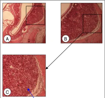
A B
C
Fig. 6. Light micrographs of the gland. These show Steno’s gland including mucinous gland (blue arrow) in the level III (original magnification;A:×40, B:×100, C:×200).
A B
Fig. 7. Light micrographs of lymphoid tissue. These show nasal- associated lymphoid tissue (NALT) in the level IV (original magnification;A:×40, B:×200).
A B
C
Fig. 8. Light micrographs of the special organ. These show vo- meronasal organ in the level II (original magnification;A:×40, B:×100, C:×200).
관을 통해 점막 표면으로 분비하는 관소포(tubuloalveolar) 선이고 장액성선이다. 여기서 호흡상피와 후각상피의 차이점 은 후각상피는 키가 크고 배상 세포가 없고, 기저층이 구별 되지 않고, 보우만선이 있다는 점이다.
1)Hebel
1)은 흰쥐 코 의 해부학적 단면도에서 앞쪽은 후각상피가 존재하지 않고 중층편평상피세포와 호흡상피로만 되어 있다고 기술하였으 나, 본 연구에서는 코의 앞쪽인 level Ⅰ에서 코의 천정부위 서에 후각상피를 볼 수 있었다. level Ⅱ, level Ⅲ, level Ⅳ의 점막상피는 level Ⅱ의 아래쪽은 편평상피 위쪽은 후각상피, 나머지는 호흡상피였다. level Ⅲ, Ⅳ에서는 아래쪽은 호흡상 피, 위쪽은 후각상피였다. 이것은 Hebel이 기술한 것과 비 슷하였다. 또한 전체적으로 호흡상피는 level Ⅰ에서 level Ⅳ 로 갈수록 점점 줄어들었고, 후각상피는 level Ⅰ에서 level Ⅳ 로 갈수록 점점 늘어났다.
흰쥐의 주요 분비선은 주로 상악동 주위에 분포한다. 상 악동의 내강은 전체가 위중층섬모원주상피세포로 되어 있고 점막아래 두꺼운 분비층으로 되어 있다. 상악동 주위의 분 비선은 상악동선(maxillary sinus gland)과 외측비선(lateral nasal gland, Steno ’ s gland)으로 되어 있는데, 내측과 위 쪽은 상악동선이고 아래쪽과 바깥쪽의 분비선의 군집이 외측 비선이다. Bojen-Moller
2)는 상악동 주위의 분비선에 점액성 과 장액성 분비선의 혼합형은 존재하지 않는다고 기술하였 다. 그러나 Vidic
7)은 외측분비선은 점액선이고 상악동선은 장 액선이라고 하였다. 본 연구에서는 상악동 주위에서 분비선의 군집을 확인 할 수 있었다. 특히 level Ⅲ에서 상악동이 가 장 크게 보이고 주위 분비선도 가장 잘 확인 할 수 있었다.
서비기관은 쌍을 이루는 상피성 관으로 비전정에서 앞니관까 지 약 10 mm 퍼져있고 관의 단면은 반달모양으로 내측에는 후 각상피세포가 외측에는 호흡상피세포가 있다.
8)9)본 연구에서는 level Ⅰ, Ⅱ에서 관찰되었으나 level Ⅱ에서 가장 잘 관찰되었다.
NALT는 비중격이 열려있는 비인두에서부터 호흡인두에 걸쳐서 양쪽 내측면에 존재하고 T와 B세포를 포함하는 여포 로 구성되어 있고, 상피는 구형 핵을 가진 섬모 입방세포로 되어 있으며 배상세포는 드물다.
10)11)본 연구에서는 level Ⅲ 와 Ⅳ에서 가장 잘 관찰 되었다. NALT의 크기의 변화, 여포 수의 변화, 림프구의 수 변화를 관찰하는 것이 비부비동염 모델에 큰 도움이 될것으로 생각된다.
위에서 흰쥐 비부비강의 각 부위에 따른 점막상피, 분비 선, 림프조직 및 서비기관의 분포를 과거 문헌과 비교하며 관찰하였다. 비부비동염을 관찰하는데 있어서는 균의 감염정 도가 중요하겠지만, 상피의 분포와 상피의 파괴 정도, 염증 세포의 침윤정도, 분비선의 분포정도를 관찰하는 것이 비부 비동염의 발생기전 및 병태 생리학적인 변화를 이해하는데 필수적일 것이다. 이러한 변화를 잘 관찰하려면 호흡상피의
분포가 명확하고 분비선이 잘보이며 림프조직이 가장 잘 관 찰 되는 곳이 흰쥐의 비부비동염 모델 실험에 가장 적합한 부위가 될 것이다. 따라서 각 절단부위 중에서는 호흡상피와 후각상피가 뚜렸하며 상악동, 상악동선과 외측비선을 볼 수 있고 NALT와 같은 림프조직을 볼 수 있는 level Ⅲ가 흰쥐 비부비동염 실험 연구에 가장 도움이 될 것으로 생각된다.
결 론
흰쥐 비부비강의 축상면 연속절편을 얻어 횡단면 구조를 관찰하고, 그 조직학적인 특성으로, 점막상피, 분비선, 림프 조직 및 서비기관의 분포를 살펴보았다. 각 절단부위 중에 서는 사골동과 상악동을 가장 잘 볼 수 있고 호흡상피와 후 각상피가 뚜렷하며 분비선의 분포가 가장 많고 NALT와 같 은 림프조직을 볼 수 있는 level Ⅲ가 흰쥐 실험동물 연구에 가장 도움이 될 것으로 생각된다. 이러한 결과는 향후 흰쥐 를 이용한 부비동염의 발생기전 및 병태 생리학적인 변화 연 구에 기초 자료가 될 것으로 생각한다.
중심 단어:흰쥐·코·조직학.
REFERENCES
1) Hebel R, Stromberg MW. Anatomy of the laboratory rat. Waverly press;1976. p.55-61.
2) Bojsen-Moller F. Topography of the nasal glands in rats and some other mammals. Anat Rec 1964;150:11-24.
3) Kim HS, Jeon SY, Ahn SK, Kim JP, Park, JJ, Jung JH, et al. A rat model of acute bacterial rhinosinusitis induced by Staphylococcus.
Korean J Otolaryngol 2005;48:735-40.
4) Jeon SY, Kim JP, Kim EA, Ahn SK, Kim BG. Rat model of platelet- activating factor-induced rhinosinusitis. Ann Oto Rhinol Laryngol 2005;114:393-8.
5) Bang BG, Bang FB. A comparative study of the vertebrate nasal chamber in relation to upper respiratory infection; I comparative morphology of the nasal chambers of the three commonly used la- boratory animals: chiken, rat and ferret. Bull. Johns Hopkins Hosp 1959;104:107-25.
6) Bojsen-Moller F. Demonstration of terminalis, olfactory, trigerminal and perivascular nervers in the rat nasal septum. J Comp Neurol 1975;159:245-56.
7) Vidic B, Greditzer HG. The histochemical and microscopical differ- entiation of the respiratory glands around the maxilary sinus of the rat. Am J Ant 1971;132:491-513.
8) Adams DR. Fine structure of the vomeronasal and septal olfactory epithelia and of glandular structure. Microsc Res Tech 1992;23:86-97.
9) Doving KB, Trotier D. Structure and function of the vomeronasal organ. J Exp Bio 1998;201:2913-25.
10) Spit BJ, Hendriksen EGJ, Bruijntjes JP, Kuper CF. Nasal lymphoid tissue in the rat. Cell Tissue Reserch 1989;255:193-8.
11) Kuper CF, Koornstra PJ, Hameleers DMH, Biewenga J. Spit BJ, Adrian MD, et al. The role of nasopharyngeal lymphoid tissue.
Immunology Today 1992;13:219-24.