- 42 -
Endoscopic Orbital Decompression for Dysthyroid Orbitopathy
Seok-Joo Koh, M.D. and Hun-Jong Dhong, M.D.
ABSTRACT
Since 1957, when Walsh and Ogura introduced transantral orbital decompression, various surgical approaches have been int- roduced for the treatment of dysthyroid orbitopathy. With the development of endoscopic sinus surgery, orbital decompression can now be approached transnasally by endoscope alone. We reviewed the medical records of 10 patients who had received endoscopic orbital decompression. Of the 10 patients, four eyes from three patients were managed for decreased visual acuity, while 13 eyes from seven patients were managed for exophthalmos. Three patients who had initially complained of decreased vision demonstrated eventual improvement. An initial mean proptosis of 19.3 mm decreased to 16.5 mm. Six eyes with abnor- mal color vision were resolved after the decompression. Four patients with diplopia complained of continued diplopia after the decompression and were managed with extraocular muscle surgery or prism glasses. Compared to the conventional transantral approach, endoscopic orbital decompression features less morbidity and comparable ophthalmic results.
KEY WORDS:Endoscopic orbital decompression·Dysthyroid orbitopathy.
INTRODUCTION
Graves’ disease most often accompanies orbital sy- mptoms occurring bilaterally. The disease occurs frequ- ently in people in their 20s and 30s and more often in women than in men. The exophthalmos associated with Graves’ disease is reportedly due to an autoimmune pr- ocess and bears little relation to the degree of the thyroid abnormalities. Therefore, even when the hyperthyroidism is treated, the orbital symptom is not eliminated, which makes treatment difficult.
1)Only 5% of exophthalmos patients require medical treatment for decreased vision,
2)which is caused by optic nerve compression or exposure keratitis.
Surgical and nonsurgical therapies have been used to prevent visual loss. Among them, the orbital decompre- ssion approach, an operative method, has been widely used since its introduction by Walsh and Ogura in 1957.
3)They performed a two-wall inferior and medial decom- pression through a Caldwell-Luc maxillary antrostomy.
Another method, the transorbital approach introduced by Anderson et al.,
4)has not been widely used. More rece- ntly Trokel et al.,
5)introduced the orbital fat removal through a subciliary incision. With the advent of intr- anasal endoscope, Kennedy et al., introduced transnasal approach for medial and inferior orbital wall decompr- ession, avoiding Caldwell-Luc approach.
6)For the last three years, we have been applying, thr- ough nasal endoscopy, the orbital decompression appr- oach on patients with exophthalmos caused by thyroid gland dysfunction. This report is our analysis of the me- dical records and includes the procedure and results of the surgery.
MATERIALS AND METHODS Subjects
The subject pool comprised 17 eyes of 10 patients with thyroid gland dysfunction, which was treated with endoscopic orbital decompression in the otorhinolary- ngology department of Samsung Medical Center from January 1995 to October 1998. Medical records were an- alyzed in the retrospective method. The patients consisted of three males and seven females, with the patients’ ages ranging from 27 to 74 years (average age of 43.3). The chief complaints of the patients consisted of decreased vision (four eyes in three patients) and exophthalmos
Department of Otorhinolaryngology, Sungkyunkwan UniversitySchool of Medicine, Samsung Medical Center, Seoul, Korea Address correspondence and reprint requests to Hun-Jong Dh- ong, M.D., Department of Otorhinolaryngology, Sungkyunkwan University School of Medicine, Samsung Medical Center, 50 Il-wondong, Kangnamku, Seoul 135-710, Korea
Tel:82-2-3410-3579, Fax:82-2-3410-3879 E-mail:[email protected] Accepted for publication on February 19, 1999
(13 eyes in seven patients). One patient had exposure keratitis in both eyes before surgery. Color vision abn- ormality was found in six eyes, and extraocular muscle abnormality was found in nine eyes. At the time of sur- gery, eight patients were euthyroid while two were hy- pothyroid.
Eyesight before and after surgery was evaluated rel- ative to corrected visual acuity, and the degree of exo- phthalmos was measured in millimeters with a Hetel exophthalmometer. Color vision was tested with an Is- hihara color vision test. Before treatment, the degree of exophthalmos in the 17 eyes ranged from 13 mm to 24 mm and averaged 19.0 mm. The mean visual acuity ra- nged from 0.03 to 1.5 with a mean of 0.69.
Surgical technique
Seven patients underwent surgery on both eyes on the same day while three patients were treated with orbital decompression on one eye only. Under general anesth- esia, the patient is placed in a supine position with the head tilted up approximately 30 degrees. The mucosa in the nasal cavity is packed with a cotton pledget conta- ining lidocaine and epinephrine, and an infiltration ane- sthesia mixed with 1% lidocaine and 1:100,000 epi- nephrine is applied to the same area as of endoscopic sinus surgery. Following the removal of the uncinate pr- ocess, a complete ethmoidectomy is conducted. The fovea ethmoidalis and the lamina papyracea are exposed in the upward and lateral direction, respectively. The maxillary ostium should be generously enlarged to provide optimal exposure of the orbital floor. Bone is removed in a pos- terior direction to the level of the back wall of the sinus.
Anterior removal stops at the thick bone of the frontal process of the maxilla, which protects the nasolacrimal duct. The ostium is enlarged superiorly to the level of the orbital floor and inferiorly to the root of the inferior tu- rbinate. The skeletonized lamina papyracea is gently pe- netrated with a small spoon curette. The bony fragments of the lamina papyracea are lifted in a medial direction.
Bone of the lamina papyracea is removed in a superior direction to the level of the ethmoid roof. As dissection continues in a posterior direction toward orbital apex, the annulus of Zinn is encountered, representing the poste- rior limit of dissection. The infraorbital nerve is identi- fied laterally within the maxillary sinus and defines the lateral limit for removal of the orbital floor. Once the periorbita is fully exposed, multiple full thickness par-
allel linear incisions are made with a sickle knife in a postero-anterior direction. This is performed at the lat- eral side of periorbita initially so that prolapse of orbital fat does not obscure the surgeon’s subsequent view. Care must be taken to keep the tip of the blade superficial so as not to injure the underlying orbital contents.
RESULTS
After the operation, 15 eyes measured with an exop- hthalmometer produced readings ranging from 9 mm to 19 mm and averaging 16.5 mm. The degree of exopht- halmos improved in 13 eyes, but two eyes showed no improvement. In the 15 eyes that could be measured post-operatively, the mean of proptosis checked before the operation was 19.3 mm. The reduction of proptosis was statistically significant (p<0.05) according to the paired t test of 15 eyes.
All six eyes with abnormal color vision showed im- provement after surgery. The four eyes treated for decr- eased vision also showed significant improvement, from an average of 0.15 before surgery to an average of 0.78 after surgery.
Acute sinusitis occurred in two patients after surgery.
In patient 3, acute maxillary sinusitis occurred on the left side two months after surgery. With two days of intrav- enous antibiotics producing no signs of improvement, an inferior meatal antrostomy was conducted. This proce- dure also was unsuccessful in eliminating the condition, so a Caldwell-Luc operation was performed after seven days. In patient 10, sinusitis occurred in the frontal sinus four months after surgery and was successfully treated with antibiotic under hospitalization.
In Patient 6, who underwent a septoplasty at the same time as the orbital decompression, a nasal septal abscess occurred 40 days after surgery. The abscess was treated with incision and drainage.
Six patients complained diplopia before surgery. After surgery, the condition disappeared in two cases but re- mained in four. Among them, one patient indicated that the condition had actually become more intense after su- rgery. To treat the diplopia, the ophthalmology department prescribed prism glasses or Min’s glasses, or performed a medial rectus recession on both eyes.
Patient 1 suffered from progressively declining visual
acuity in the right eye after surgery, so irradiation was
applied from December 1995 to January 1996. Visual
acuity conducted on August 1996 improved to 1.0 in
both eyes.
In four cases, including three cases that showed dec- reased vision, steroid had been used before or after su- rgery (Table 1).
DISCUSSION
Dysthyroid orbitopathy is an autoimmune disease ch- aracterized by inflammation, edema and secondary fib- rosis.
7)The orbitopathy is caused by hypertrophy of the extraocular muscles resulting from hydrophilic mucop- olysaccharides and immune complexes deposition in the extraocular muscles and retrobulbar fat. The expansion occurring inside the orbit increases intraocular pressure, causing compressive optic neuropathy. If the expansion continues, blindness may result. The increased intraoc- ular pressure can also result in impaired extraocular mu- scles function, which may lead to diplopia or strabismus.
Dysthyroid orbitopathy is most commonly associated with Graves’ disease, although reports also link the co- ndition with Hashimoto’s thyroiditis, thyroid cancer and primary hyperthyroidism.
8)The clinical symptoms of Gr- aves’ disease include exophthalmos, eyelid edema, lid retraction, conjunctival injection, chemosis, exposure ke- ratitis, corneal ulceration, extraocular motility abnorm- alities, optic neuropathy and blindness. In most cases, the symptoms progress very gradually and then disapp- ear, but in 2-7% of cases, the condition develops into serious ophthalmopathy, which requires medical treat- ment.
9)Most patients show improvement after medical treat- ments such as systemic steroid, local eye care and anti- thyroid drugs. A high dose steroid can be administered
in cases of acute congestive orbitopathy but is difficult to use independently. Steroid treatment also requires long- term period and when the dosage is stopped, the symp- toms reappear. Therefore, the administration of steroid is used only as a temporary treatment of optic neurop- athy.
External beam radiation is applied as a dose of 20 Gy (2000 rad) divided into ten sessions. This method is ef- fective in the initial, acute stages, demonstrating exce- llent performance in the treatment of inflammation in soft tissues, but once the tissue has developed into fibr- osis, the method is no longer productive.
10)The orbital decompression can be performed to pre- vent blindness in cases where the treatment of compr- essive optic neuropathy with a steroid or external beam radiation has been unsuccessful or contra-indicated.
11)Other indications include exposure keratitis or corneal ulceration due to severe exophthalmos, extraocular mu- scles dysfunction or when the exophthalmos hurts one’s outlook. For these cases, the orbital decompression is indicated to enable blinking of eyelids and improve oc- ular movements.
In the early 20th century, Dollinger attempted an or- bital decompression by decompressing orbital contents into the infratemporal fossa through the lateral orbitot- omy.
12)Since then, several approaches, including the an- terior cranial fossa approach
13)and the external ethmoi- dectomy approach,
14)have been attempted but the method introduced by Walsh and Ogura in 1957, which is thro- ugh the maxillary sinus and the nasal cavity, is now most widely used.
In 1990, Kennedy et al., conducted the first orbital decompression incorporating the use of an endoscope,
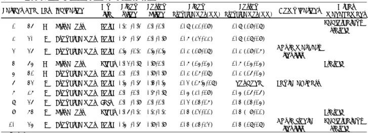
Table 1. Summary of endoscopic orbital decompression Patient Age Sex Indication Op*
site
Preop vision
Postop Vision
Preop exophthalmos (mm)
Postop
exophthalmos (mm) Complications Other management 1 46 M Visual loss Both 0.2 /0.3 1.0/1.0 108 (21/19) 108 (19/18) Radiotherapy,
steroid 2 30 F Exophthalmos Both 0.7 /0.6 1.0/0.9 107 (23/22) 108 (18/19)
3 36 F Exophthalmos Both 1.5 /1.2 1.5/1.5 102 (19/18) 102 (19/17) Acute maxillary sinusitis
4 65 M Visual loss Right 0.03/0.8 0.9/1.0 107 (15/15) 107 (15/15) Steroid 5 41 M Exophthalmos Both 1.0 /1.0 0.9/0.9 102 (17/18) 102 (16/15)
6 43 F Exophthalmos Both 0.5 /0.6 0.4/0.5 103 (17.5/18) not done Septal abscess 7 27 F Exophthalmos Both 1.0 /1.0 0.7/0.8 105 (20/19) 105 (16/16)
8 36 F Exophthalmos Left 1.0 /0.9 1.0/1.0 103 (14/17) 104 (14/15)
9 74 F Visual loss Right 0.06/0.2 0.2/0.2 104 (13/11) 104 ( 9/11) Steroid 10 35 F Exophthalmos Both 0.5 /0.6 0.9/0.9 104 (24/23) 104 (18/18) Acute frontal
sinusitis
Radiotherapy, steroid
*Operation
which allowed access to the nasal cavity and removal of some parts of the medial and inferior walls of the or- bit. They reported that in five orbits, the exophthalmos decreased by 4.7 mm. Unlike other transantral or exte- rnal approaches, endoscopic orbital decompression ma- kes sublabial and skin incisions unnecessary, thus eli- minating the possibility of such complications as facial edema, scarring, infraorbital nerve injury or tooth injury.
The merits of this method also include a bright and wide view to the posterior orbit, which allows sufficient dec- ompression in the medial orbital wall up to the orbital apex. However, the technique does not yield sufficient exposure of the orbit's anterior and lateral wall so a lat- eral orbitotomy may be required.
Despite the fact that decompression of the orbital fl- oor by the endoscopic route compared to the transantral route is limited, the reduction of proptosis by the endo- scopic approach is comparable to the transantral route.
This is probably because the limited orbital floor deco- mpression is offset by a good decompression of the me- dial wall.
15)Neugebauer et al., reported that the degree of exoph- thalmos reduced by an average of 3 mm after endoscopic orbital decompression on 21 patients.
16)In a report by Metson et al., 22 orbits that underwent endoscopic orb- ital decompression showed an average 3.2 mm decrease in exophthalmos and an average 5.6 mm decrease when the treatment was accompanied by lateral decompression conducted through the external approach.
15)Koay et al., reported an average 3.9 mm decrease in exophthalmos in 30 orbits that underwent endoscopic orbital decomp- ression.
17)In Korea, Cho et al.,
18)reported an average 3.5 mm decrease in exophthalmos after conducting both the transantral and endoscopic approaches in ten orbits.
Lee et al.,
19)reported an average 2.8 mm decrease in 14 orbits after orbit decompression using endoscope only.
The results of our study are similar to these findings:
we obtained an average 2.8 mm decrease using endos- cope only. Despite sufficient orbital decompression, ac- hieved by removing much of the bony lamina, the de- creases in proptosis were different than those reported by the authors from other country. The discrepancy may be attributable to a difference in the size of the nasal cavity and maxillary sinus through which the orbital co- ntents prolapsed. In cases of decreased vision, another possible reason is the smaller volume of orbital fat due to severe hypertrophy of the extraocular muscles. In the two eyes where the exophthalmos was unsuccessful
in this study, one patient underwent surgery due to dec- reased vision and the other had a very narrow maxillary sinus and nasal cavity.
But it is also true that there are some limitations when decompressing the orbital floor frontal and lateral to the infraorbital nerve with an endoscope only. Accordingly, Graham et al.,
20)maintain that sufficient decompression of the medial and inferior walls is possible by combining the endoscopic approach and the subciliary approach. The subciliary approach, however, carries a higher chance of damaging the infraorbital nerve when compared to the endoscopic approach.
Contra-indications of endoscopic decompression are cases involving inflammatory disease in the paranasal sinus, underdevelopment of the ethmoid and maxillary sinuses, bony thickening in the orbital wall and severe nasal septal deviation. When a bony thickening exists in the medial orbital wall, access becomes difficult. In these situations, access via conjunctiva provides a wide view of the inferior orbital wall and is relatively safe, since removal of the orbital wall is in the direction running from the orbit to the nasal cavity.
The complications of endoscopic orbital decompres- sion are similar to those of endoscopic sinus surgery. Of these, sinusitis caused by the prolapsed orbital contents is particularly noteworthy.
21)During medial wall deco- mpression, it is important that the bony support of the frontal recess be maintained. Preservation of the bony support prevents postoperative obstruction of the frontal sinus due to the escaped orbital fat. As well, the middle meatal antrostomy should be made as wide as possible.
In our study, two patients demonstrated an occurrence
of acute sinusitis after decompression:one in the ma-
xillary sinus and the other in the frontal sinus. The ma-
xillary sinus and ethmoid of the patient with maxillary
sinusitis were found to be very small, raising the poss-
ibility of obstruction of the sinus. Another patient had
undergone a septoplasty at the same time as the orbital
decompression due to a nasal septal deviation, and exh-
ibited an occurrence of a nasal septal abscess one month
after surgery. The nasal packing after the operation was
made loose as it exerted compression to the orbit, and
this may have allowed a dead space between the muco-
perichondrial flap and the septal cartilage. Accordingly,
when a nasal septal deviation is severe, making endos-
copic orbital decompression difficult, it is recommended
that a septoplasty be conducted first and the endoscopic
orbital decompression be performed two or three weeks
afterwards.
CONCLUSION
For operators accustomed to using endoscopes, the en- doscopic approach to orbital decompression is considered to be an alternative to the traditional transantral and su- bciliary approaches employed in ophthalmology.
REFERENCES
1) Jacobson DII, Gorman CA. Diagnosis and management of end- ocrine ophthalmopathy. Med Clin North Am 1985;69:73-87.
2) Hole JF, Holt GR. Surgery for exophthalmos. In: Bailey BJ editor:
Head Neck Surgery-Otolaryngology. 1st ed. Philadelphia: Lippin- cott;1993. p.2055-69.
3) Walsh TE, Ogura JH. Transantral orbital decompression for ma- lignant exophthalmos. Laryngoscope 1957;67:544-9.
4) Anderson RL, Linberg JV. Transorbital approach to decompression in Graves’ disease. Arch Ophthalmol 1981;99:120-4.
5) Trokel S, Kazim M, Moore S. Orbital fat removal: Decompression for Graves’ orbitopathy. Ophthalmology 1993;100:674-82.
6) Kennedy DW, Goodstein ML, Miller NR, Zinreich SJ. Endosco- pic transnasal orbital decompression. Arch Otolaryngol 1990;116:
275-82.
7) Weisman RA, Savino PJ. Management of endocrine orbitopathy.
Otolaryngol Clin North Am 1988;21:93-102.
8) Fries PD. Thyroid dysfunction: Managing the ocular complications of Graves’ disease. Geriatrics 1992;47:58-65.
9) Warren JD, Spector JG, Burde R. Long-term follow-up and recent observations on 305 cases of orbital decompression for dysthyroid orbitopathy. Laryngoscope 1989;99:35-40.
10) Kriss JR, Peterson IA, Donaldson SS, McDougall IR. Supervoltage orbital radiotherapy for progressive Graves’ ophthalmopathy: Re- sult of a 20-year experience. Acta Endocrinologica 1989;121 (Su- ppl):154-9.
11) Shorr N, Seiff SR. The four stages of surgical rehabilitation of the patient with dysthyroid ophthalmopathy. Ophthalmology 1986;93:
476-83.
12) Dollinger J. Die Drickentlastlung der Augenhokle durch Entfur- nung der aussern Orbitalwand bei hochgradiegen Exophthalmos und Koneskutwer Hornhauterkronkung. Dtsch Med Wochenschr 1911;4:149-63.
13) Naffziger HC, Jones OW. The surgical treatment of progressive ex- ophthalmos following thyroidectomy. J Am Med Assoc 1932;99:
638-42.
14) Sewall EC. Operative control of progressive exophthalmos. Arch Otolaryngol 1936;24:621-4.
15) Metson R, Dallow RL, Shore JW. Endoscopic orbital decompres- sion. Laryngoscope 1994;104:950-7.
16) Neugebauer A, Nishino K, Neugebauer P, Konen W, Michel O.
Effects of bilateral orbital decompression by an endoscopic endo- nasal approach in dysthyroid orbitopathy. Br J Ophthalmol 1996;
80:58-62.
17) Koay B, Bates G, Elston J. Endoscopic orbital decompression for dysthyroid eye disease. J Laryngol Otol 1997;111:946-9.
18) Cho JS, Kim YD, Byun JY, et al. Orbital decompression in dysth- yroid ophthalmopathy using combined transantral and transnasal endoscopic approach. Kor J Otolaryngol 1998;41:750-4.
19) Lee CH, Kang BS, Oh SJ, et al. Orbital decompression for dysth- yroid orbitopathy. Kor J Otolaryngol 1998;41:1557-61.
20) Graham SM, Carter KD. Combined endoscopic and subciliary or- bital decompression for thyroid-related compressive optic neuro- pathy. Rhinology 1997;35:103-7.
21) Bough ID, Huang JJ, Pribitkin EA. Orbital decompression for Gr- aves’ disease complicated by sinusitis. Annal Otol Rhinol Laryn- gol 1994;103:988-90.