저작자표시-비영리-변경금지 2.0 대한민국 이용자는 아래의 조건을 따르는 경우에 한하여 자유롭게
l 이 저작물을 복제, 배포, 전송, 전시, 공연 및 방송할 수 있습니다. 다음과 같은 조건을 따라야 합니다:
l 귀하는, 이 저작물의 재이용이나 배포의 경우, 이 저작물에 적용된 이용허락조건 을 명확하게 나타내어야 합니다.
l 저작권자로부터 별도의 허가를 받으면 이러한 조건들은 적용되지 않습니다.
저작권법에 따른 이용자의 권리는 위의 내용에 의하여 영향을 받지 않습니다. 이것은 이용허락규약(Legal Code)을 이해하기 쉽게 요약한 것입니다.
Disclaimer
저작자표시. 귀하는 원저작자를 표시하여야 합니다.
비영리. 귀하는 이 저작물을 영리 목적으로 이용할 수 없습니다.
변경금지. 귀하는 이 저작물을 개작, 변형 또는 가공할 수 없습니다.
이학석사학위논문
애기장대에서 DEMETER와 상호작용하는 인자의 선별
Screening for Interaction Partners of DEMETER in Arabidopsis thaliana
2019년 8월
서울대학교 대학원 생명과학부
김 효 진
i
ABSTRACT
Screening for Interaction Partners of DEMETER in Arabidopsis thaliana
Hyojin Gim School of biological science The Graduate School Seoul National University
DNA methylation and demethylation plays significant roles in regulating gene expression on variable stress reactions and TEs silencing. A DEMETER (DME), an active DNA demethylase in plants, is expressed in the central cell of the female gametophyte in Arabidopsis required for seed development and has glycosylase activity that can actively remove 5-mC that is replaced by cytosine via base excision pathway. DME induces maternal allele expression of the imprinted MEDEA (MEA) polycomb gene and the DNA glycosylase activity of DME leads to the DNA demethylation on its targets. In plants, there is no massive methylation reprogramming in embryo like mammal but instead, the companion cells whose DNA contents are not inherited to the next generation go through global demethylation and the hypomethylated state is maintained in late endosperm stage in which DME is no longer expressed.
ii
Despite all these distinct significances, little is known about the interactors that associate with DME. Here, to identify the interacting partners of DME, I used Bimolecular Fluorescence Complementation (BiFC). From 83 genes that have been confirmed by the Yeast Two-Hybrid system that interact with DME, I primarily chose 18 candidates to test interaction. While examining these 18 candidates by BiFC, AT5G37930, AT5G60980, AT1G70620, AT5G23090 and the C terminal part of AT1G20960 showed fluorescent signals when it was co-transfected with DME in Arabidopsis Col-0 protoplasts. These interactors contain either E3-ubiquitin ligase activity, RNA binding motif, homologous feature of transcription factor which is related to histone acetylation, or are related to RNA splicing.
To further understand its relation with DME in plants one candidate, At1g20960 that is related to RNA splicing, was chosen and its mutant was crossed with dme mutant for phenotyping analyses. dme mutant allele transmission, segregation and seed abortion ratio shown in dme single mutant were not changed in double mutants. Therefore, by doing further experiments, using different candidates, this study would give some specific and clear perspectives and contribute to widen the knowledge by identifying a novel protein involved in the DME demethylation pathway in plants.
Keywords : DEMETER(DME), DNA demethylation pathway, Arabidopsis, Epigenetics, BiFC
Student Number : 2017-20959
iii
TABLE OF CONTENTS
ABSTRACT ... ⅰ TABLES OF CONTENTS ... ⅲ LIST OF FIGURES ... ⅴ LIST OF TABLES ... ⅵ
BACKGROUNDS ... 1
1. DNA Methylation in Arabidopsis ... 1
2. DNA demethylation and DEMETER ... 4
3. Purpose of this Study ... 7
CHAPTER ONE: Interaction Partner Screening for DEMETER using Bimolecular Fluorescence Complementation(BiFC) Assay ... 8
1. Introduction ... 9
2. Materials and methods ... 12
2.1. Plant material and growth conditions ... 12
2.2. Previous yeast two-hybrid data ... 12
2.3. List-up for BiFC assay ... 15
2.4. Cloning for BiFC assay ... 15
2.5. BiFC assay ... 21
3. Results and discussion ... 23
3.1. Cloning for BiFC assay ... 23
3.2. 5 genes were identified as interactors of DME through BiFC ... 25
iv
CHAPTER TWO: Phenotypic Study for the DME interactors ... 31
1. Introduction ... 32
2. Materials and methods ... 35
2.1. Plant material and growth conditions ... 35
2.2. Seed-set analysis and whole-mount clearing ... 35
3. Results and discussion ... 38
3.1. EMB/emb1507-1 seed phenotype ... 38
3.2. EMB/emb1507-1;DME/dme-2 double heterozygote mutant seed phenotype ... 38
3.3. Transmission of emb and dme alleles in EMB/emb1507-1;DME/dme-2 mutant plants ... 41
CONCLUSION ... 43
REFERENCE ... 46
ABSTRACT IN KOREAN ... 52
v
LIST OF FIGURES
Figure 1. Schematized active CG demethylation by DNA glycosylase…...………6
Figure 2. Schematic description of BiFC assay in this study.………...………...11
Figure 3. Map of pSAT4-nEYFP C1 vector………..………...17
Figure 4. Map of pSAT4-cEYFP C1 B vector………...………..…..18
Figure 5. Schematic description of DME and AT1G20960 CDS used for BiFC assay…..……….24
Figure 6. Confocal laser scanning microscopy of BiFC assay shows that DME physically interacts with AT1G20960 C terminal……….………26
Figure 7. Confocal laser scanning microscopy of BiFC assay shows that DME physically interacts with AT5G37930, AT5G60980, AT1G70620 and AT5G23090...29
Figure 8. DME and AT5G37930 interaction varied whether the E3 ubiquitin ligase activity of AT5G37930(E3 ubiquitin-protein ligase SINA-like 10) is active or not……...………....30
Figure 9. Gametogenesis and development in Arabidopsis……….…………..34
Figure 10. EMB/enb1507-1 seed abortion phenotypes………..…39
Figure 11. EMB/emb1507-1;DME/dme-2 seed abortion phenotypes……….…………...40
Figure 12. Seed viability and allele transmission ratio of EMB/emb1507-1;DME/dme-2……...42
vi
LIST OF TABLES
Table 1. List of genes showed physical interaction with the N terminal fragment of
DME(AT5G04560.1) confirmed with yeast two-hybrid screnning………....13
Table 2. List of 18 candidate genes for further analysis using BiFC assay……...16
Table 3. List of primers used for PCR amplification in constructing BiFC clones………...19
Table 4. List of the information used to construct for BiFC clones....………….………...20
Table 5. T-DNA insertion alleles and genotyping primers used in this study……….……37
1
BACKGROUND 1. DNA methylation in Arabidopsis
Epigenetics is a study of heritable but reversible phenotypic changes in organisms caused by modification of gene expression rather than alteration of the genetic code itself and its inheritance.
DNA methylation is a major research topic to understand epigenetic phenomena along with histone modifications. DNA methylation is one of the most well-conserved epigenetic makers and has significance in development and stress reaction of plant and animal (Reik et al., 2001; Law and Jacobsen, 2010; Wu and Zhang, 2010; Zhang and Zhu, 2012). Unlike many other popular model organisms, Arabidopsis has retained a multi-layered methylation system that contributes to gene and transposon silencing, imprinting, and genome stability and many of the findings are applicable to other eukaryotes (Gehring and Henikoff, 2008). Cytosine can be methylated at the carbon five position, and in plants this can occur on any cytosine regardless of the sequence context (CG; CHG; CHH) (Gehring and Henikoff, 2008). In general, 5-methylcytosine (5mC) is associated with transcriptional silencing.
The Arabidopsis genome contains methylation at 24% of CG sites, 6.7% of CHG and 1.7% of CHH (Cokus et al., 2008) and DNA methylation tends to prefer being located at repetitive DNA sequences. The tendency of methylation prefering repetitive DNA sequences suggests that one of methylation’s primary functions is to silence the transcription of transposable elements(TEs) (Zilberman and Henikoff, 2004; Gehring and Henikoff, 2007). In Arabidopsis, transposons are generally methylated throughout their length at cytosines in all sequence contexts, although distinct patterns do emerge at individual loci (Lippman et al., 2003; Lippman et al., 2004; Zhang et al., 2006; Zilberman et al., 2007). TEs invade genomes and increase in copy number, with strong potential for damaging the host. All organisms have adopted mechanisms to keep TEs silent, including RNA-based chromatin silencing, histone modifications, DNA methylation, or a combination thereof (Slotkin and Martienssen, 2007).
2
DNA methyltransferase is an enzyme that donates methyl group to the carbon 5 position of cytosine and all known cytosine 5-methyltransferases belong to a single family with several subfamilies (Gehring and Henikoff, 2008). There are three subfamilies of DNA methyltransferases in Arabidopsis: CG maintenance methyltransferases that have a role in maintaining 5-mC of CG methylation context, chromomethylases that involved in maintaining 5mC of CHG context, and the de novo methyltransferases that have to construct a new 5mC in CHH context since there is no complementary 5-mC to be used as a template for maintaining the methylation. Multiple genes exist for each enzyme class, but only one enzyme of each type appears to be active; the other genes are either expressed at low levels or contain stop codons in various backgrounds, and none have been recovered in mutant screens (Gehring and Henikoff, 2008).
METHYLTRANSFERASE1 (MET1) is the CG maintenance methyltransferase in Arabidopsis.
This designation is based on sequence similarity to Dnmt1, the orthologous mammalian maintenance methyltransferase, and on the effect mutations in the gene have on DNA methylation (Finnegan and Kovac, 2000). More than half of the regions that are methylated in wild type are lost in met1. At some repetitive sequences significant amounts of CHG and CHH methylation are also lost (Gehring and Henikoff, 2008). Many TEs become transcriptionally active in a met1 mutant (Kato et al., 2003; Lippman et al., 2003; Zhang et al., 2006; Zilberman et al., 2007).
Mutations in MET1 and antisense-directed MET1 silencing cause various phenotypes from gametogenesis onwards since MET1 is essential to maintain methylation patterns during the haploid gametophyte stage of the plant life cycle (Saze et al., 2003). In the sporophyte, met1 phenotypes include abnormal embryo patterning, narrow leaves, homeotic transformations of floral organs, altered flowering time, and reduced fertility (Finnegan et al., 1996; Kankel et al., 2003; Saze et al., 2003; Xiao et al., 2006; Mathieu et al., 2007). MET1 might also have de novo methyltransferase activity since de novo methylation of CG sites is impaired in met1 mutants (Aufsatz et al., 2004).
CMT3 (CHROMOMETHYLASE3) is another methyltransferase unique to plants containing a
3
chromodomain which makes it possible to bind to methylated lysines in histone tails (Henikoff and Comai, 1998). CMT3 maintains methylation in the CHG sequence context. cmt3 single mutants do not have any morphological phenotypes, but shows severe defects when combined with a null met1 allele (Xiao et al., 2006; Zhang and Jacobsen, 2006). The additional loss of CHG methylation in met1 cmt3 mutants might push the genome over a methylation threshold such that the remaining methylation, in whatever sequence context, is insufficient to accomplish its functions (Gehring and Henikoff, 2008).
DRMs (DOMAINS REARRANGED METHYLTRANSFERASES) were identified as de novo methyltransferases based on homology to the mammalian de novo methyltransferases Dnmt3a and Dnmt3b (Cao and Jacobsen, 2002). DRM2 appears to be the only functional enzyme in Arabidopsis. CHH methylation must be maintained after DNA replication in a de novo manner since it is not symmetric between complementary DNA strands. DRM2 is required for establishing methylation at all loci examined and for maintenance, it has locus-specificity on asymmetric methylation (CHH). In in vitro assays, tobacco DRM preferentially methylates CHH and CHG sites, with far less activity at CG sites, and prefers unmethylated DNA over hemimethylated DNA (Wada et al., 2003). Loss of drm2 and cmt3 has little overall effect on the distribution of methylation genome-wide (Zhang et al., 2006) since non-CG methylation is always found in the vicinity of CG methylation and CG methylation is the most abundant context for cytosine methylation. Also, drm2 single mutant does not have significant morphological phenotypes but drm2 cmt3 mutant plants shows multiple phenotypes including small size, twisted leaves, and reduced fertility (Cao and Jacobsen, 2002). Additionally, drm2 cmt3 mutants retain some CHG and CHH methylation, particularly in pericentromeric heterochromatin (Cokus et al., 2008). This suggests that another enzyme(s) like MET1 as speculated above has de novo methylation activity.
4
2. DNA demethylation and DEMETER
DNA demethylation can be achieved by two different manners: Passive and active demethylation. Passive demethylation does not require some enzymes to play a role in elimination of methyl group from 5-mC. Instead, DNA methylation gets diluted by going through multiple DNA replications in an absence or reduction of the influence of methyltransferases like MET1 which have critical role in maintaining DNA methylation (Zhu, 2009). In the pollens of flowering plants TEs were found to be unexpectedly reactivated only in the vegetative cell, which does not deliver DNA to the fertilized zygote (Slotkin et al. 2009). In Arabidopsis, reduced expression of the RNA-directed DNA methylation (RdDM) pathway components during male gametogenesis results into passive DNA demethylation in the vegetative cell (Slotkin et al. 2009). Similarly, during female gametophyte development passive DNA demethylation may also occur in the central cell which becomes endosperm and does not provide DNA to the next generation (Li et al., 2018). Transcriptional repression of MET1 was found to be associated with genome-wide DNA demethylation in the central cell (Jullien et al., 2008). However, results from a recent study argue against decreased MET1 expression in the central cell (Park et al., 2016), making the involvement of passive DNA demethylation in female gametogenesis controversial.
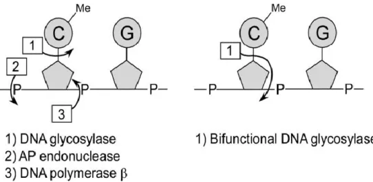
Unlike a passive DNA demethylation lowering methylated DNA level in a replication- dependent manner, in active DNA demethylation some enzymes do the role to remove 5-mC that is replaced by unmethylated cytosine. In plant, HhH-GPD (helix-hairpin-helix-Gly/Pro/Asp) DNA glycosylases play a role as DNA demethylases using the base excision repair (BER) (Gehring and Henikoff, 2008). In Arabidopsis, DEMETER (DME); REPRESSOR OF SILENCING1 (ROS1); DME LIKE2 (DML2); DME LIKE3 (DML3), that have both glycosylase and AP lyase activities can directly cleave the glycosidic bond between the base and sugarphosphate backbone of the 5-mC then the DNA polymerase fill in the abasic site (AP site) with unmethylated cytosine (Fig. 1) (Penterman et al., 2007; Zhu 2009).
5
DME is expressed primarily in the central cell of the female gametophyte and highly involved in the early female gametophyte development and seed development (Choi et al., 2002). The significance of DME in female gametophyte development and seed development will be discussed later in CHAPTER TWO.
As an active DNA demethylase, DME cannot cleave the unmethylated cytosine but the 5-mC (Morales-Ruiz et al., 2006) in all context but the activity is most efficient in 5meCG whether the DNA strands are both methylated or hemi-methylated (Gehring et al., 2006; Morales-Ruiz et al., 2006). DME has been the most extensively biochemically characterized in vitro along with ROS1, although the details from different groups are not always identical. While HhH-GPD DNA glycosylases are the largest class of glycosylases among all organisms, the DME family appears to be unique to the plant lineage (Gehring and Henikoff, 2008). HhH-GPD DNA glycosylases are characterized by a conserved aspartic acid residue and an invariant lysine. When either of these residues is mutated in recombinant DME, 5-mC DNA glycosylase activity is lost (Gehring et al., 2006; Morales-Ruiz et al., 2006).
6
Figure 1. Schematized active CG demethylation by DNA glycosylase (Kress et al., 2006).
To eliminate 5-mC by using base excision repair, the DNA glycosylase activity that can cleave the bond between base and sugarphosphate and the AP nuclease activity that can remove the sugarphosphate are necessary. In Arabidopsis the enzymes that works as DNA demethylases have DNA glycosylase and AP lyase activity at the same time. The AP site that has been emerged by the bi-functional enzyme activity then repaired by DNA polymerase so the 5-mC is replaced with cytosine.
7
3. Purpose of this study
In Arabidopsis, the studies related to DNA methylation and demethylation have been focused on the enzymes and their regulations in molecular and organismal levels or the phenotypic analysis related to their genetic and epigenetic consequences. DNA methylation and demethylation play a key role in epigenetic regulation on gene expressions through their reversibility and can keep the organisms or species from being severely defected by TEs despite going through alternation of generations by silencing them effectively. Especially, in gametogenesis and onward developmental process, there is a possibility that hypomethylation in companion cells like central cell or vegetative cell whose DNA contents are not delivered to the next generation can reinforce the silencing of the egg cell and sperm cells which become a zygote after fertilization and deliver their DNA contents to the next generation. In this way, plants do not have to take a risk of being damaged by having massive global demethylation in egg cell or sperm cells and go through a safer way to achieve TE silencing.
Despite all these significances that DNA methylation and demethylation have had, physical or direct interactors of have not been studied. Therfore, in this study, I will focus on uncovering the physical interactors of DME so that it can contribute to understand more about the active DNA demethylation pathway of DME. To do so, I used BiFC assay to in vivo screen the interactors of DME from the candidates selected by yeast two-hybrid (Y2H) screen and observed the interactors’
influences on dme mutant by using double heterozygous mutants. Doing this research can ultimately give a cue to understand the epigenetic significance and value started from the gametogenesis and seed development in plant more clearly.
8
CHAPTER ONE:
Interaction partner screening for DEMETER using Bimolecular
Fluorescence Complementation(BiFC) Assay
9
Ⅰ. Introduction BiFC assay
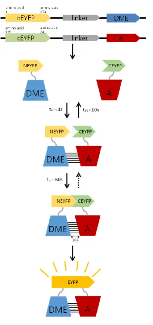
BiFC (Bimolecular Fluorescence Complementation) is an effective experimental tool that can visualize protein-protein interactions in living cells. In BiFC, fragmented fluorescent proteins attach to each of the proteins of interest recover their original 3-dimensional conformation and shows fluorescent signal if the two proteins physically interact (Fig.2). In E. coli, fluorescence complementation was first detected using fragments of a green fluorescent protein (GFP) variant fused to artificial peptide sequences designed to form an anti-parallel coiled coil (Ghosh, et al., 2000). In tobacco, onion and Arabidopsis thaliana, interactions between many different types of proteins have been visualized by introducing expression vectors encoding the fusion proteins using Agrobacterium infiltration or particle bombardment (Kerppola, 2006)
The BiFC approach provides several unique advantages for the investigation of molecular complexes in living cells. Examining the interaction in living cells make it possible to observe the interaction in a very similar context of the actual interaction conditions (Kerppola, 2006). Also it gives an easier way to observe the changes followed by varying the given interaction conditions.
The BiFC approach is applicable for the visualization of a wide range of molecular interactions proteins from different structural classes in a variety of cell types and species. And as the interaction is visualized, the cellular localization can also be detected. In addition, the intensity of fluorescent signal indicates the intensity of protein-protein interaction itself so the interactions can be compared qualitatively (Kerppola, 2006).
Based on these he advantages, BiFC-based screening is a powerful tool in that the interactions can be detected within the cell, and the effects of stimuli on the interaction can be directly tested.
One limitation of BiFC-based screens however is that differences in protein expression levels are likely to influence the partners that can be identified (Keppola, 2006). Nevertheless, BiFC analysis has the potential to identify partners that interact with a protein of interest under specific
10
cellular conditions. BiFC analysis can also be used to identify synthetic molecules or cellular factors that can modulate protein interactions.
In this study, I used Enhanced yellow fluorescent (EYFP) protein fragments to make fusion proteins. DME was fused at the 3’ end of the N terminal fragment of EYFP and the genes of interest were fused at the 3’ end of the C terminal fragments of EYFP each by using pSAT4 vector system (Fig. 2). To verify the interaction clearly, I used nEYFP-SSRP1N with cEYFP-SPT16C and nEYFP-cDME with cEYFP-SPT16C as positive controls that were previously reported as interactors for transfection efficiency and used confirmed non-interactors of DME which is constructed in a pSAT4-cEYFP vector as negative controls.
11
Figure 2. Schematic description of BiFC assay in this study.
The blue boxes indicate either CDS of DME of full DME proteins and the red ones indicates the gene of interest or the protein. If the nEYFP-DME fusion protein and cEYFP-candidat fusion protein get close enough for the EYFP fragments to resume their original 3D structure because of the physical interaction of DME and the candidate, EYFP signal would be detected by confocal laser scanning microscopy. The entire BiFC reaction to emit the EYFP signal needs at least 6 hours to be expressed.
12
Ⅱ. Materials and methods
Plant Material and Growth Conditions
Arabidopsis thaliana Columbia-0 (Col-0) was used as the wild type. Plants were grown in an environmentally-controlled chamber with a long photoperiod (16 hr light and 8 hr dark) at 22°C.
The transgenic lines and plasmids were obtained from the Arabidopsis Biological Resource Center (ABRC, Columbus, OH)
Previous yeast two-hybrid data
The yeast two-hybrid (Y2H) library screening using yeast mating (MatchmakerTM Gold Yeast Two-Hybrid System User Manual) was performed to identify physical interactors of partial fragment of DME. A cDNA library which was constructed in an activation domain (AD) vector of GAL4 was made from mRNA extracted from inflorescence meristem of Arabidopsis Columbia (col-0) wild type plant and the construct was mated with 667 amino acid-long N terminal fragment of DME(AT5G04560.1) of function is unknown cloned in GAL4 DNA-binding domain (BD).
After plating the culture on DDO at 30℃ for 6 to 12 days, the selected colonies tested on QDO plate and the plasmids were extracted and sequenced.
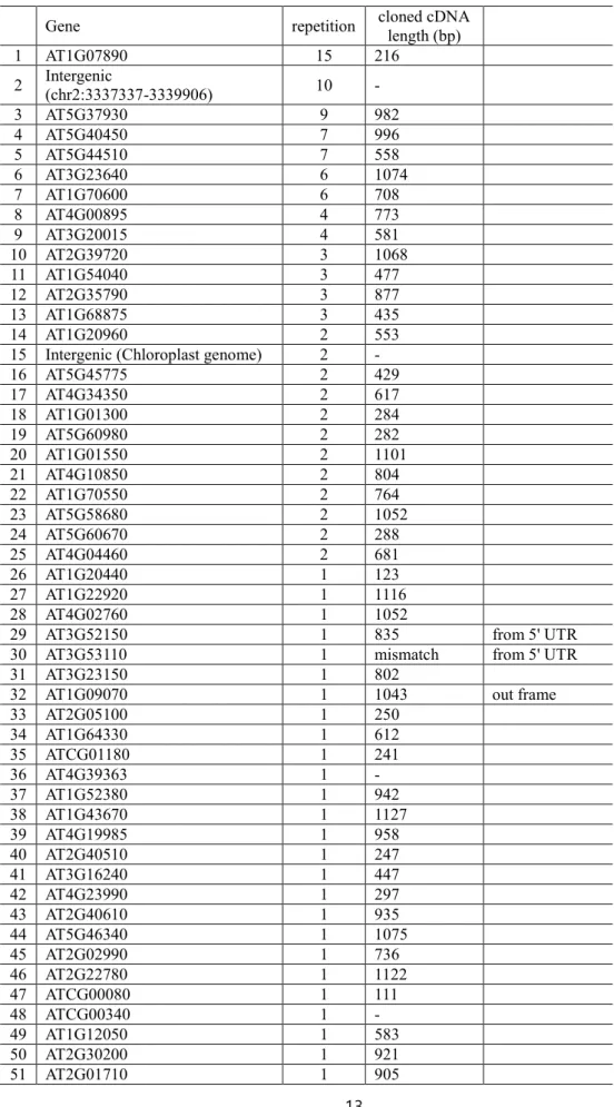
Through these previous experiment, the mating efficiency was about 10% and 239 clones were screened from QDO. After the sequencing 83 genes were found to be physical interactors of DME (Table 1).
13
Table 1. List of genes showed physical interaction with the N terminal fragment of DME(AT5G04560.1) confirmed with yeast two-hybrid screening
Gene repetition cloned cDNA
length (bp)
1 AT1G07890 15 216
2 Intergenic
(chr2:3337337-3339906) 10 -
3 AT5G37930 9 982
4 AT5G40450 7 996
5 AT5G44510 7 558
6 AT3G23640 6 1074
7 AT1G70600 6 708
8 AT4G00895 4 773
9 AT3G20015 4 581
10 AT2G39720 3 1068
11 AT1G54040 3 477
12 AT2G35790 3 877
13 AT1G68875 3 435
14 AT1G20960 2 553
15 Intergenic (Chloroplast genome) 2 -
16 AT5G45775 2 429
17 AT4G34350 2 617
18 AT1G01300 2 284
19 AT5G60980 2 282
20 AT1G01550 2 1101
21 AT4G10850 2 804
22 AT1G70550 2 764
23 AT5G58680 2 1052
24 AT5G60670 2 288
25 AT4G04460 2 681
26 AT1G20440 1 123
27 AT1G22920 1 1116
28 AT4G02760 1 1052
29 AT3G52150 1 835 from 5' UTR
30 AT3G53110 1 mismatch from 5' UTR
31 AT3G23150 1 802
32 AT1G09070 1 1043 out frame
33 AT2G05100 1 250
34 AT1G64330 1 612
35 ATCG01180 1 241
36 AT4G39363 1 -
37 AT1G52380 1 942
38 AT1G43670 1 1127
39 AT4G19985 1 958
40 AT2G40510 1 247
41 AT3G16240 1 447
42 AT4G23990 1 297
43 AT2G40610 1 935
44 AT5G46340 1 1075
45 AT2G02990 1 736
46 AT2G22780 1 1122
47 ATCG00080 1 111
48 ATCG00340 1 -
49 AT1G12050 1 583
50 AT2G30200 1 921
51 AT2G01710 1 905
14
52 ATCG00730 1 224
53 AT3G42800 1 1089
54 AT3G10912 1 934
55 AT5G51140 1 176
56 AT3G58500 1 976
57 AT3G13510 1 710
58 AT3G56740 1 1052
59 AT5G06130 1 1086
60 AT4G18810 1 1071
61 AT1G11910 1 703
62 AT4G20830 1 1015
63 AT1G07240 1 376
64 AT1G55560 1 565
65 AT5G16730 1 1073
66 AT1G51060 1 637
67 AT5G37310 1 960
68 AT5G61900 1 1015
69 AT1G70620 1 815
70 Intergenic
(chr2:16028595-16031261) 1 -
71 AT3G20060 1 713
72 AT1G05780 1 230
73 AT3G06850 1 1081
74 AT1G56190 1 950
75 ATMG00020 1 1046
76 AT1G15230 1 405
77 AT4G34320 1 973
78 AT1G53542 1 185
79 AT4G20360 1 235
80 AT5G23090 1 787
81 AT1G70680 1 871
82 AT2G05632 1 382
15
List-up for BiFC assay
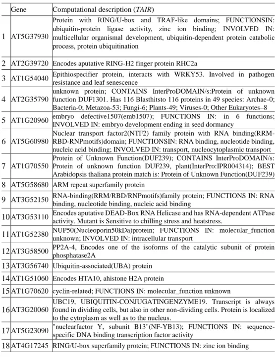
From the 83 genes confirmed as potential interactors for DME in Arabidopsis by Y2H assay, I listed them up to 18 candidates as a priority for a further analysis using BiFC (Table 2). These 18 candidate genes were selected by their molecular, structural, physiological validities with DME.
Most of them 1) have either RNA or DNA binding motifs, 2) related to the epigenetic modification of DNA or histone proteins or 3) related to protein degradation.
Cloning for BiFC assay
For the BiFC assay, the 18 candidates and DME(AT5G04560.2) were constructed as partial EYFP-fused proteins. pSAT4-nEYFP-C1 vector was used to clone nEYFP-DME construct and pSAT4-cEYFP-C1-B vector was used to clone cEYFP-candidate gene constructs (Fig. 3; Fig. 4).
15~42bp-long multiple cloning site(MCS) sequence of the vector between partial EYFP sequence and inserted gene was used as a linker. All genes were amplified as their full CDS forms except for DME and AT1g20960 (Table 3; Table 4) using cDNA synthesized from mRNA extracted from Arabidopsis thaliana col-0 inflorescence meristem as a PCR template.
16
Table 2. List of 18 candidate genes for further analysis using BiFC assay Gene Computational description (TAIR)
1 AT5G37930
Protein with RING/U-box and TRAF-like domains; FUNCTIONSIN:
ubiquitin-protein ligase activity, zinc ion binding; INVOLVED IN:
multicellular organismal development, ubiquitin-dependent protein catabolic process, protein ubiquitination
2 AT2G39720 Encodes aputative RING-H2 finger protein RHC2a
3 AT1G54040 Epithiospecifier protein, interacts with WRKY53. Involved in pathogen resistance and leaf senescence
4 AT2G35790
unknown protein; CONTAINS InterProDOMAIN/s:Protein of unknown function DUF1301. Has 116 Blasthitsto 116 proteins in 49 species: Archae-0;
Bacteria-0; Metazoa-53; Fungi-6; Plants-49; Viruses-0; Other Eukaryotes–8 5 AT1G20960 embryo defective1507(emb1507); FUNCTIONS IN: in 6 functions;
INVOLVED IN: embryo development ending in seed dormancy 6 AT5G60980
Nuclear transport factor2(NTF2) family protein with RNA binding(RRM- RBD-RNPmotifs)domain; FUNCTIONSIN: RNA binding, nucleotide binding, nucleic acid binding; INVOLVED IN: transport, nucleocytoplasmic transport 7 AT1G70550
Protein of Unknown Function(DUF239); CONTAINS InterProDOMAIN/s:
Protein of unknown function DUF239, plant(InterPro:IPR004314); BEST Arabidopsis thaliana protein match is: Protein of Unknown Function(DUF239) 8 AT5G58680 ARM repeat superfamily protein
9 AT3G52150 RNA-binding(RRM/RBD/RNPmotifs)family protein; FUNCTIONS IN: RNA binding, nucleotide binding, nucleic acid binding
10 AT3G53110 Encodes aputative DEAD-Box RNA Helicase and has RNA-dependent ATPase activity. Mutant is Sensitive to chilling stress and heatstress.
11 AT1G52380 NUP50(Nucleoporin50kDa)protein; FUNCTIONS IN: molecular_function unknown; INVOLVED IN: intracellular transport
12 AT3G58500 PP2A-4, Encodes one of the isoforms of the catalytic subunit of protein phosphatase2A
13 AT3G56740 Ubiquitin-associated(UBA) protein 14 AT1G51060 Encodes HTA10, ahistone H2A protein
15 AT1G70620 cyclin-related; FUNCTIONS IN: molecular_function unknown 16 AT3G20060
UBC19, UBIQUITIN-CONJUGATINGENZYME19. Transcript is always found in dividing cells, but also in other non-dividing cells. Protein is localized to the cytoplasm as well as to the nucleus.
17 AT5G23090 "nuclearfactor Y, subunit B13"(NF-YB13); FUNCTIONS IN: sequence- specific DNA binding transcription factor activity
18 AT4G17245 RING/U-box superfamily protein; FUNCTIONS IN: zinc ion binding
17 Figure 3. Map of pSAT4-nEYFP C1 vector
pSAT4-nEYFP-C1 vector has two duplicated CaMV 35S promoters and 5’ UTR in front of 174 amino acid-long N terminal fragment of EYFP. MCS lies 3’ end of the nEYFP coding sequence.
18 Figure 4. Map of pSAT4-cEYFP-C1-B vector
pSAT4-cEYFP-C1-B vector has two duplicated CaMV 35S promoters and 5’ UTR in front of 67 amino acid-long C terminal fragment of EYFP. MCS lies 3’ end of the cEYFP coding sequence.
19
Table 3. List of primers used for PCR amplification in constructing BiFC clones Primer label Sequence (5' to 3')
1 cDME infu F GAATTCTGCAGTCGACATGAATTCGAGGGCTGATCC 2 cDME infu R TTTTGCGGACTCTAGATTAGGTTTTGTTGTTCTTCAA 3 berkeley cDME infu F TTTTGCGGACTCTAGATTAGGTTTTGTTGTTCTTCAA
4 berkeley cDME infu R* CTGCAGAATTCGAAGCGGTTTTGTTGTTCTTCAATTTGCTCG 5 AT2G35790 infu F AATTCTGCAGTCGACATGGGAAGATCAGCTCTGAT
6 AT2G35790 infu R TTTGCGGACTCTAGATTACAAGTTCTTGAAAGCAG 7 AT1G70550 infu F AATTCTGCAGTCGACATGTGTTTAATAGGTTTGTT 8 AT1G70550 infu R TTTGCGGACTCTAGATCATGGACATCTAGGGTTTT 9 AT5G37930 infu F GACTCAGATCTCGAGggATGGCGAGATTCTCAGTT 10 AT5G37930 infu R TGCAGAATTCTCACGAATGAACAAAGATCC 11 AT2G39720 infu F AATTCTGCAGTCGACATGGCTTCTGGATCTTACTG 12 AT2G39720 infu R TTTGCGGACTCTAGATCACGCTAGCCAATTTCGTC 13 AT1G54040 infu F AATTCTGCAGTCGACATGGCTCCGACTTTGCAAGG 14 AT1G54040 infu R TTTGCGGACTCTAGATTAAGCTGAATTGACCGCAT 15 AT1G20960C infu F GGACTCAGATCTCGAGaaGACTTGCAACCTCTCCCAGT 16 AT1G20960 infu R TCGACTGCAGAATTCTCATTCTTCCATGCGATCTC 17 AT5G60980 infu F AATTCTGCAGTCGACATGGCACAGCAGGAAGCTAG 18 AT5G60980 infu R TTTGCGGACTCTAGATCAAGATGAACCACCACCTC 19 AT5G58680 infu F GACTCAGATCTCGAGggATGGCGAATCACAACAGTTT 20 AT5G58680 infu R TCGACTGCAGAATTCTTATCTCTCGTTGTCGTTAG 21 AT3G53110 infu F AATTCTGCAGTCGACATGGCGGATACGGTAGAGAA 22 AT3G53110 infu R TTTGCGGACTCTAGATCACTCGTCCAGCAGGCCAG 23 AT1G52380 infu F AATTCTGCAGTCGACATGGGTGACTCGGAAAACGT 24 AT1G52380 infu R TTTGCGGACTCTAGATCAAGTATCTGTAGCTGTTG 25 AT3G56740 infu F AATTCTGCAGTCGACATGAACGGCGGTCCCTCCGG 26 AT3G56740 infu R TTTGCGGACTCTAGATTAGTGGGACTGTGCTTCGA 27 AT1G51060 infu F AATTCTGCAGTCGACATGGCGGGTCGTGGTAAAAC 28 AT1G51060 infu R TTTGCGGACTCTAGATCAATCGTCTTCAGCAGATG 29 AT3G20060 infu F AATTCTGCAGTCGACATGGCGACGGTTAATGGGTA 30 AT3G20060 infu R TTTGCGGACTCTAGATCATGCGTTTAAAGGCTTGT 31 AT5G23090 infu F AATTCTGCAGTCGACATGGATCCAATGGATATAGT 32 AT5G23090 infu R TTTGCGGACTCTAGATTAGCTTTGCGGACTTCTCT 33 AT4G17245 infu F AATTCTGCAGTCGACATGCGGCTGCTGCTATCATC 34 AT4G17245 infu R TTTGCGGACTCTAGACTAAGGTGTACTCGTTAGTG 35 AT3G52150 Infu F GACGGTACCGCGGggATGGCGACTTTCCTAACAAA 36 AT3G52150 infu R CAGGTGGATCCCGGGCTAAGCCTTATTCACCCGAA 37 AT3G58500 infu F GACGGTACCGCGGggATGGGCGCGAATTCGCTTCC 38 AT3G58500 infu R CAGGTGGATCCCGGGTCAAAGGAAATAGTCAGGTG 39 AT1G70620 infu F GACGGTACCGCGGggATGGACGCGTACCAGCCACC 40 AT1G70620 infu R CAGGTGGATCCCGGGTCAGACCAGTTTGCTAGAGT
41 AT1G20960C NLS F GGACTCAGATCTCGAGaaAGGCAGAGTAAGAGGCGGCGTCT AAGGGAAGAGGGAGGATCCGACTTGCAACCTCTCCCAGTG 42 noncatalytic E3 N R GGACAATCAAGAACATCTGGATCC
43 noncatalytic E3 C F GATAGGTTATGTTCGTTGCAG
20
Table 4. List of the information used to construct for BiFC clones
GENE CDS
length used enzyme site competent
cell vector 1 AT5G37930 1050bp EcoRⅠ XhoⅠ DH10b pSAT4-cEYFP-C1-B 2 AT2G39720 1206bp SalⅠ XbaⅠ DH10b pSAT4-cEYFP-C1-B 3 AT1G54040 1026bp SalⅠ XbaⅠ DH10b pSAT4-cEYFP-C1-B 4 AT2G35790 717bp SalⅠ XbaⅠ DH10b pSAT4-cEYFP-C1-B 5 AT1G20960C* 2538bp EcoRⅠ XhoⅠ DH10b pSAT4-cEYFP-C1-B 6 AT5G60980 1383bp SalⅠ XbaⅠ DH10b pSAT4-cEYFP-C1-B 7 AT1G70550 1398bp SalⅠ XbaⅠ DH10b pSAT4-cEYFP-C1-B 8 AT5G58680 1074bp EcoRⅠ XhoⅠ DH10b pSAT4-cEYFP-C1-B 9 AT3G52150 762bp SacⅡ XmaⅠ DH10b pSAT4-cEYFP-C1-B 10 AT3G53110 1491bp SalⅠ XbaⅠ DH10b pSAT4-cEYFP-C1-B 11 AT1G52380 1323bp SalⅠ XbaⅠ DH10b pSAT4-cEYFP-C1-B 12 AT3G58500 942bp SacⅡ XmaⅠ DH10b pSAT4-cEYFP-C1-B 13 AT3G56740 882bp SalⅠ XbaⅠ DH10b pSAT4-cEYFP-C1-B 14 AT1G51060 399bp SalⅠ XbaⅠ DH10b pSAT4-cEYFP-C1-B 15 AT1G70620 2874bp SacⅡ XmaⅠ DH10b pSAT4-cEYFP-C1-B 16 AT3G20060 546bp SalⅠ XbaⅠ DH10b pSAT4-cEYFP-C1-B 17 AT5G23090 480bp SalⅠ XbaⅠ DH10b pSAT4-cEYFP-C1-B 18 AT4G17245 501bp SalⅠ XbaⅠ DH10b pSAT4-cEYFP-C1-B 19 +48 DME.1** 5190bp HindⅢ - DH10b pSAT4-nEYFP-C1 20 +111 DME.2*** 6075bp SalⅠ XbaⅠ DH10b pSAT4-nEYFP-C1
*) cEYFP-AT1G20960C construct has 2538bp long-C terminal fragment of original CDS
**) nEYFP-+48 DME.1 construct has no termination codon on its original termination site so extra 15 amino acids of pSAT4-nEYFP- C1 vector MCS sequence would be added until it meets the first termination codon on the vector.
***) nEYFP-+111 DME.2 construct has 2nd intron (111bp) so has longer insert product length(6075bp) then a original CDS length(5964bp). The 2nd intron was expected to be spliced out when the fusion protein is translated.
21
BiFC Assay
(1) Protoplast preperation
To perform BiFC assay, 15 to 20 leaves (width: 2 cm, length: 5 cm) were collected from 3 to 4-week-old plants grown under optimal light (ca. 150 μE·m-2·s-1) conditions. Selected leaves were used in a 'Tape-Arabidopsis Sandwich' experiment (Wu et. Al., 2009). The upper epidermal surface was stabilized by affixing a strip of Time tape (Time Med, Burr Ridge, IL) while the lower epidermal surface was affixed to a strip of Magic tape (3 M, St. Paul, MN). The Magic tape was then carefully pulled away from the Time tape, peeling away the lower epidermal surface cell layer. The peeled leaves still adhering to the Time tape, were transferred to a Petri dish containing 20 mL of enzyme solution [1% cellulase R-10, 0,2% macerozyme R-10, 20mM KCl, 20mM MES- KOH, 0.4M mannitol, 0.1% BSA, and 10mM CaCl2, pH 5.7]. The leaves were incubated in 28℃
for 1 hour then gently shaken (40 rpm on a platform shaker) for 5 to 10 minutes until the protoplasts were released into the solution. The protoplasts were centrifuged at 850rpm for 5 minutes washed twice with 10 and 5 mL each time of pre-chilled modified W5 solution [154 mM NaCl, 125 mM CaCl2, 5 mM KCl, 5 mM glucose, and 2 mM MES, pH 5.7] and incubated on ice for 30 min in dark. The protoplasts were then centrifuged and resuspended in modified MaMg solution [0.4 M mannitol, 15 mM MgCl2, and 4 mM MES, pH 5.7] to a final concentration of 2 to 5 × 10⁵ cells/mL.
(2) DNA-PEG-Calcium transfection
Approximately 5 × 10⁴ protoplasts (2 × 10⁴ to 1 × 10⁵) in 0.2 mL of MaMg solution were mixed with 20 μg each of plasmid DNA (pSAT4-nEYFP-cDME and pSAT4-cEYFP-candidate gene) at room temperature. An equal volume of a freshly-prepared solution of 40% (w/v) PEG (MW 4000; Fluka) with 0.1 M CaCl2 and 0.2 M mannitol was added, and the mixture was
22
incubated at room temperature for 15 minutes. After incubation, 0.8 mL of W5 solution was added slowly, the solution was mixed, and protoplasts were pelleted by centrifugation at 1000rpm for 2 minutes. The protoplasts were resuspended gently in 0.2 mL of W5 and were incubated at 22℃
for 12 to 16 hours in dark.
(3) Confocal laser scanning microscopy
Protoplasts were observed with a Zeiss LSM700 META laser scanning confocal microscope using 20×/0.8 Plan-Apochromat, 40×/1.2 W C-Apochromat or 63×/1.4 Oil Plan-Apochromat in multi-track channel mode. Excitation wavelengths and emission filters were 488 nm/band-pass 505-530 nm for YFP and 488 nm/band-pass 650-710 nm for chloroplast auto-fluorescence. Image processing was performed using Zen lite (Carl Zeiss, blue edition).
23
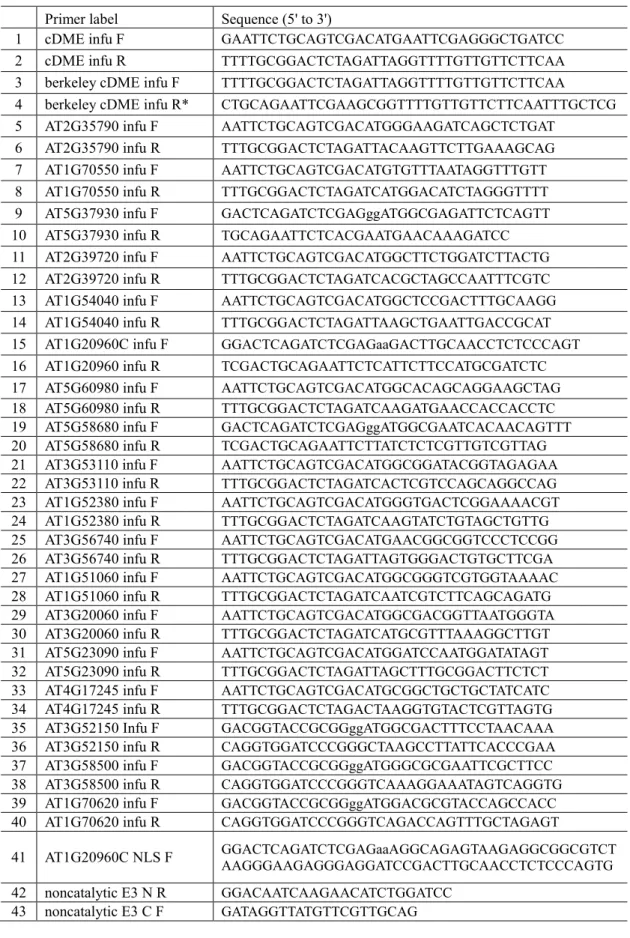
Ⅲ. Results and discussion Cloning for BiFC assay
As making the constructs for the BiFC assay, there were a few changes to confirm before examine the microscopy results. In pSAT4-nEYFP-DME construct for the general BiFC assay (+111 DME.2) there is 111bp-long 2nd intron of DME gDNA but it is expected to be spliced out when the construct is expressed in the protoplast. Also, in pSAT4-nEYFP-+48 DME.1 which is used to compare the interactions related to DME with AT5G37930, the DME.1 lose its own stop codon, thereby it has 48 more base-pairs. Thus when this construct is expressed in the protoplast, 15 more amino acids extended before it gets to the nearest stop codon of the pSAT4 MCS.
Additionally, pSAT4-cEYFP-At1g20960C and pSAT4-cEYFP-+NLS At1g20960C constructs were used to verify its interaction with DME. These constructs do not have full CDSs but a 2538bp-long partial At1g20960 C terminal fragment and pSAT4-cEYFP-+NLS At1g20960C has NLS that lies on the N terminal part of At1g20960 with a 6 base-pair-long linker sequence. (Fig.
5).
Except for these constructs, all the other constructs that were generated for the initial interaction screening for BiFC were made properly with their main CDS forms (Table 3).
24
Figure 5. Schematic description of DME and AT1G20960 CDS used for BiFC assay
(A) CDS of DME. First two strands are the original CDS form of DME and the lower two strands refers to be an experimentally used ones. +111 DME.2 has 111 base-pair-long 2nd intron in its N terminal and +48 DME.1 has 48 base-pair-long additional sequence that originates from the MCS of pSAT4-nEYFP vector. The total length is longer than the original form with 111bp and 48bp each.
(B) CDS of At1g20960. The CDS of At1g20960 is 6517bp long but in this study the 2538 base-pair-long C terminal part of At1g20960 had been used. There are two different constructs that have the same C terminal part of At1g20960 but one in the very bottom has NLS on its 5’ end. The NLS was from its own NLS sequence located in the N terminal part and was linked with the C terminal part of At1g20960C with 6-base-pair-long linker sequence.
25
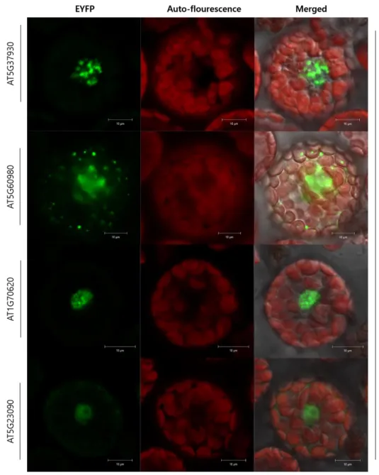
5 genes were identified as interactors of DME through BiFC
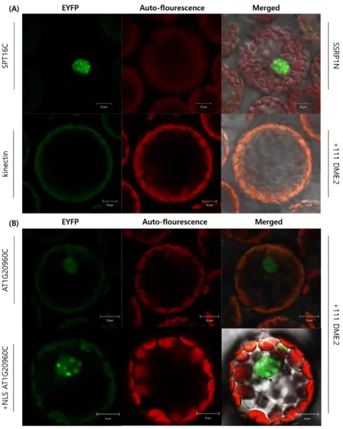
Using pSAT4-nEYFP-+111 DME.2, 18 candidate genes (Table 3) were analyzed with BiFC and 5 of them showed positive EYFP signals: AT1G20960; AT5G37930; AT5G60980;
AT1G70620; AT5G23090 (Fig. 6; Fig. 7).
AT1G20960C physically interacts with DME regardless of NLS. But with the NLS, DME and AT1G20960C were co-localized in the cell nucleus and the signal intensity was clear and strong (Fig. 6). Also the transfected cell ratio that with positive fluorescent signals were higher in +NLS AT1G20960C and DME interaction (Data not shown). In addition, the EYFP pattern differed when AT1G20960C has NLS. Without NLS, the EYFP signal was faint and blurry but when the NLS is added, the EYFP signals were localized more specifically in the nucleus and had spot-like patterns instead (Fig. 6).
AT1G20960 is U5 small nuclear ribonucleoprotein helicase also known as BRR2a, components of the spliceosome and highly conserved in eukaryotes (TAIR). Arabidopsis BRR2a is ubiquitously expressed in all analyzed tissues and involved in the processing of flowering time gene transcripts, mostly FLC (Mahrez et al., 2016). In addition, BRR2a showed physical interaction with yeast PRP6-like splicing factor STA1 which was screened to be a suppressor of ros1 (Dou et al., 2013). Thereby it is plausible that BRR2a involved in the DNA demethylation and regulating gene expression by physical interaction with DME.
26
Figure 6. Confocal laser scanning microscopy of BiFC assay shows that DME physically interacts with AT1G20960 C terminal.
(A) Controls for BiFC assay. For a positive control, nEYFP-SSRP1N and cEYFP-SPT16C had been co-transfected into the protoplast and as a negative control for DME cEYFP- kinectin was introduced into the protoplast.
(B) AT1G20960C interacts with DME whether it has NLS or not. But with the NLS, the co- localization seemed to be more clear and strong in the nucleus and the EYFP pattern differed like spot-like signals.
27
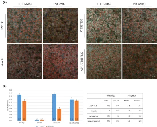
Another physical interactor of DME that had been screened by BiFC is AT5G37930 which described as E3 ubiquitin-protein ligase SINA-like 10. The EYFP signal patterns of AT5G37930 and DME interaction were variable and scattered (Fig. 7; Fig. 8). More than a half of transfected cells showed irregular patterns of EYFP signals whether cEYFP-AT5G37930 were co-transfected with nEYFP-+111 DME.2 or nEYFP-+48 DME.1(Fig. 8A).
As AT5G37930 has the E3 ligase activity, to verify if the E3 ligase activity may affect the interaction of DME and AT5G37930 itself by protein degradation, non-catalytic form of AT5G37930 which lost its RING-domain catalytic activity thereby cannot catalyze E3 ubiquitin ligase activity was constructed by changing a single histidine of the well-conserved RING-domain into tyrosine. There were two assumptions in setting this BiFC analysis: 1) If the only one form of DME is major in Arabidopsis, the other minor form of DME might be degraded by ubiquitin- proteasome pathway or at least, affected by it. 2) If the protein degradation pathway is fast enough, the complemented fluorescent signal may not be emitted even if the two proteins physically binds to each other in vivo.
As shown in Figure. 8, ubiquitin ligase activity of AT5G37930 affects the interaction between DME and AT5G37930 itself but not as distinct as all or none. Compared to the control groups, +111 DME.2 showed slightly lower transfection ratio when the AT5G37930 lost its catalytic activity (Fig 8B). But in +48 DME.1, the transfection ratio was elevated as much as the ratio of +111 DME.2 level when the catalytic activity of AT5G37930 is lost (Fig 8B). In other way, the catalytic activity of AT5G37930 seemed to affect the interaction of DME and AT5G37930 especially in the DME.1. If the catalytic activity of AT5G37930 has gone, the transfection ratio which can indicate the degree of some physical interaction got recovered as much as that of DME.2 and AT5G37930.
Previously, DME.2 was reported as a major form of DME in general. Combined with this result, the major form, DME.2 is less affected by the catalytic activity of AT5G37930 so it does not be degraded in Arabidopsis in vivo. However, the minor form, DME.1 has higher possibility of going
28
through an ubiquitin-associated protein degradation pathway.
The other interactors of DME in vivo are Nuclear transport factor 2 (NTF2) family protein with RNA binding (RRM-RBD-RNP motifs) domain-containing protein AT5G60980, cyclin-like protein AT1G70620 and nuclear factor Y, subunit B13 (NF-YB13) AT5G23090.
AT5G60980 is a NTF2 family protein which mediates the nuclear import of Ran-GDP (Stewart, 2000). Since NTF2 should import Ran-GDP into the cell nucleus and then have to go back to the cytosol as it is dissociated from Ran-GTP, AT5G60980 does not have any NLS in its CDS. So it makes sense that the BiFC signals of AT5G60980 seems blurry and faint but accumulated quite a few in the nucleus (Fig. 7). Most importantly, AT5G60980 has a RRM (RNA-recognition motif)- RBD (RNA binding domain)-RNP(Ribonucleoprotein) motif so it can recognize and bind to RNA.
AtMBD6, a methyl CpG binding domain protein, was previously reported to physically interact with AtNTF2 and the ntf2 mutant showed decreased DNA methylation at miRNA/siRNA producing loci, pseudogenes and some targets of RdDM like MOP9.1 and SDC (Parida et al., 2017). Therefore, AT5G60980 would be one of the interesting interactor to be further studied.
AT1G70620 shows distinct spot-like EYFP signal patterns specifically located in the cell nucleus (Fig. 7). These spots repeatedly shown in DME interactors’ BiFC microscopy (Fig. 6B;
Fig. 7) may possibly indicate heterochromatic regions (Lungu et al., 2017). AT1G70620 is described as a cylin-like protein but little is known about this particular gene or protein. However, as the intensity and the transfection ratio of the recovered EYFP signal are the highest amongst, the physical interaction itself is the most distinct in vivo.
AT5G23090 is a homolog of transcription factor protein Dr1. Human Dr1 has DNA binding activity and transcriptional corepressor activity, and it is involved in chromatin remodeling and histone H3 acetylation (Vaquerizas et al., 2009). AT5G23090 shows low intense EYFP signal compared to the other interactors of DME that found in this study (Fig.7) but the molecular property is plausible to associate with DME in epigenetic context, so further studies in the plants’
phenotypic level are necessary.
29
Figure 7. Confocal laser scanning microscopy of BiFC assay shows that DME physically interacts with AT5G37930, AT5G60980, AT1G70620 and AT5G23090.
AT5G37930 showed scattered and irregular patterns of EYFP signals when it was co-transfected with DME. AT5G60980 had lower tendency of being located in a nucleus but showed diffused fluorescent signals in cytosol as well. AT1G70620 showed spot-like signal patterns like +NLS AT1G20960C. AT5G23090 showed weak EYFP signals but those were constantly located in the nucleus.
30
Figure 8. DME and AT5G37930 interaction varied whether the E3 ubiquitin ligase activity of AT5G37930(E3 ubiquitin-protein ligase SINA-like 10) is active or not.
(A) Confocal laser scanning microscopy image of +111 DME.2 and +48 DME.1 interacting with AT5G37930 and non-catalytic AT5G37930. The HΔYAT5G37930, a non-catalytic form of AT5G37930 was made by a point mutagenesis that changed 131H of RING- domain into Y.
(B) As compared to the control groups, +111 DME.2 showed slightly lower transfection ratio when the AT5G37930 lost its catalytic activity. But in +48 DME.1, the transfection ratio is elevated about 1.675 times when the catalytic activity of AT5G37930 is lost. In other way, the catalytic activity of AT5G37930 seemed to affect the interaction of DME and AT5G37930.
31
CHAPTER TWO:
Phenotypic Study for the DME interactors
32
Ⅰ. Introduction
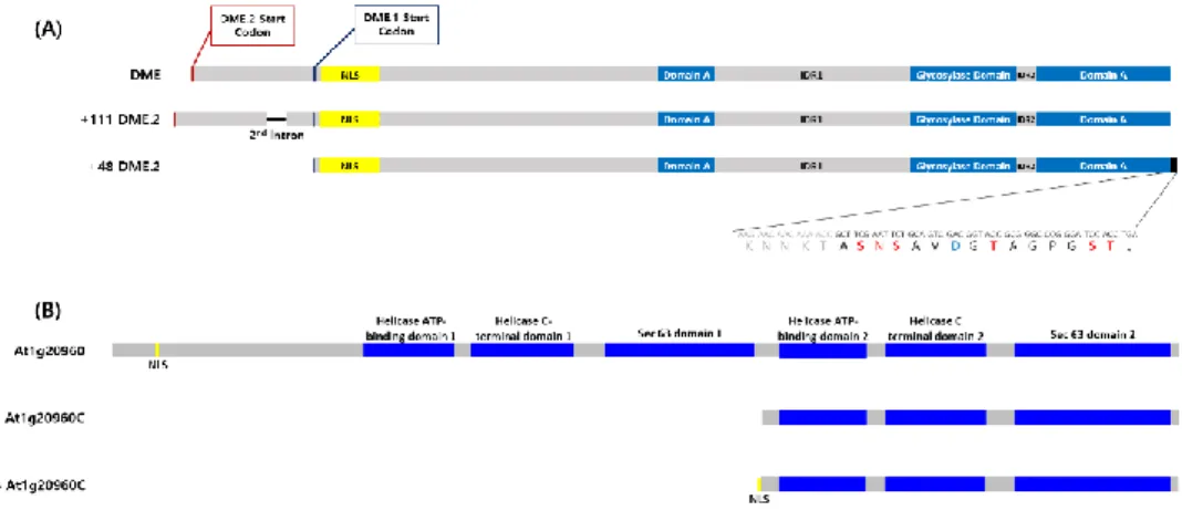
Role of DNA demethylation in gametogenesis and seed development
Formation of the male gametophyte in flowering plants consists of two distinct sequential phases, microsporogenesis and microgametogenesis(Borg et al., 2009). Microsporocytes undergo a meiotic division to produce a tetrad of four haploid microspores. During microgametogenesis, the released microspores undergo a highly asymmetric division, called Pollen Mitosis I (PMI), to produce a bicellular pollen grain with a small germ cell engulfed within the cytoplasm of a large vegetative cell (Fig 9A). While the vegetative cell exits the cell cycle, the germ cell undergoes a further mitotic division at Pollen Mitosis II (PMII) to produce twin sperm cells (Fig 9A).
Female gametogenesis also starts with a meiosis, a single haploid cell, usually the basal (chalazal) cell, enlarging and generating the functional megaspore while the remaining products of meiosis degenerate (Skinner and Sundaresan, 2018). This haploid megaspore will go through three mitotic divisions accompanied by nuclear movement to create a defined pattern at each division (Fig 9B). From stage FG4, the large vacuole (blue) separates the nuclei along the chalazal-micropylar axis. At FG5, the polar nuclei (red) migrate to meet each other and eventually fuse. At FG6/FG7, the mature female gametophyte has seven cells: two synergids, egg cell, central cell with large diploid nucleus (central cell nucleus), and three antipodal cells (Skinner and Sundaresan, 2018).
During the double fertilization of Arabidopsis, the fertilization of the central cell(2n) and a sperm cell(n) generates triploid endosperm which does not deliver its DNA contents to the next generation and the fertilization of the egg cell(n) and a sperm cell(n) generates diploid embryo.
DME is expressed in the vegetative cell nucleus of the male gametophyte (Schoft et al., 2011;
Park et al., 2017) and central cell of the female gametophyte during the early gametogenesis and development (Choi et al., 2002).
DME induces hypomehtylation of maternal effect gene MEDEA (MEA) Polycomb gene (PcG)
33
in the central cell before fertilization by demethylation so that the MEA got transcriptionally activated to produce MEA proteins (Gehring et al., 2006). After fertilization, the FIE-MEA PcG complex activated by maternal DME binds to the paternal MEA making it to be silenced (Gehring et al., 2006). So if there are mutations in maternal dme, maternal MEA cannot be transcriptionally activated so the phenotype of which is identical to mea mutant: Endosperm overproliferation;
embryo arrest; seed abortion (Choi et al., 2002; Grossniklaus et al., 1998; Kiyosue et al., 1999;
Luo et al., 1999).
Meanwhile, in mammal, there reported a genomewide methylation reprogramming by massive demethylation and re-methylation process that take places in preimplantation embryo (Reik et al., 2001). In plants, however, there is no massive methylation reprogramming in embryo but instead, there reported that the companion cells whose DNA contents are not inherited go through global demethylation and the hypomethylated state is maintained in late endosperm stage in which DME is no longer expressed (Hsieh et al., 2009; Ibarra et al., 2012 Park et al., 2016; Kim et al., 2019).
Based on these researches, the hypothesis that massive TEs demethylation in the central cell makes TE transcriptions highly activated and the small RNAs generated from them may be delivered to the neighboring egg cell or to the embryo so that the TE repressions are reinforced in the embryo by RdDM pathway was developed (Gehring, 2019).
34
Figure 9. Gametogenesis and development in Arabidopsis
(A) Development of the male gametophyte (Borg et al., 2009). DME is expressed in the vegetative cell nucleus. From the mature pollen, two sperm cells participate in the double fertilization and generate embryo and endosperm.
(B) Development of the female gametophyte (Skinner and Sundaresan, 2018). DME is expressed in the central cell nucleus (CCN). Mature female gametophyte generates egg cell and central cell. After fertilization, the central cell fertilized with a pollen become an endosperm and the egg cell fertilized with a pollen become an embryo that delivers DNA contents to the next generations.
35
Ⅱ. Materials and methods
Plant Material and Growth Conditions
Arabidopsis thaliana Columbia-0 (Col-0) was used as the wild type. Plants were grown in an environmentally-controlled chamber with a long photoperiod (16 hour light and 8 hour dark) at 22°C. The transgenic lines were obtained from the Arabidopsis Biological Resource Center (ABRC, Columbus, OH).
Seed-set analysis and whole-mount clearing
The T-DNA insertion knock-out mutant alleles used in this study are emb1507-1 (Stock name:
CS16090, ABRC) and dme-2 (Table 5).
Heterozygous EMB/dme1507-1 plants were first sowed on the MS only plates. To observe seed abortion, siliques (DAP 8 to 10) were dissected on a dissecting microscope (Stemi DV4, Carl Zeiss) and aborted seeds and undeveloped ovules were counted. For whole mount clearing, siliques (DAP 8 to 10) were dissected and mounted in clearing solution [2.5g chloral hydrate;
0.3ml 100% glycerol; 0.7ml distilled water] for 1 hour. Then the samples were observed using an Axio Imager A1 microscope (Carl Zeiss) under DIC optics and were photographed using an AxioCam HRc camera (Carl Zeiss).
After checking the single heterozygotic phenotype of emb1507-1, to examine the phenotypic changes in double heterozygous mutant, EMB/emb1507-1 was crossed with dme/dme-2 homozygous mutant allele. To obtain the double heterozygote mutant, EMB/emb1507-1 buds of FG7 stage were dissected and emasculated. The emasculated pistils were placed in 22℃ long day condition growth chamber for 24 hours for maturation then crossed with dme/dme-2 used as a pollen donor. Fully mature fertilized seeds were harvested and the F1 sowed on MSbasta plate for selection. Confirming their genotypes with PCR amplifications (Table 5), check the seed abortion and embryo defects with the same processes used in examining heterozygous EMB/dme1507-1
36
mutant. F2 of these double heterozygote mutants were genotype and in addition, reciprocal cross using Col-0 as a wild type was done to verify the segregation ratio changes.
37
Table 5. T-DNA insertion alleles and genotyping primers used in this study Gene Name Back
ground insertion location primers for genotyping
AT1G 20960
emb
1507-1 col pCSA
104luc exon
EMB1507-1 F GGGATAAATGATG AGGATGC
EMB1507-1 R CTCTATCAGCAACA TCTCTCC
pCSA104 RB3
TAACAATTTCACAC AGGAAACAGCTAT GAC
AT5G
04560 dme-2 col-gl pSKI15 exon
B33F CACTTGTTCCCTAT
GAGAGC
B33R CACTGATTGTGATG
TTCCAC
SKI015 LB3 TTGACCATCATACT CATTGCTG
38
Ⅲ. Results and Discussion EMB/emb1507-1 seed phenotype
EMB/emb1507-1 showed constant abortion ratio near 25% (Fig. 10A). It seems that emb/emb1507-1 would arrested in the globular stage of embryo and eventually aborted (Fig. 10B).
DME/dme-2 shows 50% abortion because maternal inheritance of mutant dme allele causes seed abort while paternal dme did not affect. EMB/emb1507-1 single heterozygote mutant plants are expected to follow the Mendelian genetics and traditional segregation ratio.
EMB/emb1507-1;DME/dme-2 double heterozygote mutant seed phenotype
To examine the effect of BRR2a on DME, double mutant was generated. Since DME is a maternal effect gene, if the dme-2 allele is derived from maternal allele, the F2 seeds would be all aborted. So using dme/dme-2 mutant as paternal pollen donor, EMB/emb1507-1 was crossed to make EMB/emb1507-1;DME/dme-2 mutant.
EMB/emb1507-1;DME/dme-2 varied seed abortion ratio from 40 to 68% (Fig. 11A). Expected seed abortion ratio is calculated as 62.5%. The lowest seed abortion lines tend to have highest ovule abortion ratio so it is hard to consider that the seed abortion phenotype of dme-2 was rescued.
As examining the arrested seed phenotypes, detectable aborted seeds’ ratio is almost 66%, a bit higher than expected. Among the 66% detectable arrested seeds, half of them are arrested in the hear stage which is the seed phenotype of dme-2 and the other half is arrested in the globular stage which is the seed abortion phenotype of emb1507-1. And 5% of seeds were proceeded to torpedo stage.
39 Figure 10. EMB/enb1507-1 seed abortion phenotypes.
(A) Seed abortion ratio of EMB/emb1507-1 mutant plants. About 25% of the seeds are aborted (lowest: 20%, highest:27%) while WT siliques showed no seed abortion and slightly lower ovule abortions than EMB/emb1507-1. And overall plants, minor ovule abortions were constantly observed.
(B) DIC image of mounted-clearing image of aborted seeds. Most of the aborted seeds of EMB/emb1507-1 (right) are seemed to be arrested in the globular stage (red arrow) while the WT seeds were almost fully mature. Scale bar=100μm