저작자표시-비영리-변경금지 2.0 대한민국 이용자는 아래의 조건을 따르는 경우에 한하여 자유롭게
l 이 저작물을 복제, 배포, 전송, 전시, 공연 및 방송할 수 있습니다. 다음과 같은 조건을 따라야 합니다:
l 귀하는, 이 저작물의 재이용이나 배포의 경우, 이 저작물에 적용된 이용허락조건 을 명확하게 나타내어야 합니다.
l 저작권자로부터 별도의 허가를 받으면 이러한 조건들은 적용되지 않습니다.
저작권법에 따른 이용자의 권리는 위의 내용에 의하여 영향을 받지 않습니다. 이것은 이용허락규약(Legal Code)을 이해하기 쉽게 요약한 것입니다.
Disclaimer
저작자표시. 귀하는 원저작자를 표시하여야 합니다.
비영리. 귀하는 이 저작물을 영리 목적으로 이용할 수 없습니다.
변경금지. 귀하는 이 저작물을 개작, 변형 또는 가공할 수 없습니다.
수의학박사학위논문
유전자 편집 기법으로 VEGF와 LEF1을 발현시킨 중간엽 줄기세포의
심근경색 치료효과
Therapeutic Effects of Genome-edited Mesenchymal Stem Cells Expressing VEGF and
LEF1 on Myocardial Infarction
2020년 2월
서울대학교 대학원
수의학과 수의생명과학 전공(수의생화학)
조 현 민
유전자 편집 기법으로 VEGF와 LEF1을 발현시킨 중간엽 줄기세포의
심근경색 치료효과
Therapeutic Effects of Genome-edited Mesenchymal Stem Cells Expressing VEGF and LEF1 on Myocardial Infarction
지도 교수 조 제 열
이 논문을 수의학박사학위논문으로 제출함 2019년 11월
서울대학교 대학원
수의학과 수의생명과학 전공 (수의생화학) 조 현 민
조현민의 수의학박사 학위논문을 인준함 2019년 12월
위 원 장 (인) 부위원장 (인) 위 원 (인) 위 원 (인) 위 원 (인)
Therapeutic Effects of Genome-edited Mesenchymal Stem Cells Expressing
VEGF and LEF1 on Myocardial Infarction
Under the supervision of Professor Je-Yoel Cho
DISSERTATION
Presented in Partial Fulfillment of the Requirement for the Degree of DOCTOR OF PHILOSOPHY
By
Hyun-Min Cho
Major in Veterinary Biomedical Sciences (Veterinary Biochemistry)
Department of Veterinary Medicine The Graduate School
Seoul National University
February 2020
i
ABSTRACT
Therapeutic Effects of Genome-edited Mesenchymal Stem Cells Expressing
VEGF and LEF1 on Myocardial Infarction
Hyun-Min Cho Major in Veterinary Biomedical Sciences Department of Veterinary Medicine The Graduate School Seoul National University
Cardiovascular disease is one of the leading causes of death worldwide, and according to the 2016 World Health Organization (WHO) statistics, more than 9 million patients die each year from different forms of this disease. Among these, myocardial infarction is a serious disease in which cardiomyocytes that do not receive blood due to coronary artery obstruction die, and the number of patients is increasing the fastest among cardiovascular diseases. The major therapy for myocardial infarction is stent implantation, which expands the endoscopic diameter by inserting the stent into the blocked vessel. Although, it is a fundamental
ii
treatment for the survival of myocardial tissues, side effects such as reperfusion injury may occur, and since cardiomyocytes have very low regenerative capacity, additional treatment strategies are needed to regenerate damaged myocardial tissues after surgery.
In this regard, human umbilical cord blood-derived mesenchymal stem cells (hUCB-MSCs) have received special attention. These cells have fast self-renewal and differentiation into cardiomyocytes, and secrete various cytokines such as HGF, bFGF, VEGF-D, and PDGF to help repair damaged areas in the heart and form new blood vessels. However, despite these advantages, hUCB-MSCs are limited in that less than 3% of cells survive after transplantation and they secrete very little VEGF-A, which has the most important effect on angiogenesis even after successful transplantation. These limitations raise the need for the induction of VEGF-A and the enhancement of cell proliferation in therapies using hUCB-MSCs.
In this study, we applied VEGF-A and LEF1 genes to the AAVS1 locus, a safe harbor site in stem cells, using the TALEN and CRISPR / Cas9 systems. The effect of restoring heart function was confirmed, thus suggesting a new strategy for treating myocardial infarction of stem cells into which therapeutic genes have been introduced through target genomic engineering.
In CHAPTER I, the vascular endothelial growth factor (VEGF-A) was used to improve the angiogenic capacity of hUCB-MSCs. After coronary artery occlusion, neovascularization is essential to replenish heart tissue and slow the progression of
iii
myocardial necrosis. To this end, mesenchymal stem cells (VEGF / hUCB-MSCs) secreting therapeutic levels of VEGF-A were produced using the TALEN system, a second-generation gene editing technology, and the therapeutic effect of these cells was confirmed in the myocardial infarction model.
VEGF-A can induce angiogenesis and form new blood vessels for therapeutic purposes, but excessive amounts of VEGF-A can cause abnormal blood vessel formation and hemangioma growth. Therefore, in order to utilize this for therapeutic purposes, a device capable of controlling its expression was required.
For this purpose, the vector was designed to only produce VEGF-A protein in the presence of doxycycline. In addition, to solve the problems of low gene transfer efficiency, stability, and specific expression, which are limitations of conventional gene therapy, the TALEN gene editing technology was used to introduce the VEGF expression vector cassette in the AAVS1 locus.
Thus, the VEGF/hUCB-MSCs produced secreted VEGF-A only upon doxycycline treatment, and consequently increased the expression of genes associated with angiogenesis and improved angiogenesis in animal models. Finally, when VEGF/hUCB-MSCs were implanted into the rat myocardial infarction model and doxycycline was supplied via negative water, cardiac function was restored, MI size and fibrosis decreased, and the recovery effect of the heart damaged by myocardial infarction could be confirmed.
iv
In CHAPTER II, hUCB-MSCs incorporating the LEF1 gene were studied for their proliferation and survival effects. It has previously been noted that there is potential for therapeutic applications, but hUCB-MSCs have certain limitations in their application to the treatment of myocardial infarction because of their low post-transplantation survival rate. Accordingly, there is demand for a treatment strategy using stem cells into which a therapeutic gene has been introduced. In this section, hUCB-MSCs (LEF1/hUCB-MSCs) incorporating the LEF1 gene were constructed using the CRISPR/Cas9 system and applied to the myocardial infarction model.
Through In silico literature surveys, LEF1 was identified as a therapeutic gene, and it was overexpressed in hUCB-MSCs to confirm cell proliferation and survival.
After fully confirming the possibility of applying the treatment to the actual disease model, a rat myocardial infarction model was prepared by introducing stem cells (LEF1/hUCB-MSCs) into which the LEF1 gene had been incorporated into the AAVS1 locus via the CRISPR/Cas9 system. Upon subsequent transplantation of LEF1/hUCB-MSCs survival after myocardial infarction improved, and myocardial protective effects such as recovery of cardiac function, fibrosis and reduction of infarct area was later noted.
In conclusion, human mesenchymal stem cells expressing VEGF-A and LEF1 by targeted genome editing (VEGF/hUCB-MSCs and LEF1/hUCB-MSCs) were generated in the present study. Each of these cells showed enhanced angiogenesis
v
and cell proliferation. Furthermore, when implanted into the myocardial infarction model, the cardiovascular recovery was improved. Through this, we proposed a new cell therapy strategy that overcomes the limitations of existing cell therapies.
Keywords: Myocardial infarction, Mesenchymal stem cells, Targeted genome engineering, Vascular endothelial growth factor, Lymphocyte enhancer-binding factor 1, Recovery of cardiac function
Student Number: 2012-21532
vi
CONTENTS
ABSTRACT ... ⅰ
CONTENTS ... ⅵ
LIST OF FIGURES ... ⅷ
ABBREVIATIONS ... ⅹ
LITERATURE REVIEW ... 1
1. MYOCARDIAL INFARCTION
...
12. STEM CELL THERAPY IN MI
...
53. VEGF AND LEF1
...
124. TALEN AND CRISPR/CAS9
...
17vii
CHAPTER 1
Targeted Genome Engineering to Control VEGF Expression in Human Umbilical Cord Blood-Derived Mesenchymal Stem Cells: Potential Implications for the Treatment of Myocardial Infarction
1. INTRODUCTION ... 22
2. MATERIAL AND METHODS ... 26 3. RESULTS ... 31 4. DISCUSSION ... 56
CHAPTER 2
Transplantation of human mesenchymal stem cells genome- edited with LEF1 improves cardio-protective effects in myocardial infarction
1. INTRODUCTION ... 63
2. MATERIAL AND METHODS ... 67 3. RESULTS ... 73 4. DISCUSSION ... 99
REFERENCES ... 103
ABSTRACT IN KOREAN ... 118
viii
LIST OF FIGURES
CHAPTER 1
Figure 1-1.
Figure 1-2.
Figure 1-3.
Figure 1-4.
Figure 1-5.
Figure 1-6.
Figure 1-7.
Figure 1-8.
Figure 1-9.
Generation of VEGF/hUCB-MSCs
Confirmation of conditional VEGF secretion
Effect of VEGF secretion from VEGF/hUCB-MSCs on the gene expression of neo-angiogenesis markers and cell cycle regulators
The induction of VEGF expression by VEGF/hUCB- MSCs transplanted on mouse skin and neo- angiogenesis by secreted VEGF in Matrigel plugs
Engraftment of VEGF/hUCB-MSCs Representative echocardiogram images Recovery of cardiac function
Representative images of heart sections
Evaluation of MI size, LV fibrosis and wall thickness after the transplantation of VEGF/hUCB-MSCs
ix Figure 1-10.
Figure 1-11.
Immunohistochemical staining of VEGF, blood vessel formation by VEGF/hUCB-MSCs
Prolonged cell survival of transplanted human VEGF/hUCB-MSCs
x
CHAPTER 2
Figure 2-1.
Figure 2-2.
Figure 2-3.
Figure 2-4.
Figure 2-5.
Figure 2-6.
Figure 2-7.
Selection process of therapeutic target gene LEF1 and cell proliferation effects of LEF1 in hUCB- MSCs
LEF1 overexpression protects hUCB-MSCs from oxidative stress induced apoptosis
Experimental strategy of therapeutic hUCB-MSC transplantation system
Transplantation of LEF1/hUCB-MSCs recovered cardiac function in MI rat
The LEF1/hUCB-MSC transplantation greatly reduced MI size, fibrosis and restored the LV wall thickness
Immunohistochemical staining confirmed the cells surviving and expressing LEF1 from the engrafted MI+ LEF1/hUCB-MSCs
LEF1 triggered growth factors (HGF, IGF, VEGF) and IL-8 expressions in MI+ LEF1/hUCB-MSCs
xi
ABBREVIATIONS
bFGF CRISPR
CAD DSBs EF FS HGF HDR IL IHD LEF1 MSCs MI TALEN VEGF ZFN
Basic fibroblast growth factor
Clustered regularly interspaced short palindromic repeats
Coronary artery disease Double-stranded breaks Ejection fraction
Fractional shortening Hematocyte growth factor Homology-directed repair Interleukin
Ischemic heart disease
Lymphoid enhancer-binding factor 1 Mesenchymal stem cells
Myocardial infarction
Transcription activator-like effector nuclease Vascular endothelial growth factor
Zinc finger nuclease
1
LITERATURE REVIEW
1. Myocardial infarction
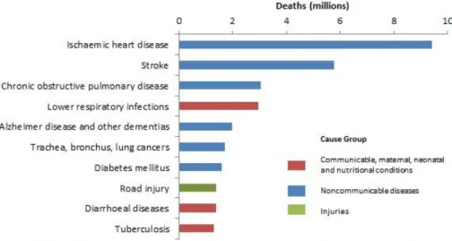
Ischemic heart disease (IHD) is one of the leading causes of death. According to the World Health Organization (WHO) statistics in 2016, more than 9 million patients die from this disease each year (Nowbar et al., 2019) (Figure B-1). The main cause of this disease is the blockage of the coronary artery due to an atherosclerotic plaque, which causes disruptions in blood flow and oxygen supply.
Severe or prolonged ischemia causes death of surrounding tissues (Mills et al., 2009, Avery et al., 2010).
Myocardial infarction (MI) is a type of ischemic heart disease (Alpert, 2009), and it refers to the irreversible death of the cardiac tissue (Figure B-2). The main therapeutic strategy for MI is reperfusion therapy through primary percutaneous coronary intervention (PPCI) (Frohlich et al., 2013). This treatment is a non- surgical procedure that reopens the blocked coronary artery by inserting a catheter into the blood vessel. Although this therapeutic process is a pre-requisite for the survival of myocardial tissue, it can lead to paradoxical phenomena termed
‘myocardial reperfusion injuries,’ including additional intravascular and myocardial damage and death of remaining cardiomyocytes (Carden and Granger, 2000).
2
In order to overcome this problem, combined therapies using anti-platelet drugs such as prasugrel, abciximab and anti-thrombotic agents such as bivalirudin have been tried (Showkathali and Natarajan, 2012, Metharom et al., 2015). However, it has proven insufficient in restoring myocardial reperfusion injury, and since cardiomyocytes have a very low regenerative capacity, there is a need for additional measures to regenerate damaged myocardium after the intervention (Kikuchi and Poss, 2012).
3
Figure B-1. Top 10 global causes of deaths, 2016.
Of the 56.9 million deaths, more than half (54%) were due to the top 10 causes.
Cardiovascular diseases are the world’s biggest killers, accounting for a combined 9.3 million deaths in 2016. These diseases have remained the leading cause of death globally in the last 10 years.
Adapted from Geneva, World Health Organization et al., 2018
4 Figure B-2. Myocardial infarction.
Myocardial infarction (MI) occurred by atherothrombotic coronary artery disease (CAD) and usually precipitated by plaque disruptions (rupture or erosion).
Prolonged atherosclerosis causes thrombosis, and dynamic thrombus components eventually lead to necrosis of cardiomyocytes.
Adapted from Thygesen K et al., 2018
5
2. STEM CELL THERAPY IN MI
After myocardial infarction, more than 1 billion cardiomyocytes die. The lost cells are replaced by fibrotic connective tissue through the process called ‘cardiac fibrosis’. This process causes scarring and stiffness in the cardiac muscle, and ultimately leads to thinning of the heart wall and weakness of heart function (Talman and Ruskoaho, 2016). Since cardiomyocytes are terminally differentiated cells and their regenerative capacity is so limited, additional strategies based on stem cells are required (Foglia and Poss, 2016). Stem cells have the potential to proliferate and differentiate into various types of cells, including cardiomyocytes (Slack, 2007, Slack, 2013). In this respect, the therapeutic strategies utilizing stem cells have sufficient potential to be applied to MI. Different types of stem cells in various administration methods have been investigated, and promising results have been shown in many preclinical studies. Particularly Mesenchymal stem cells (MSCs), with their proliferative capacity and ability to be differentiated into various lineage cells, are promising candidates for MI treatment (Kubo et al., 2009, Shafei et al., 2017) (Table B-1).
MSCs were first discovered in 1970. According to the international society for cellular therapy, MSCs are defined as self-renewing cells that express CD90 and CD73 but do not express CD45, CD34, CD14, CD11b, CD79a, or CD19 cell surface markers. MSCs can be derived from a variety of sources, including bone marrow, adipose tissue, umbilical cord blood, dental pulp, etc (Kern et al., 2006).
Each type of MSC has a different isolation protocol and culture conditions, and due
6
to their various sources, each has its own unique properties.
Recently, human umbilical cord blood derived mesenchymal stem cells (hUCB- MSCs) have received special attention in regenerative medicine because they form a connection between embryonic and adult stem cells, and have the advantages of both (Han et al., 2019). Compared to adult stem cells, hUCB-MSCs have a higher proliferative and differentiative capacity, enabling better clinical applications. On the other hand, when compared to embryonic stem cells, hUCB-MSCs have a lower potential for teratoma formation, thus enabling safer treatment. Especially regarding cardiovascular diseases, hUCB-MSCs have shown positive results in various preclinical studies due to their various advantages such as fast self-renewal, immuno-modulation, or capacity to differentiate to various lineages. Additionally, hUCB-MSCs could encourage neovascularization and anti-fibrosis (Lee, 2018, Bagno et al., 2018) (Figure B-3).
The major function of hUCB-MSCs involves their paracrine effects in MI.
Transplanted cells secrete various cytokines that could restore cardiac metabolism.
These are cell chemotactic factors that assist cell recruitment to the ischemic region, thereby reducing the scar formation. They also promote angiogenesis, alleviate the symptoms caused by limb ischemia, and restore blood supply that has been interrupted. hUCB-MSCs produce high levels of angiogenic growth factors such as hematocyte growth factor (HGF), basic fibroblast growth factor (bFGF), VEGF-D, and platelet-derived growth factor (PDGF) (Majka et al., 2017) (Figure B-4).
However, the interesting thing is that they produce very little VEGF-A (Akimoto et al., 2013). These facts indicate that the cells can induce angiogenesis independently
7
of VEGF-A, and thus there is no need for induction of VEGF-A in cell therapy using hUCB-MSCs. In addition to neovascularization, the immunomodulatory properties of hUCB-MSCs targeted to inflammation are thought to be responsible for additional paracrine effects. HUCB-MSCs produce interleukin (IL)-6, which promotes the acquisition of tolerogenic phenotypes, and prostaglandin E2 (PGE2), which suppresses cytotoxicity of NK cells and proliferation of CD4+/CD8+ T-cells (Cortes-Araya et al., 2018). In addition, hUCB-MSCs secrete anti-inflammation cytokines, such as interferon-a, identifying these cells as the major regulators of early inflammation. Transplantation of hUCB-MSCs has a positive effect on the innate immune system, as well as on the adaptive immune system. Transplanted cells produce IL-6 and HGF-rich microenvironments, which primes the monocytes to produce IL-10 (Wang et al., 2009).
With regards to cardiac regeneration, the introduction of hUCB-MSCs for cell therapy showed various effects such as anti-fibrotic, anti-inflammatory and pro- angiogenic effects. This has been demonstrated in many preclinical studies and offers ample potential for clinical studies (Singh et al., 2016). Nevertheless, there remains many limitations to overcome for application in clinical treatment, including the low accuracy of delivered cells to the damaged areas and low survival rates of transplanted hUCB-MSCs due to harsh conditions of infarcted regions (Yu et al., 2008). To overcome these limitations, a variety of additional strategies have been attempted, including combinations with other cell types (e.g., CSCs or macrophages), additional pharmaceutical approaches, and enhancing specific cellular functions through gene overexpression. However, the therapeutic
8
effects are short-lived or incomplete. Therefore, continuous and diverse efforts should be encouraged to develop the best treatment strategy using hUCB-MSCs.
9
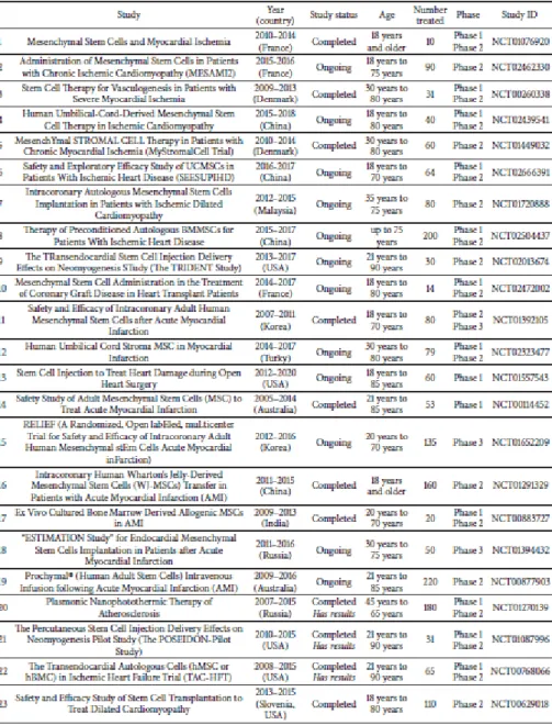
Table B-1. 23 Clinical trials using MSCs in relation to MI.
Clinical Trials related to MSCs in myocardial infarction.
Adapted from Seung Taek Ji et al., 2017
10
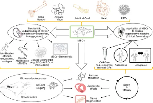
Figure B-3. MSCs are an attractive cellular platform for cardiac regenerative medicine.
MSCs can be isolated from various tissues, including bone, adipose tissue, umbilical cord, or they can be originated from pluripotent stem cells. Ongoing research, examining the relationship of MSCs to heart regeneration, will lead to the development of more innovative strategies, such as isolating cardiac MSC lineages, generating genetically engineered MSCs with various growth factors and exosome secretomes, and optimizing combination strategies of MSCs with biomaterials similar to heart tissue.
Adapted from Luiza Bagno et al., 2018
11
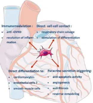
Figure B-4. Therapeutic effect of mesenchymal stem cells in myocardial infarction.
Mesenchymal stem cells mechanisms of action in myocardial infarction.
Abbreviation: GVHD, graft-versus-host disease.
Adapted from Marcin Majka et al., 2017
12
3. VEGF AND LEF1
Vascular endothelial growth factor (VEGF) is a special mitogen that induces vascular endothelial cells to begin mitosis. It is one of the most actively studied angiogenic growth factors, and is known to play major roles in vascular formation during embryonic development, injury, bone formation and even cancers (Shibuya, 2011).
In 1989, Henzel and Ferrara first purified and cloned the mitogen from endothelial cells, and began to name this factor VEGF. The factor can be divided into VEGF-A, B, C, E and F depending on the structure, and each interacts with different receptors. Each member has a variety of roles, such as angiogenesis, embryonic angiogenesis and development of lymphatic vasculature, depending on the interacting receptors (Ferrara, 2016).
Among them, VEGF-A is a protein which has two 23kDa homodimer subunits.
The gene encoding this protein in humans is located on p21.1 of chromosome 6 and its size is 16,304 bases. VEGF-A is closely associated with angiogenesis of most organs during vertebrate development, and is associated with angiogenesis during organ remodeling in adults (Figure B-5). Endothelial cell differentiation is highly dependent on the expression of this gene, so deletion of this gene during development could be embryonic lethal. VEGF-A has various isoforms due to alternative splicing. Five isoforms are produced in humans (VEGF-A 121, 145, 165, 183, 189, and 206), and the most expressed isoform is VEGF-A 165 (Holmes and Zachary, 2005) (Figure B-6).
13
Lymphoid enhancer-binding factor 1 (LEF1) is a protein in the nucleus of pre-B and T cells, and has a molecular weight of 48kDa (Milatovich et al., 1991). In humans, it is translated from the LEF1 gene at position q25 of chromosome 4. It is known that this growth factor attaches to T cell receptor-alpha enhancers and induces maximum enhancer activity and is also expressed at the early stages of differentiation in B-cells thus playing a key role in cell survival and division. In addition, it is considered to be an important mediator of the Wnt/β-Catenin signaling pathway, one of the factors of the T-cell factor (TCF)/LEF1 transcription factor family (Cadigan and Waterman, 2012). Currently, seven LEF1 isoforms, including LEF1A, LEF1B and LEF1DN, have been identified through alternative promoter usage and alternative splicing. In addition, through computational mapping, three more potential isoforms are also suspected to exist.
LEF1 has received much attention in recent cardiac regeneration research. In 2009, it was confirmed that LEF1 is necessary for the development of major blood vessels and heart tissue through xenopus tropicalis models (Roel et al., 2009). In 2019, LEF1, along with other TCF families, was reported to induce cardiac maturation through temporal and spatial control in cardiac development through Wnt/β-Catenin signaling (Ye et al., 2019). LEF1 was mainly expressed in the valvular region. Furthermore, stem cell research continues to report that LEF1 promotes self-renewal. According to a 2010 study by Huang C, LEF1 increased the activity of the Oct4 promoter and physiologically interacted with Nanog to help maintain mouse embryonic stem cells (Huang and Qin, 2010b). In addition, it has been reported that the proliferation of adult hippocampal precursor cells as well as
14 Pro-B cells is also promoted by LEF1.
Considering these studies and results, we can expect that LEF1 plays a major role in heart development and promoting cell proliferation. However, studies related to cell proliferation and survival of LEF1 in hUCB-MSCs have not been reported yet. Experimental confirmation of these functions and application of LEF1 overexpressed MSCs to the MI to confirm its therapeutic applicability seems to be necessary.
15 Figure B-5. Growth factors of the VEGF family.
The three signaling receptors of the VEGF family (VEGFR-1, VEGFR-2, and VEGFR-3) and the accessory isoform specific receptors neuropilin-1, neuropilin-2 are displayed with structural features.
Adapted from Masoumi Moghaddam S et al., 2012
16 Figure B-6. Various isoforms of the VEGF.
Vascular endothelial growth factor (VEGF) mRNA generates four different isoforms, VEGF121, VEGF165, VEGF189, or VEGF206 after alternative splicing.
Of these, VEGF121 is a nonheparin-binding acidic protein, which is freely diffusible.
Adapted from Chloe J. Peach et al., 2018
17
4. TALEN AND CRISPR/CAS9
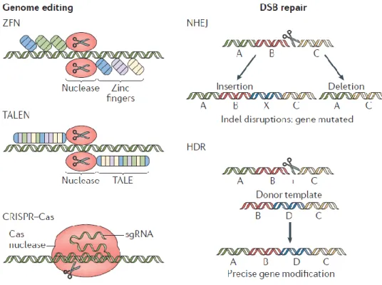
Genome editing technology is an emerging therapeutic strategy that manipulates eukaryotic genomes using specific nucleases. These nucleases are composed of restriction enzymes and DNA binding domains engineered to bind to specific sequences and produce double-stranded breaks (DSBs). There are many known genome-editing nucleases, but they share the same platforms. After the nucleases cut a specific sequence on the genome, the DSB can be repaired by the following two endogenous DNA repair systems: non-homologous end joining (NHEJ) and homology-directed repair (HDR) with Donor DNA (Cox et al., 2015, Lieber et al., 2003).
NHEJ is efficient, but often causes errors during the process. Thus, the repeated break and repair process at the same genomic locus results in deletions or small insertions (indels) in the desired site. These indels may result in mRNA degradation or production of nonfunctional proteins. Therefore, programmed NHEJ can be used to permanently disrupt target genes (Hilton and Gersbach, 2015). In contrast, HDR requires donor DNA to be exogenously delivered. The donor DNA contains homologous sequences with DSBs and foreign sequences need to be inserted between the homologous sequences. After a DSB, the donor DNA makes an accurate modification at the locus, and in the process the external sequence can be integrated into the endogenous locus. Thus, HDR-based genome editing is used to knock in target genes at specific locus to induce expression patterns of the genes (Lee et al., 2018) (Figure B-7). There are three major platforms for genome-editing
18
nucleases to induce DSBs in the genome: zinc finger nuclease (ZFN), transcription activator-like effector nuclease (TALEN), and clustered regularly interspaced short palindromic repeats (CRISPR/Cas9).
ZFNs and TALENs generate site recognition specificity via protein–DNA interactions. TALEN consists of a transcription activator-like effector (TALE) domain that binds to DNA and a FOKⅠnuclease, which is a cleavage domain.
Although additional molecular cloning is still required, TALEN has high specificity as well as potency since it can be designed quickly. Unlike the two conventional nucleases, the CRISPR/Cas9 system consists of a guide RNA molecule, which forms a base pair with the target sequence, and a CAS nuclease (Jiang and Doudna, 2017). The system requires a simple manufacturing process because it can assign target sequences on various genomes, by simply changing the guide RNA sequences (Figure B-8). For this reason, the recent application of this system to gene therapy has had a tremendous impact.
19
Figure B-7. The mechanisms of genome editing.
Zinc-finger nuclease (ZFN), transcription activator-like effector (TALE) nuclease (TALEN) and CRISPR–Cas systems induce double-strand breaks (DSBs) in DNA.
There are two mechanisms repair the DSB for genome editing: non-homologous end joining (NHEJ) and homology-directed repair (HDR). Among these, NHEJ disrupts the target genome region through insertions or deletions, whereas HDR inserts donor DNA template into the target genomic region to install insertions, deletions or alterations of genomic sequences.
Adapted from Hao Yin et al., 2017
20
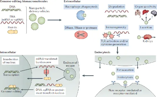
Figure B-8. Barriers to delivery of genome-editing components.
Genome-editing bio macromolecules (DNA, RNA, protein) or nanoparticles containing these bio macromolecules should avoid extracellular barriers. These barriers include phagocytosis, degradation through enzymatic means, and the induction of an immune response and the cytokines.
Adapted from Hao Yin et al., 2017
21
CHAPTER I
Targeted Genome Engineering to Control VEGF Expression in Human Umbilical Cord Blood-Derived Mesenchymal Stem Cells:
Potential Implications for the
Treatment of Myocardial Infarction
22
INTRODUCTION
Myocardial infarction (MI) is a serious health and economic problem and a primary cause of mortality in developed countries. A primary therapy for this disease is cardiac regeneration, which represents a difficult and immense challenge. During the process of MI, the coronary artery is occluded after plaque rupture. After a few weeks, the ischemic region is replaced with a thin fibrotic area that finally causes cardiac dysfunction (Vallee et al., 2012). The cardiomyocytes in the ischemic site have a limited capacity for self-regeneration, and their repopulation is almost impossible (Segers and Lee, 2008). Thus, other sources of cells and factors are required to regenerate the infarcted region of the heart.
Stem cell-based therapeutic approaches have attracted interest for heart regeneration and cardiac repair after MI due to their regenerative potency. Various trials have treated the injured myocardium using diverse mesenchymal stem cell populations (Kang et al., 2014, Karam et al., 2012, Shi and Li, 2008). Some of these studies showed restoration of damaged cardiomyocytes, enhancement of cardiac function and a reduced infarct size to certain degrees. Cell therapy using human umbilical cord blood-derived mesenchymal stem cells (hUCB-MSCs) has also shown positive effects in pre-clinical trials (Kang et al., 2014). However, stem
23
cell therapy has encountered problems, such as low survival and proliferation rates because the MI region is localized in a hostile microenvironment and the oxygen supply is low. Therefore, to acquire efficient and rapid therapeutic efficacy of stem cells, providing an intrinsic vascularization stimulus by growth factors are demanded.
Vascular endothelial growth factor (VEGF) is an important angiogenic factor that promotes the survival of endothelial cells (EC) and prevents EC apoptosis (Ferrara et al., 2003). VEGF can be used as a therapeutic reagent in ischemic injuries. Rat mesenchymal stem cells expressing VEGF transiently by adenovirus induced angiogenesis and protected the remaining cardiomyocytes in MI animal models (Matsumoto et al., 2005). VEGF protein with HGF has also been demonstrated to have a cardio-protective effect by increasing the tolerance of cardiomyocytes to ischemia and reducing cardiomyocyte apoptosis (Deuse et al., 2009). However, transient gene expression and protein injection are limited for long-term therapeutic effect accomplishment in myocardial infarction. Thus, for long-term gene expression, retrovirus-mediated gene delivery has been attempted.
However, gene delivery through a retroviral vector for cell transduction also carries a risk of stem cell neoplastic transformation and oncogene activation by the LTR promoter (Baum et al., 2003). Therefore, this retrovirus system has a limit for therapeutic application and may cause tumors and insertional mutagenesis via the incorporation of the growth factor gene into a critical region of the host chromosome. Moreover, high and uncontrollable level of VEGF itself also has the potential to cause abnormal blood vessel formation and hemangiomas (Ozawa et al.,
24 2004).
One way to overcome these issues is to use a controllable system for the transgene and to ensure integration into a safe harbor site in the chromosome. The tetracycline (Tet)-controlled system is commonly used for gene regulation in mammalian cells (Gossen and Bujard, 1992). In this system, rtTA binds to the Tet- On promoter in the presence of doxycycline (Dox) and activates Tet-On/CMV- driven gene transcription (Watanabe et al., 2007). For safe gene insertion into a safe harbor site of human chromosome, genome editing technologies have been used; zinc-finger nucleases (ZFN), Transcription activator-like effector nucleases (TALEN) and recent clustered regulatory interspaced short palindromic repeat (CRISPR)/Cas9 (Gaj et al., 2013). TALENs are targetable nucleases composed of a FokI nuclease domain and customizable DNA binding domain (Bogdanove and
Voytas, 2011). The DNA-binding domain has conserved repeats that originate from transcription activator-like effectors (TALEs) found in Xanthomonas plant pathogens. TALENs have rapidly emerged as a genome editing technology with high efficiency and low toxicity that allow safe, targeted non-viral gene delivery to a specific chromosome locus (Joung and Sander, 2013). It is known that the human chromosome 19 AAVS1 integration site is a ‘safe harbor site’ for the targeted insertion of foreign genes by producing DNA double-strand breaks and subsequent repair via homologous recombination (HR) (Mussolino et al., 2014).
In this study, we developed inducible VEGF-secreting hUCB-MSCs by TALEN-mediated VEGF gene integration into a safe harbor site of the chromosome. These engineered stem cells were capable of secreting VEGF at
25
physiological concentration upon induction by Dox treatment. VEGF/hUCB-MSCs were implanted in a myocardial infarction rat model to assess whether they could enhance angiogenesis and provide a cardioprotective effect.
26
MATERIAL AND METHODS
Isolation and culture of human UCB-MSCs
Human UCB-MSCs were isolated as previously described (Seo et al., 2011).
Human UCB-MSC isolation was performed according to the procedure approved by the Borame Hospital Institutional Review Board and Seoul National University (IRB No. 0603/001-002-07C1). The medium was changed at 48 hr intervals and the cells were subcultured after they reached 90% confluence, unless described.
Designing new TALEN vectors and Inducible VEGF donor vectors
The left and right TALEN plasmids were newly constructed as follows. The target sequences of the chromosome 19 AAVS1-targeting TALENs were: 5ʹ- TGGAGCCATCTCTCTCCTT-3ʹ (Left) – gccagaacctctaa (spacer) - 5ʹ- GGTTTGCTTACGATGGA-3ʹ (Right). The plasmids encoding the TALENs targeting this sequence were prepared as previously described (Kim et al., 2013).
To prepare the efficient targeting donor DNA, we also designed new 800 bp homology arms that were approximately 50 bp apart and flanked both sides of the TALEN target site. The homology arms were PCR-amplified from human genomic DNA and cloned into the pGEM T-Easy vector. The left and right homology arms
27
were isolated using pairs of restriction enzymes (KpnI/AgeI/NotI for the left and NotI/EcoRI/SphI for the right homology arms) and cloned into the KpnI/SphI site of the pUC19 vector.
The inducible donor vectors were constructed using three DNA fragments. The Tet-on mini-CMV promoter (Addgene® , Cambridge, MA, USA), VEGF cDNA (synthesized by Bioneer Co. Ltd) and hEF1a-rtTA-pA (Addgene® , http://www.addgene.org) were amplified by PCR using specific primers containing
the flanking sequences. Each amplified DNA fragment was cloned into the pZDonor-AAVS1-puromycin DNA vector (Sigma–Aldrich) digested with the AgeI and EcoRI restriction enzymes (New England Biolabs® ) using the In-Fusion® HD Cloning Kit (Clonetech) (Figure 1). Then, the whole insert TetO-CMV-VEGF- hEF1a-rtTA was transferred into the pUC19-AAVS1 donor vector that contained newly designed HA-L and HA-R sequences to target to the chromosome 19 AAVS1 site.
TALEN-mediated homologous recombination
For the genetic modification of hUCB-MSCs to introduce the inducible VEGF DNA construct into the safe harbor site, 8 x 10⁵ cells seeded on a 60 mm dish were transfected with TALEN left (L), right (R) (1.5 ug) and the pUC19-TetOn-CMV- VEGF-hEF1a-rtTA donor vector (3 ug). At 5 days post-transfection, the medium was changed; then, the cells were sub-cultured two times at 4.5 day intervals.
Genomic DNA was isolated from the hUCB-MSCs by adding 1 mL of the SNET extraction buffer per 106 cells (20 mM Tris-HCl [pH 8], 5 mM EDTA [pH 8], 400
28
mM NaCl, and 1% SDS) containing proteinase K (100 mg/mL; Sigma-Aldrich).
The DNA samples were incubated at 55°C for 2 hrs. Proteinase K was inactivated by incubating the DNA samples at 98°C for 10 min; then, RNase (1 mg/mL) was added and the cells were incubated at 37°C for 30 min. The DNA was precipitated and its concentration was determined by spectrophotometry. TALEN-mediated HR was detected using PCR genotyping. PCR amplification of the genomic DNA was performed using the following parameters: an initial denaturation step at 94°C for 5 min, followed by 35 cycles at 94°C for 15 s, 65°C for 45 s and 72°C for 150 s with a final extension step at 72°C for 10 min. The amplified products were analyzed on a 1% agarose gel.
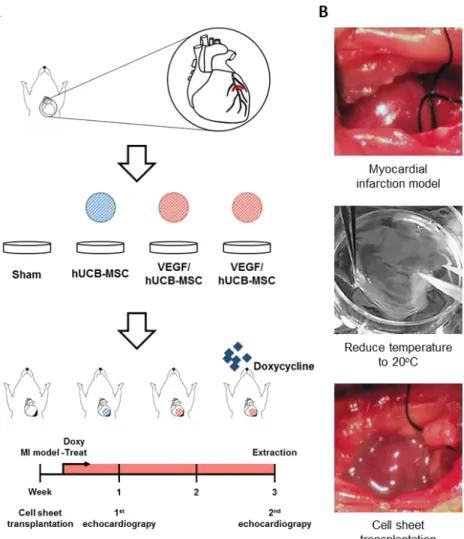
Subcutaneous transplantation of VEGF/hUCB-MSCs on cell sheets
VEGF/hUCB-MSC cell sheets were subcutaneously transplanted on the mouse
dorsal skin as previously described (Obokata et al., 2011). Briefly, VEGF/hUCB- MSCs were cultured on the UpCell surface at a density of 1x10⁶ cells per 35 mm dish. Six female mice were anesthetized and the dorsal skin was lifted and cut vertically. The cell sheet was transferred onto a PVDF membrane and the PVDF membrane was slowly removed. Doxycycline was added to the drinking water at a concentration of 1 µg/mL. Eight days after transplantation, the animals were sacrificed for analysis.
29 Cell-Matrigel transplantation into SCID mice
The animal protocol was approved by the Seoul National University Institutional Animal Care and Use Committee (IACUC) prior to the experiment. Vascular formation was evaluated in 7- to 8-week-old female SCID mice (Orient Bio, Seongnam, Korea). Matrigel was loaded with a final concentration of 1x10⁶ cells/100 µl. The injection sites were cleaned with ethanol twice prior to injection of Cell-Matrigel into each site using 1 mL syringes and 27 G needles. Eight days after the injections, the animals were sacrificed for analysis.
Myocardial infarction (MI) rat model and cell transplantation
The myocardial infarction rat models were surgically induced as previously described (Kang et al., 2014). Briefly, Male Sprague-Dawley rats weighting 260- 300 g (Orient Bio, Seongnam, Korea) were used for the MI model. All animal procedures were performed in accordance with the guidelines of the SNU IACUC.
After MI induction, the rats were divided into four treatment groups: rats that received VEGF/hUCB-MSCs (n=12, later divided into 2 groups), hUCB-MSCs (n=6), or a sham operation (Control, n=6).
Immediately after the MI induction surgery, the UpCell sheets layered with VEGF/hUCB-MSC or hUCB-MSC cells on PIPAAm-grafted surfaces were
detached from the plates by incubating at 20°C for 20 min. Then, the cell sheets were attached to polypropylene supporting membranes (Thermo Scientific) and transplanted onto the injured epicardial anterolateral area. Seven minutes after transplantation, the supporting membrane was removed and the cell sheets showed
30
stable attachment. The same procedures were repeated to make a bilayer cell sheet composed of 2x 10⁶ cells. To prevent the rat’s immune rejection of the human cells, all rats received cyclosporine as previously described (Kang et al., 2014).
Functional assessment of the infarcted myocardium
The cardiac functional assessment was performed as previously reported by our group (Kang et al., 2014). Briefly, cardiac functions were assessed by transthoracic echocardiography prior to MI surgery (normal baseline) and 1 week and 4 weeks after MI for each cell transplantation group.
Statistical Analysis
All statistical analyses were performed with SPSS version 20.0 (SPSS, Chicago, IL, USA). A Kruskal-Wallis test was used to assess differences among the groups.
The Mann-Whitney U test was performed as the post hoc test. A p value less than 0.05 was considered to be statistically significant.
31
RESULTS
Genetic engineering of an inducible VEGF-expressing DNA cassette into the hUCB-MSC chromosome by TALEN-mediated genome editing
Figure 1-1A shows the TALEN domains that consisted of specific binding sequences targeting the AAVS1 locus (safe harbor site) and the FokI non-specific endonuclease domains. As illustrated in Fig 1-1B, TALEN-mediated AAVS1 site integration of an inducible VEGF-secreting cassette was accomplished in two steps.
First, the TALEN left (L) and right (R) arms bind to their target sequences in the host chromosome and the FOKI endonuclease induces a DNA double-strand break.
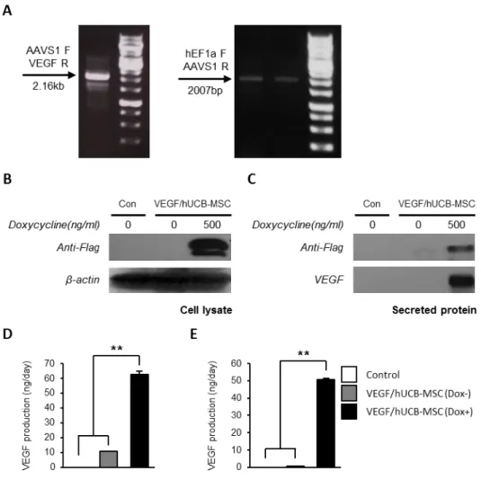
Then, the DSB is repaired by the HR machinery using a pUC19-TetO-CMV- VEGF-hEF1a-rtTA donor vector. To confirm the TALEN-mediated transgene insertion in hUCB-MSCs, the cells were co-transfected with TALEN-L/R and the pUC19-TetO-CMV-VEGF-hEF1a-rtTA donor vector. At 5 days post-transfection, the medium was changed and the cells were sub-cultured 2 times at 4.5 day intervals. After 2 weeks, the genomic DNA was extracted and PCR genotyping was performed. The amplification of 2160 bp and 2007 bp PCR fragments indicated the site-specific integration of the inducible VEGF-secreting donor cassette into the AAVS1 locus (Fig 1-2A).
32
Next, to confirm whether the inducible VEGF expression system from the VEGF/hUCB-MSCs is properly induced by treatment with Dox, western blotting
was performed on VEGF/hUCB-MSC samples. VEGF expression and secretion were detected in the cell lysates (Fig 1-2B) and secreted media (Fig 1-2C) after Dox treatment for 48 hrs. In the transient episomal plasmid transfection experiment, we observed the induction of the VEGF secretion into the media after Dox treatment by ELISA (Fig 1-2D). Similar VEGF levels were measured in the VEGF/hUCB-MSC cells with the inducible system integrated into the chromosome
by TALEN-mediated genome editing (Fig 1-2E). Too high a continuous level of VEGF is known to result in angioma formation, which is one of the main obstacles for VEGF therapy. Thus, the VEGF concentration is the main consideration for the treatment of ischemic disease with VEGF. In our study, the VEGF concentration secreted into the media following induction was 50.74 ng/106 cells/day, which was within the physiological concentration range (32.3-70 ng/106 cells/day). From these results, we confirmed that VEGF/hUCB-MSCs harboring an inducible VEGF gene in a safe harbor site secreted VEGF into the cell culture media in the physiological therapeutic concentration range.
33 Figure 1-1. Generation of VEGF/hUCB-MSCs.
(A) A schematic diagram of the TALEN binding domains in AAVS1 locus (FokI denotes the FokI endonuclease). The colored DNA sequences indicate the TALEN- binding sites (red letters and blue letters). (B) The integration of the inducible VEGF secretion DNA cassette into the AAVS1 site following the TALEN- mediated DNA double-strand break. HR (L) and HR (R), the left and right arms for
34
homologous recombination. FP and RP, forward primer and reverse primer binding sites for the PCR primers.
35
Figure 1-2. Confirmation of conditional VEGF secretion.
(A) Junction PCR analysis for the confirmation of the integrated inducible VEGF secretion cassette with two different PCR primer sets. For validation of the gene integration into the UCB-MSC genome, two sets of primers were used to detect the specific sites; a 2.16 Kb fragment with the AAVS1 (forward) and VEGF (reverse) region primers and a 2007 bp fragment with the hEF1a (forward) and AAVS1 (reverse) region primers. (B) Western blotting analysis detected inducible VEGF expression in VEGF/hUCB-MSC cell lysates and (C) inducible VEGF secretion in
36
the VEGF/hUCB-MSC culture media. The doxycycline treatment was maintained for 48 hrs. (D) Quantification of the secreted VEGF in the VEGF/hUCB-MSC (Dox+) culture media 2 days after transient transfection and (E) 2 weeks after stable transduction. The amount of VEGF production is indicated as ng/2x106 cells/day on the Y axis. The error bars indicate the SEM of three replicate measurements per group. (*: p< 0.05, **: P < 0.01).
37
Increased angiogenic markers and decreased cell cycle inhibitor expression by VEGF induction
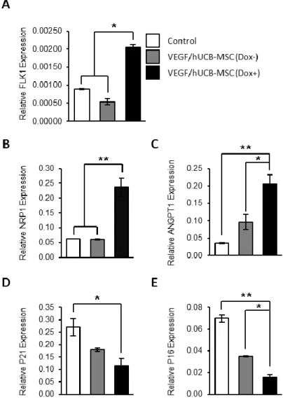
To examine the biological effects of VEGF secreted from VEGF/hUCB-MSCs, we compared the expression levels of angiogenic markers and cell cycle-related genes in each group by real-time RT-PCR. At 48 hrs after the induction of VEGF secretion by Dox, the VEGF/hUCB-MSCs showed a significant increase in the expression of the angiogenic markers FLK1, NRP1 and ANGPT1, and a reduction in the expression of the cell cycle arrest- and senescence-related genes p21 and p16 (Fig 1-3). These results indicate that the VEGF/hUCB-MSCs exhibit endothelial cell-like marker characteristics through VEGF autocrine effects.
38
Figure 1-3. Effect of VEGF secretion from VEGF/hUCB-MSCs on the gene expression of neo-angiogenesis markers and cell cycle regulators.
(A-C) Induced VEGF enhanced the expression of angiogenesis-related markers.
The mRNA levels of Flk-1, NRP1, and ANGPT1 in VEGF/hUCB-MSCs (Dox+) were evaluated by comparing to control hUCB-MSCs (Con) and VEGF/hUCB- MSCs (Dox-) using real-time RT-PCR. (D-E) Cell cycle regulation in VEGF/hUCB-MSCs in response to secreted VEGF. Secreted VEGF from
39
VEGF/hUCB-MSCs (Dox+) decreased cell cycle inhibitors (p21 and p16), suggesting that VEGF had a positive effect on hUCB-MSC survival. The experiments were performed in triprlicates, and the data were presented as the means±SE. (*: P < 0.05, **: P < 0.01, n=3).
40
Functional assessment of transplanted VEGF/hUCB-MSCs in a mouse model Prior to the application of VEGF/hUCB-MSCs to the MI animal model, we first tested: 1) whether VEGF was secreted properly by the VEGF/hUCB-MSCs upon Dox treatment in vivo and 2) whether VEGF secreted from VEGF/hUCB-MSCs could enhance neo-angiogenesis.
To confirm VEGF secretion by the implanted VEGF/hUCB-MSCs, we divided the mice into 3 groups: the hUCB-MSC cell only group, the VEGF/hUCB-MSC without Dox group and the VEGF/hUCB-MSC with Dox group. Using an UpCell transplantation system, hUCB-MSC or VEGF/hUCB-MSC cell sheets were transplanted onto the skin on the backs of the mice. For the VEGF/hUCB-MSC (Dox+) group, the mice were freely administered Dox in their drinking water (1 ug/mL) for 4 days starting one day after UpCell transplantation of VEGF/hUCB- MSCs (Fig 1-4A). VEGF expression was detected in the transplanted subcutaneous tissues in the Dox(+) group mice by Western blot analysis using anti-Flag and anti- VEGF antibodies (Fig 1-4B). The amount of secreted VEGF from the extracted subcutaneous tissue was quantified by ELISA. Significantly higher levels of VEGF were detected in the VEGF/hUCB-MSC (Dox+) group mice (15.4 ng/mg) compared to the Dox- and cell only groups (Fig 1-4C).
To test the angiogenetic function of the VEGF/hUCB-MSCs, we subcutaneously injected the mice with Matrigel mixed with VEGF/hUCB-MSCs (Fig 1-4D). The injected Matrigel plugs were removed 8 days after implantation. The plugs of the Dox-fed mice showed a more reddish color compared with the group not provided Dox, indicating greatly enhanced angiogenesis (Fig 1-4E). Immunostaining of the
41
Matrigel with an anti-VEGF antibody (Fig 1-4F) and an anti-vWF antibody (Fig 1- 4G) showed high levels of VEGF secretion and higher levels of blood vessel formation in the doxycycline-treated group. These results demonstrated that the inducible VEGF-secreting hUCB-MSC cells secreted VEGF within normal physiological ranges and induced neo-vascularization in vivo following doxycycline treatment.
42
43
Figure 1-4. The induction of VEGF expression by VEGF/hUCB-MSCs transplanted on mouse skin and neo-angiogenesis by secreted VEGF in Matrigel plugs.
(A) In vivo test design for the transplantation of the UpCell seeded with hUCB- MSCs and VEGF/hUCB-MSCs. (B-C) Western blot and ELISA analysis for VEGF secreted at the UpCell transplantation site. Error bars indicate the SEM of three replicate measurements per clone or population. (**: P < 0.01, n=3). (D) Design of the blood vessel induction test in Matrigels with VEGF/hUCB-MSCs. (E) Blood vessels formed in the Matrigel containing VEGF/hUCB-MSCs without doxycycline (Dox-) and with doxycycline (Dox+). (F-G) Immunohistochemical staining for VEGF and newly formed vessels by vWF in Matrigel with VEGF/hUCB-MSCs (Dox- or Dox+). (Scale bar: 100 μm).
44
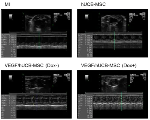
Enhanced cardiac function by VEGF secretion from VEGF/hUCB-MSCs in the rat MI model
Next, we tested whether the VEGF/hUCB-MSCs could enhance cardiac function upon VEGF induction in the rat myocardial infarction model. After MI generation in 6 rats each group total 24 rats including sham control, hUCB-MSCs or VEGF/hUCB-MSCs grown on the UpCell cell sheets were transplanted into the
infarcted region of the rat hearts; a Sham operation without MI generation was also performed as a control. Echocardiography was performed to evaluate the therapeutic efficacy of VEGF secretion in combination with VEGF/hUCB-MSC cells in the rat MI-induced heart. Healthy heart function indicators (i.e., ejection fraction (EF), fractional shortening (FS), left ventricle inner diameter at diastole (LVIDd) and left ventricle inner diameter at systole (LVIDs)) were measured 1 week and 3 weeks after the coronary artery ligation and implantation (Fig 1-5A &
B). One week after transplantation of VEGF/hUCB-MSCs (Dox+), the values of LVIDd, LVIDs, EF and FS showed improved heart function compared with the hUCB-MSC alone and VEGF/hUCB-MSC (Dox-) groups. Further improved cardiac functions were observed 3 weeks after the treatment. LV dilation and dysfunction was reduced in the VEGF/hUCB-MSC (Dox+) group compared to the control and other groups (Fig 1-6). Three weeks after implantation, the ejection fraction in the VEGF/hUCB-MSC (Dox+) group (65.39 ± 3.5%) was much higher than the ejection fraction in the control (35.31 ± 4.10%), hUCB-MSC (49.95 ± 4.19%), and VEGF/hUCB-MSC (Dox-) (49.24 ± 2.92%) groups (Fig 1-7A).
Similarly, fraction shortening in the VEGF/hUCB-MSC (Dox+) group (31.87 ±
45
1.94%) was significantly higher compared to the control (14.61 ± 1.88%), hUCB- MSC (22 ± 1.87%), and VEGF/hUCB-MSC (Dox-) (21.80 ± 2.28%) groups (Fig 1- 7B). Although the LVIDd values in the hUCB-MSC alone (8.71 ± 0.32 mm) and VEGF/hUCB-MSC (Dox-) (8.43 ± 0.87 mm) groups were slightly reduced compared to the control group (9.83 ± 0.61 mm), the VEGF/hUCB-MSC (Dox+) group (6.42 ± 0.26 mm) showed a significant reduction (p<0.01) (Fig 1-7C & D).
46
Figure 1-5. Engraftment of VEGF/hUCB-MSCs.
(A) Schematic illustration of treatment of MI by VEGF/hUCB-MSCs.
Echocardiography measurements performed one week and 3 weeks after MI induction and cell transplantation. (B) The image of an MI and the transplantation process of the UpCell system.
47
Figure 1-6. Representative echocardiogram images.
Representative echocardiogram images in the MI site after 3 weeks. The infarcted rat heart served as the control for transplantation with hUCB-MSCs, VEGF/hCUB- MSC (Dox-), and VEGF/hUCB-MSC (Dox+). In the quantitative analysis, treatment of VEGF/hUCB-MSCs with doxycycline significantly enhanced the cardiac function in one week and 3 weeks. The VEGF/hUCB-MSC (Dox+) group showed.
48 Figure 1-7. Recovery of cardiac function.
(A) an increase in the ejection fraction (EF) and (B) fractional shortening (FS) and (C) a decrease in the left ventricle inner diameter at diastole (LVIDd) and (D) left ventricle inner diameter at systole (LVIDs). (*: P < 0.05, **: P < 0.01, n=6 each group). All values represent the mean ± SEM.
49
Reduced MI size and fibrosis but thicker left ventricle due to VEGF secretion by VEGF/hUCB-MSCs in the rat MI model
We conducted histological and histomorphometric analyses to better understand the improvement of cardiac function by Masson’s trichrome staining of the rat hearts at 3 weeks after implantation. Fibrosis due to MI appears blue and the preserved myocardium appears red in Masson’s trichrome staining. Dramatic left ventricle (LV) wall thinning was detected in the MI model in the hUCB-MSC alone group. The VEGF/hUCB-MSC (Dox-) group showed a minor protective effect. However, a large anti-fibrosis effect was observed in the LV wall in the rats treated with VEGF/hUCB-MSCs (Dox+) (Fig 1-8). The MI size was greatly reduced in the VEGF/hUCB-MSC (Dox+) group compared to the MI control, hUCB-MSC alone, and VEGF/hUCB-MSC (Dox-) groups (Fig 1-9). The ventricular fibrosis rate was significantly reduced in the VEGF/hUCB-MSC (Dox+)-treated rats compared with the other groups (Fig 1-9). However, the LV wall thickness of the VEGF/hUCB-MSC (Dox+) group was significantly thicker than the MI, hUCB-MSC alone, and VEGF/hUCB-MSC (Dox-) groups (Fig 1-9).
Immunohistochemical staining was performed to investigate Dox-induced VEGF secretion in the infarcted area following treatment with VEGF/hUCB-MSC + Dox.
Heart tissues were stained with a VEGF antibody. The infarcted area in the VEGF/hUCB-MSC (Dox+) group showed high expression of VEGF, which was
not observed in the control, hUCB-MSC, and VEGF/hUCB-MSC (Dox-) groups (Fig 1-10A). These results suggested that VEGF was secreted properly by the VEGF/hUCB-MSC cells upon doxycycline treatment. Furthermore, anti-von
50
Willibrand factor (vWF) staining showed that the VEGF/hUCB-MSC (Dox+) group had significantly increased vessel density in the infarcted area, whereas the hUCB-MSC alone and VEGF/hUCB-MSC (Dox-) groups showed small increases in vessel density compared to the control group (Fig 1-10B). These results suggest that VEGF secreted from VEGF/hUCB-MSCs greatly enhanced neo-angiogenesis compared to stem cell therapy alone by directly and indirectly stimulating angiogenic factors or EC cell-like factors.
51
Figure 1-8. Representative images of heart sections.
Representative images of heart sections stained with Masson’s trichrome show fibrosis and wall thinning at the infarcted area. Fibrotic areas are colored in blue and the viable myocardium is colored in red.
52
Figure 1-9. Evaluation of MI size, LV fibrosis and wall thickness after the transplantation of VEGF/hUCB-MSCs.
Infarct size, percentage of LV fibrosis and LV wall thickness were compared among the different groups. (*: P < 0.05, **: P < 0.01). All values represent the mean ± SEM.
53
Figure 1-10. Immunohistochemical staining of VEGF, blood vessel formation by VEGF/hUCB-MSCs.
MI-induced rats were divided in to 4 group. Control (MI alone), treated with hUCB-MSCs, VEGF/hUCB-MSCs (Dox-), and VEGF/hUCB-MSCs (Dox+). (A) VEGF staining were performed at the infarcted area. High levels of VEGF were detected only in the VEGF/HUCB-MSC (Dox+) group. (B) Generation of blood vessels in the MI regions. More blood vessels were observed in VEGF/hUCB-MSC (Dox+) group than other groups.
54
Prolonged cell survival of transplanted human VEGF/hUCB-MSCs in the infarcted region
To determine the effects of VEGF secreted from VEGF/hUCB-MSCs (Dox+) on the survival of transplanted human stem cells, we detected transplanted VEGF/hUCB-MSC in rat hearts via immunohistochemical staining. An anti-lamin
A+C antibody was used to detect human cells in the infarcted region. Lamin A+C- positive cells were not detected in the MI control group (Fig 1-11). Significant numbers of transplanted human cells were detected above the myocardium of the hUCB-MSC alone and VEGF/hUCB-MSC (Dox-)-transplanted groups. However, in the VEGF/hUCB-MSC (Dox+) group, a large number of Lamin A+C-positive cells were detected above the myocardium (Fig 1-11). These results indicate that secreted VEGF also has a positive effect on stem cell survival and thus leads to the induction of enhanced paracrine effects and improved protection from cardiac damage.
55
Figure 1-11. Prolonged cell survival of transplanted human VEGF/hUCB- MSCs.
An anti-human Lamin A+C antibody was used to visualize the transplanted human stem cells. More human Lamin A+C positive cells were observed in rat hearts transplanted with VEGF/hUCB-MSC (Dox+). (Scale bar = 100 μm).
56
Discussion
Stem cell-based therapy has been widely investigated for the treatment of myocardial infarction. In recent studies, stem cell therapy using MSCs offered a positive result for the treatment of myocardial infarction due to paracrine effects and revascularization (Williams and Hare, 2011). However, the application of cell therapy faces many obstacles due to the low survival of the implanted cells and insufficient oxygen and nutrients. Moreover, cardiomyocytes have a limited regeneration capacity (Mangi et al., 2003). To overcome these obstacles with ischemic disease, combined therapy using vascular stimulating growth factor and MSCs is required. In our previous study, VEGF protein delivery with stem cells showed a therapeutic effect on ischemic disease (Kim et al., 2014). However, the protein therapy highlighted the limitation of the application periods due to the short half-life of the VEGF proteins (Henry et al., 2003). To overcome this limitation, several studies reported genetic modifications using retroviruses for the integration of transgenes into the host chromosome to induce their long-term expression (Misteli et al., 2010, Chen et al., 2005). However, retroviruses can produce oncogene activation or tumorigenesis due to the random and unpredictable integration of the transgene, which is a major side effect. Furthermore,
57
uncontrolled VEGF expression also causes side effects such as hemangioma and nonstandard blood vessel formation.
To address these issues, we generated an inducible VEGF-secreting stem cell using TALEN-mediated transgene integration into a specific safe harbor site by AAVS1 locus-directed homologous recombination. With this TALEN-mediated safe harbor gene delivery strategy, the human cord blood-derived mesenchymal stem cells were able to secrete VEGF upon doxycycline treatment, protect the host from cardiac damage and improve cardiac function in a rat myocardial infarction model.
In this study, we used TALENs technology to introduce an inducible VEGF- 165a gene cassette into the AAVS1 safe harbor site in human umbilical cord blood mesenchymal stem cells. The initial targeting vector with the left and right homology arm (HA-L & HA-R) cassette containing the inducible VEGF gene did not produce a sufficient number of cells integrated with the gene cassette.
Therefore, we redesigned several TALEN-L/R targeting vectors along with several HA-Ls and HA-Rs in donor vectors and tested their efficiency. Finally, we could generate a quite efficient combination of TALEN-L/R targeting vector and HA-L and HA-R donor vector cassette for the AAVS1 locus by giving a 50 bp space apart between the homology arms and the TALEN target sites. With these newly designed vectors, we could generate efficient integration of the VEGF gene cassette that resulted in the production of a physiologically relevant concentration of VEGF.
Successful stem cell-based cell transplantation combined with vascular growth factors such as VEGF would induce vascularization to treat ischemic or vascular
58
disease (Vunjak-Novakovic et al., 2011). However, excessive amounts of VEGF can induce unwanted effects. Thus, the regulation of VEGF expression is important.
The proper VEGF concentration for therapeutic angiogenesis was reported to be below 100 ng per 106 cells per day in vitro (Ozawa et al., 2004). Our VEGF/hUCB- MSCs secreted VEGF at a concentration of approximately 50 ng/106 cells/day when exposed to doxycycline (Fig 1-2E), thereby providing a safe therapeutic ranges of VEGF against vascular diseases.
In addition to the long-term expression of VEGF, the secreted VEGF has to be biologically functional and capable of binding to the VEGF receptor (Flk-1) to induce angiogenic gene expression and prevent cell cycle arrest (Yla-Herttuala and Alitalo, 2003). Our results showed that VEGF secreted from VEGF/hUCB-MSCs by Dox induction increased the expression of angiogenesis markers and down- regulated cell cycle arrest factors (Fig 1-3). These results indicate that the VEGF secreted by hUCB-MSCs gave endothelial cell-like characteristics to these cells by autocrine stimulation. This approach may assist in the neovascularization of vascular disease. We also demonstrated that VEGF secreted by hUCB-MSCs significantly enhanced ne