저작자표시-비영리-변경금지 2.0 대한민국 이용자는 아래의 조건을 따르는 경우에 한하여 자유롭게
l 이 저작물을 복제, 배포, 전송, 전시, 공연 및 방송할 수 있습니다. 다음과 같은 조건을 따라야 합니다:
l 귀하는, 이 저작물의 재이용이나 배포의 경우, 이 저작물에 적용된 이용허락조건 을 명확하게 나타내어야 합니다.
l 저작권자로부터 별도의 허가를 받으면 이러한 조건들은 적용되지 않습니다.
저작권법에 따른 이용자의 권리는 위의 내용에 의하여 영향을 받지 않습니다. 이것은 이용허락규약(Legal Code)을 이해하기 쉽게 요약한 것입니다.
Disclaimer
저작자표시. 귀하는 원저작자를 표시하여야 합니다.
비영리. 귀하는 이 저작물을 영리 목적으로 이용할 수 없습니다.
변경금지. 귀하는 이 저작물을 개작, 변형 또는 가공할 수 없습니다.
의학박사 학위논문
난소암 세포주에서 항암제와 insulin-like growth factor 1과 insulin-like growth factor 1
수용체에 대한 small interfering RNA들의 항암치료효과 증강에 관한 연구
Small interfering RNAs targeting insulin-like growth factor 1 and insulin-like growth factor
1 receptor potentiate the cytotoxic effect of chemotherapy in ovarian caner cell lines
2 0 1 2년 6월
서울대학교 대학원
의학과 산부인과학 전공
한 승 수
난소암 세포주에서 항암제와 insulin-like growth factor 1과 insulin-like growth factor 1
수용체에 대한 small interfering RNA들의 항암치료효과 증강에 관한 연구
지도교수 김 석 현
이 논문을 의학박사 학위논문으로 제출함
2 0 1 2년 6월
서울대학교 대학원
의학과 산부인과학 전공
한 승 수
한 승 수의 의학박사 학위논문을 인준함
2 0 1 2년 6월
위 원 장 ( 인)
부위원장 ( 인)
위 원 ( 인)
위 원 ( 인)
위 원 ( 인)
Small interfering RNAs targeting insulin-like growth factor 1 and insulin-like growth factor
1 receptor potentiate the cytotoxic effect of chemotherapy in ovarian caner cell lines
by
Se ungSuHa n
A ThesisSubmittedinPartialFulfillmentoftheRequirements fortheDegreeofDoctorofPhilosophyinMedicine
(Obstetrics& Gynecology)
attheSeoulNationalUniversityCollegeofMedicine
June2012
Approvedbythesiscommittee:
Professor Chairman Professor ViceChairman Professor
Professor Professor
i
Abstract
Small interfering RNAs targeting insulin-like growth factor 1 and insulin- like growth factor 1 receptor potentiate the cytotoxic effect of chemotherapy
in ovarian cancer cell lines.
Seung Su Han College of Medicine, Department of Obstetrics & Gynecology The Graduate School Seoul National University
Objectives: To determine the synthetic therapeutic effects of small interfering RNAs targeting IGF1 and IGF1R with chemotherapy in ovarian cancer cell lines.
Material and methods: We used three ovarian cancer cell lines OVCAR3, SKOV3 and SNU119 in this study. Small interfering RNAs (siRNAs) targeting human IGF1 and IGF1R mRNAs were transfected in each cell line. We firstly investigated the potency of siRNAs by real-time RT-PCR in each cell line. Cytotoxicity of IGF1or IGF1R siRNAs was measured by MTT assay in each ovarian cancer cell line. The therapeutic effect of combination of IGF1/IGF1R siRNAs and paclitaxel/carboplain was also assessed by MTT assay. Cell invasion assay was determined by Matrigel test
Results: An effective gene silencing of both IGF1 and IGF1R by siRNAs occurred in SNU119:
63% silencing of IGF1 and 73% silencing of IGF1R. OVCAR3 showed 78.5% silencing efficacy of IGF1 siRNA. The silencing efficacy of IGF1R siRNA was 62.1% in SKOV3. Cell viability was 58.1±5.6%, 87.1±9.7%, and 45.2±6.5% respectively after 72hrs of long term treatment IGF1 siRNA, IGF1R siRNA, and IGF1 siRNA + IGF1R siRNA in SNU119. Cell viability was decreased to 34.9±5.4% and 45.9±2.4% with 48hrs treatment of paclitaxel and carboplatin in SNU119, respectively. Cell viability after 48hrs was more decreased to 6.9±3.5%
ii
by IGF1 siRNA + paclitaxel and 13.8±5.9% by IGF1R siRNA + paclitaxel in SNU119. Cell viability after treatment of IGF1 siRNA was decreased to 40.3±9.1% in OVCAR3. Concurrent treatment of IGF1 siRNA and paclitaxel/carboplatin showed more cytotoxic effect. Synthetic treatment of IGF1R siRNA and paclitaxel/carboplatin also proved to be more cytotoxic in SKOV3. Cell invasion assay showed that cell invasion rate was decreased to 55±8.9% after treatment of IGF1 siRNA + IGF1R siRNA compared with IGF1 siRNA (92.5±15.5%) and IGF1R siRNA (81.7±7.9%).
Conclusions: IGF1 and IGF1R gene expression were well expressed in ovarian cancer cell lines.
Single IGF1 or IGF1R siRNA showed effective cytotoxicity. Concurrent silencing of IGF1 and IGF1R genes more potently sensitized cancer cells to paclitaxel or carboplatin-induced cytotoxicity in ovarian cancer cell line. Treatment of IGF1 or IGF1R siRNA also was effective in decreasing cell invasion.
Key words: siRNA, IGF1, IGF1R, ovarian cancer, carboplatin, paclitaxel
Student Number: 2008-30548
iii
List of Tables
Table 1. Primers of IGF1 and IGF1R for real time PCR primer design---8 Table 2. Design of IGF1 and IGF1R siRNA in ovarian cancer cell lines---10 Table 3. The reduction rate of IGF1 and IGF1R by siRNAs in each ovarian cancer cell line
---11 Table 4. Cell viability after paclitaxel and carboplatin treatment---12 Table 5. Mean cell viability after IGF1/IGF1R siRNAs transfection or anticancer drugs in
ovarian cancer cell lines---16
iv
List of Figures
Figure 1.
Ct values of IGF1 and IGF1R in each ovarian cancer cell line---9
Figure 2. Cytotoxicity assay after IGF1/IGF1R siRNAs transfection or anticancer drugsin SNU-119: (a) cytotoxicity after IGF1/IGF1R siRNAs transfection (b) cytotoxicity after anticancer drugs treatment (c) cytotoxicity after both IGF1/IGF1R siRNAs transfection and anticancer drugs treatment---13 Figure 3. Cytotoxicity assay after IGF1 siRNA transfection or anticancer drugs in OVCAR3:
(a) cytotoxicity after IGF1 siRNA transfection (b) cytotoxicity after anticancer drugs treatment (c) cytotoxicity after both IGF1 siRNA transfection and anticancer drugs treatment---14 Figure 4. Cytotoxicity assay after IGF1R siRNA transfection or anticancer drugs in SKOV3:
a) cytotoxicity after IGF1R siRNA transfection (b) cytotoxicity after anticancer drugs treatment (c) cytotoxicity after both IGF1R siRNA transfection and anticancer drugs treatment---15 Figure 5. Cell invasion assay after IGF1/IGF1R siRNAs transfection in SNU-119---18
v
List of Abbreviations
IGF1: insulin-like growth factor 1
IGF1R: insulin-like growth factor 1 receptor siRNA: small interfering RNA
vi
Contents
I. Introduction --- 1
II. Materials and Methods --- 3
III. Results --- 6
IV. Discussion --- 19
V. References --- 23
VI. 국문초록 --- 27
1
Introduction
Ovarian cancer is one of the most lethal gynecologic malignancies. Epithelial ovarian cancer shows a reasonable clinical response to debulking surgery and chemotherapy, but about 80% of patients experience recurrences. This chemoresistance is the cause of ovarian cancer-related deaths. Now, we have no available drugs to cure recurrent ovarian cancer. Thus, it is very important to develop new therapeutic modalities that will cure and salvage the patients with recurrent ovarian cancer.
It is very well known that both insulin-like growth factor1 (IGF1) and insulin-like growth factor1 receptor (IGF1R) are involved in the development and progression of cancer.1 IGF1R is the most prominent factor and has been extensively studied for its role in the proliferation and differentiation of cancer cells.2 IGF1 is a small, single-chain polypeptide ligand (7-8 kD) that is derived from prepropeptides in a similar way to insulin, but contains the C-peptide bridge between B and A chains.3 IGF1R is a transmembrane heterotetrameric protein, encoded by the IGF1R gene located on chromosome 15q25–q26. IGF1R is composed of two and two subunits linked by disulfide bonds. The extracellular subunit is responsible for ligand binding, whereas the subunit consists of a transmembrane domain and a cytoplasmic tyrosine kinase domain.4
Elevated plasma concentrations of IGF1 have been linked to a high risk for several types of cancers including breast, prostate, and lung cancer.1, 5, 6 Two recent prospective studies reported a higher ovarian cancer risk in the top tertile group of IGF1 level among women aged 55 or less at time of diagnosis.7, 8 High free IGF1 protein expression was independently associated with the progression of ovarian cancer and IGF1 mRNA expression was also associated with disease progression in ovarian cancer.9 Genes of IGF1 and IGF1R were overexpressed in tumors associated with an unfavorable prognosis.10 Shen et al. reported that IGF1 stimulates KCl cotransport, which is necessary for invasion and proliferation of ovarian cancer cells.11 IGF1 also induced cyclooxygenase-2 partly by enhancing vascular endothelial growth factor
2
production with the activation of PI3K, MAPK, and PKC pathways12 and stimulated the migration of SKOV-3 cells by favoring the urokinase-type plasminogen activator over the plasminogen activator inhibitor-1 through the PI3K/AKT pathway.6
The IGF1R is mainly involved in regulation of cell proliferation, antiapoptosis, differentiation and cell motility.13 There are various approaches to inhibit IGF1R such as IGF1R antisense oligonucleotides, IGF1R–blocking antibodies, and small-molecule tyrosine kinase inhibitors.
BMS-554417 (IGF1R kinase inhibitor) showed an antiproliferative effect and also induced apoptosis in OV202 cells.14 IGF1R specific antibody, designated 486/STOP, could reverse transformed phenotype of the CaOV-3 cells in vitro and inhibit tumorigenicity in vivo.15 However, the stability and delivery efficacy of these drugs seems to be limited in exerting an inhibitory effect on the targeted molecule.13
RNA interference is the sequence-specific, posttranscriptional gene-silencing method used as a research tool, and its clinical use is now being explored. The treatment of RNA-mediated interference has high efficiency and specificity and could be a powerful and widely used tool in cancer therapy.16
In this study, small interfering RNAs (siRNAs) targeting to the IGF1 gene or IGF1R gene were introduced into human ovarian cancer cell lines. We investigated their respective or synergistic effects without or with chemotherapy on cellular cytotoxicity and invasion in ovarian cancer cell lines.
3
Material and Methods
Cell lines and cell culture
We used the human ovarian cancer cell lines OVCAR3, SKOV3 and SNU119 in this study.
Cancer cell lines were purchased from the Korean Cell Line Bank (Seoul, Korea). All cell lines were cultured in Dulbecco's Modified Eagle's Medium (DMEM; Gibco BRL, Rockville, MD) supplemented with 10% fetal bovine serum (FBS) and antibiotics (penicillin, 100 units/mL and streptomycin, 100 μg/mL) in a humidified 5% CO2 atmosphere at 37˚C.
Quantification of IGF1 and IGF1R gene transcripts by quantitative Real time PCR IGF1 and IGF1R gene expression was measured by real-time RT-PCR using the IGF1 forward primer: 5’-ACCTTGCAAAAATGGTCCTG-3’, and the reverse primer: 5’- GCAGCCAAGATTCAGAGAGG-3’ and the IGF1R forward primer: 5’-
AACCCCAAGACTGAGGTGTG-3’, and the reverse primer: 5’-
TGACATCTCTCCGCTTCCTT-3’. (Table 1) For real-time PCR, 50ng of RNA was amplified in 25μL containing 10pM IGF1R and IGF1 gene specific primers and 12.5μL SYBR premix One Step SYBR ⓇPrimeScriptTM RT-PCR Kit (Perfect Real Time) (Takara Bio, Kyoto, Japan).
PCR conditions were Stage 1:Reverse Transcription, 42℃ 5 min. 95℃ 10 sec. Stage 2:PCR reaction Repeat:40 times. Human β-actin gene was amplified as an internal control to normalize the amount of RNA from each sample.
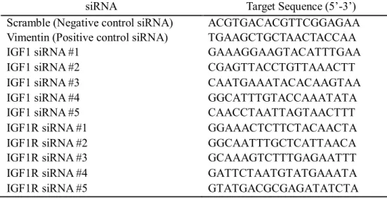
Silencing of IGF1 and IGF1R genes by siRNA in ovarian cancer cell lines
Small interfering RNAs targeting human IGF1 and IGF1R mRNAs were designed and purchased from Genolution Pharmaceuticals (Seoul, Korea). Transfection was performed with G-Fectin (Genolution Pharmaceuticals, Seoul, Korea) according to the manufacturer’s instructions. To examine the effect of each siRNA, we added 50ul of PBS and 1ul of carrier into each tube for final 10nM concentration of siRNA and mixed briefly and left at room
4
temperature for 10-15 min. Spilt cells and added 450ul of media were seeded at 1.2 x 10⁵ cells per well in 24 well plates. After 10-15 min, we just added the lipoplex to each intended plate.
The cells were harvested 24 hr after transfection. To check the level of IGF1 and IGF1R gene transcripts in cells, total RNA were isolated by Trizol extraction.
Carboplatin and paclitaxel IC50 test
Carboplatin and paclitaxel in phosphate-buffered saline (PBS) was added directly to the media at differing doses ranging from 40ng/well to 640ng/well and from 15ng/well to 240ng/well, respectively. The cells were incubated with carboplatin and paclitaxel at 37°C for 24 and 48 hours for each trial. Media was aspirated out at the end of the treatment period and replaced with complete media. Inhibitory concentration that kills 50% of the cell population (IC50) for carboplatin or paclitaxel was obtained.
Monitoring of the cytotoxicity with treatment of IGF1/IGF1R siRNAs and anti-cancer drugs by cytotoxicity assay
Cytotoxicity was measured by the 3-(4,5-dimethylthiazol-2-yl)-2,5-diphenyltetrazolium bromide (MTT;Sigma) assay in accordance with the manufacturer’s instructions. Ovarian cancer cells (5x10³ cells/well) were seeded in 96-well microplates. The next day, the cells were treated with IGF1 or IGF1R siRNA. Scheduled dose of carboplatin or paclitaxel was administered to the well plate after 24 hrs IGF1 or IGF1R siRNA transfection. MTT was added (20 mL/well of 5 g/L solution in PBS) after culture for 24, 48, and 72 hours. When incubated at 37.8C for 4 hours, the reaction was stopped by addition of 100 mL dimethylsufoxide (Sigma).
The dark-blue crystals of MTT–formazan was dissolved by shaking the plates at room temperature for 15 minutes and the absorbance of each well was measured on a Bio-Rad Microplate Reader (Bio-Rad) at a wavelength of 490 nm. Growth inhibition (%) was calculated using the following formula: [1‒ (A/B)]x100, where A was the absorbance of treated cells and B was the absorbance of untreated control cells. All samples were assayed repeatedly in six wells.
5
Cell invasion assay with treatment of IGF1 and IGF1R siRNA
Eight hours after treatments, one hundred thousand cells were added per transwell invasion chamber coated with 1-2mg/ml Matrigel (reconstituted basement membrane; BD Biosciences, Mississauga, ON). Cells were allowed to invade for 22 h. Cells were fixed for 30 minutes in methanol, stained for 30 minutes with 1% crystal violet and the number of invaded cells was counted per field of view at 20x magnification.
6
Results
Table 1 shows the primers of IGF1 and IGF1R for real time PCR primer design. The IGF1/IGF1R gene expression was confirmed by real time PCR in each ovarian cancer cell line.
(Figure 1) We made five IGF1 and IGF1R target siRNAs using si-direct program. (Table 2)
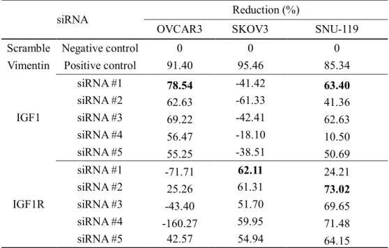
Reduction rates of IGF1 and IGF1R by siRNAs in each ovarian cancer cell line
The reduction rates of IGF1 and IGF1R siRNA were 63% and 73% in SNU119, respectively.
The silencing efficacy of IGF1 siRNA was 78% in OVCAR3. The silencing efficacy of IGF1R siRNA was 62% in SKOV3. (Table 3) The silencing efficacy of IGF1R siRNA was not significant in OVCAR3. The silencing efficacy of IGF1 siRNA was not appeared in SKOV3.
Cell viability after paclitaxel and carboplatin treatment
Cell viability was checked after 24 hrs and 48hrs treatment of paclitaxel and carboplatin.
(Table 4) IC50 values of paclitaxel and carboplatin were assessed in each ovarian cancer cell line. IC50 values of paclitaxel and carboplatin were 37.5ng/well and 300ng/well in SNU119, respectively. IC50 values of paclitaxel and carboplatin were 22.5ng/well and 160ng/well in OVCAR3, respectively. The IC50 values of paclitaxel and carboplatin in SKOV3 were same to IC50 values in OVCAR3.
Cytotoxicity assay after IGF1/IGF1R siRNAs transfection or anticancer drugs in each ovarian cancer cell line
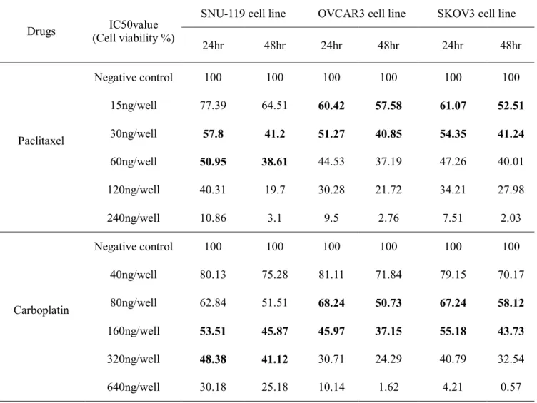
Cytotoxicity by MTT assay was sequentially achieved with treatment of IGF1 or IGF1R siRNA, paclitaxel or carboplatin, and concurrent IGF1 or IGF1R siRNA and paclitaxel or carboplatin. Time-dependent decrease of cell viabilities with transfection of IGF1 or IGF1R siRNA was shown in ovarian cancer cell lines. (Figure 2, Figure 3, Figure 4) Cell viability was 58.1±5.6%, 87.1±9.7%, and 45.2±6.5% respectively after 72hrs of long term treatment IGF1 siRNA, IGF1R siRNA, and IGF1 siRNA + IGF1R siRNA compared with negative control siRNA in SNU119 . Cell viability was decreased to 34.9±5.4% and 45.9±2.4% with 48hrs
7
treatment of paclitaxel and carboplatin in SNU119, respectively. Cell viability after 48hrs was more decreased to 6.9±3.5% by IGF1 siRNA + paclitaxel and 13.8±5.9% by IGF-1R siRNA + paclitaxel in SNU119. Cell viability after 48hrs was more decreased to 10.3±5.9% by IGF1 siRNA + carboplatin and 3.5±1.2% by IGF-1R siRNA + carboplatin in SNU119. Cell viability after 72hrs by IGF1 siRNA + paclitaxel and IGF-1R siRNA + paclitaxel was 0%. Cell viability after 72hrs by IGF1 siRNA + carboplatin and IGF-1R siRNA + carboplatin also was 0%. Cell viability after 24hrs by IGF1 or IGF1R siRNA + paclitaxel + carboplatin was 0% in SNU119.
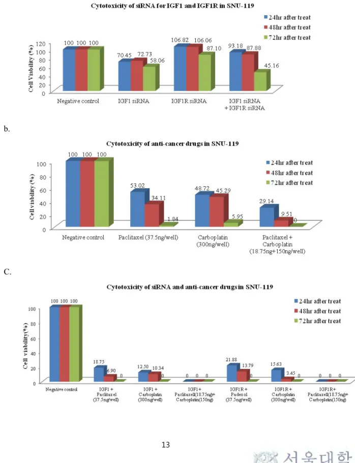
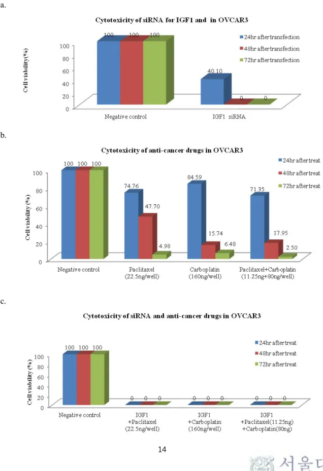
Cell viability after 24hrs and 48hrs treatment of IGF1 siRNA was decreased to 40.3±9.1% and 0% in OVCAR3, respectively. Concurrent treatment of IGF1 siRNA and paclitaxel/carboplatin showed 0% cell viability in OVCAR3. Synthetic treatment of IGF1R siRNA and paclitaxel/carboplatin also proved to be more cytotoxic than single IGF1R siRNA or paclitaxel/carboplatin in SKOV3. Table 5 summarizes mean cell viability in ovarian cancer cell lines.
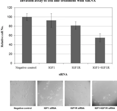
Cell invasion assay after IGF1/IGF1R siRNAs transfection in SNU-119
Cell invasion assay showed that cell invasion rate was decreased to 92.5±15.5% and 81.7±7.9% after treatment of IGF1 siRNA and IGF1R siRNA in SNU119, respectively.
Furthermore, concurrent silencing of IGF1 and IGF1R showed 55±8.9% synergistic decrease of cell invasion in SNU 119. (Figure 5)
8
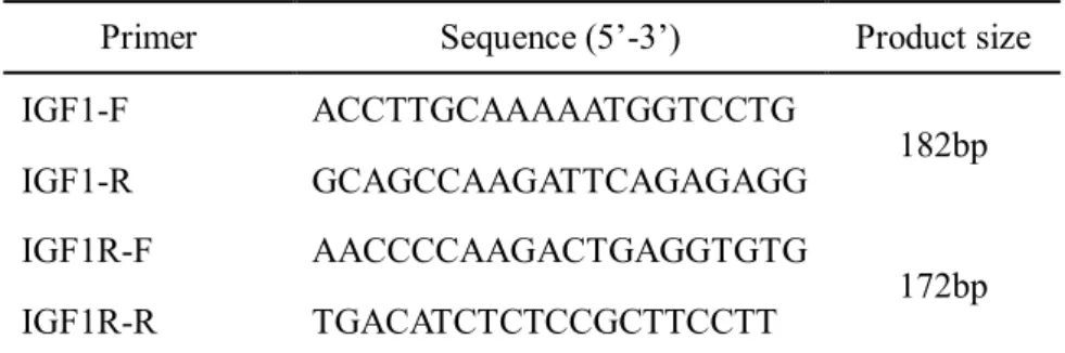
Table 1. Primers of IGF1 and IGF1R for real time PCR primer design
Primer Sequence (5’-3’) Product size
IGF1-F ACCTTGCAAAAATGGTCCTG
182bp
IGF1-R GCAGCCAAGATTCAGAGAGG
IGF1R-F AACCCCAAGACTGAGGTGTG
172bp
IGF1R-R TGACATCTCTCCGCTTCCTT
9
Figure 1. Ct values of IGF1 and IGF1R in each ovarian cancer cell line
10
Table 2. Design of IGF1 and IGF1R siRNA in ovarian cancer cell lines
siRNA Target Sequence (5’-3’)
Scramble (Negative control siRNA) ACGTGACACGTTCGGAGAA Vimentin (Positive control siRNA) TGAAGCTGCTAACTACCAA
IGF1 siRNA #1 GAAAGGAAGTACATTTGAA
IGF1 siRNA #2 CGAGTTACCTGTTAAACTT
IGF1 siRNA #3 CAATGAAATACACAAGTAA
IGF1 siRNA #4 GGCATTTGTACCAAATATA
IGF1 siRNA #5 CAACCTAATTAGTAACTTT
IGF1R siRNA #1 GGAAACTCTTCTACAACTA
IGF1R siRNA #2 GGCAATTTGCTCATTAACA
IGF1R siRNA #3 GCAAAGTCTTTGAGAATTT
IGF1R siRNA #4 GATTCTAATGTATGAAATA
IGF1R siRNA #5 GTATGACGCGAGATATCTA
11
Table 3. The reduction rate of IGF1 and IGF1R by siRNAs in each ovarian cancer cell line
siRNA Reduction (%)
OVCAR3 SKOV3 SNU-119
Scramble Negative control 0 0 0
Vimentin Positive control 91.40 95.46 85.34
IGF1
siRNA #1 78.54 -41.42 63.40
siRNA #2 62.63 -61.33 41.36
siRNA #3 69.22 -42.41 62.63
siRNA #4 56.47 -18.10 10.50
siRNA #5 55.25 -38.51 50.69
IGF1R
siRNA #1 -71.71 62.11 24.21
siRNA #2 25.26 61.31 73.02
siRNA #3 -43.40 51.70 69.65
siRNA #4 -160.27 59.95 71.48
siRNA #5 42.57 54.94 64.15
12
Table 4. Cell viability after paclitaxel and carboplatin treatment
Drugs IC50value
(Cell viability %)
SNU-119 cell line OVCAR3 cell line SKOV3 cell line 24hr 48hr 24hr 48hr 24hr 48hr
Paclitaxel
Negative control 100 100 100 100 100 100
15ng/well 77.39 64.51 60.42 57.58 61.07 52.51
30ng/well 57.8 41.2 51.27 40.85 54.35 41.24
60ng/well 50.95 38.61 44.53 37.19 47.26 40.01
120ng/well 40.31 19.7 30.28 21.72 34.21 27.98
240ng/well 10.86 3.1 9.5 2.76 7.51 2.03
Carboplatin
Negative control 100 100 100 100 100 100
40ng/well 80.13 75.28 81.11 71.84 79.15 70.17
80ng/well 62.84 51.51 68.24 50.73 67.24 58.12
160ng/well 53.51 45.87 45.97 37.15 55.18 43.73
320ng/well 48.38 41.12 30.71 24.29 40.79 32.54
640ng/well 30.18 25.18 10.14 1.62 4.21 0.57
13
Figure 2. Cytotoxicity assay after IGF1/IGF1R siRNAs transfection or anticancer drugs in SNU-119: (a) cytotoxicity after IGF1/IGF1R siRNAs transfection (b) cytotoxicity after anticancer drugs treatment (c) cytotoxicity after both IGF1/IGF1R siRNAs transfection and anticancer drugs treatment
a.
b.
C.
14
Figure 3. Cytotoxicity assay after IGF1 siRNA transfection or anticancer drugs in OVCAR3: (a) cytotoxicity after IGF1 siRNA transfection (b) cytotoxicity after anticancer drugs treatment (c) cytotoxicity after both IGF1 siRNA transfection and anticancer drugs treatment
a.
b.
c.
15
Figure 4. Cytotoxicity assay after IGF1R siRNA transfection or anticancer drugs in SKOV3: (a) cytotoxicity after IGF1R siRNA transfection (b) cytotoxicity after anticancer drugs treatment (c) cytotoxicity after both IGF1R siRNA transfection and anticancer drugs treatment
a.
b.
c.
16
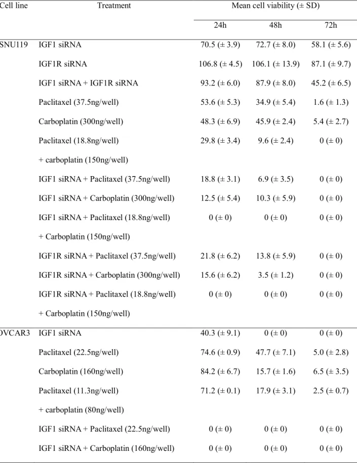
Table 5. Mean cell viability after IGF1/IGF1R siRNAs transfection or anticancer drugs in ovarian cancer cell lines.
Cell line Treatment Mean cell viability (± SD)
24h 48h 72h
SNU119 IGF1 siRNA 70.5 (± 3.9) 72.7 (± 8.0) 58.1 (± 5.6)
IGF1R siRNA 106.8 (± 4.5) 106.1 (± 13.9) 87.1 (± 9.7)
IGF1 siRNA + IGF1R siRNA 93.2 (± 6.0) 87.9 (± 8.0) 45.2 (± 6.5) Paclitaxel (37.5ng/well) 53.6 (± 5.3) 34.9 (± 5.4) 1.6 (± 1.3) Carboplatin (300ng/well) 48.3 (± 6.9) 45.9 (± 2.4) 5.4 (± 2.7) Paclitaxel (18.8ng/well)
+ carboplatin (150ng/well)
29.8 (± 3.4) 9.6 (± 2.4) 0 (± 0)
IGF1 siRNA + Paclitaxel (37.5ng/well) 18.8 (± 3.1) 6.9 (± 3.5) 0 (± 0) IGF1 siRNA + Carboplatin (300ng/well) 12.5 (± 5.4) 10.3 (± 5.9) 0 (± 0) IGF1 siRNA + Paclitaxel (18.8ng/well)
+ Carboplatin (150ng/well)
0 (± 0) 0 (± 0) 0 (± 0)
IGF1R siRNA + Paclitaxel (37.5ng/well) 21.8 (± 6.2) 13.8 (± 5.9) 0 (± 0) IGF1R siRNA + Carboplatin (300ng/well) 15.6 (± 6.2) 3.5 (± 1.2) 0 (± 0) IGF1R siRNA + Paclitaxel (18.8ng/well)
+ Carboplatin (150ng/well)
0 (± 0) 0 (± 0) 0 (± 0)
OVCAR3 IGF1 siRNA 40.3 (± 9.1) 0 (± 0) 0 (± 0)
Paclitaxel (22.5ng/well) 74.6 (± 0.9) 47.7 (± 7.1) 5.0 (± 2.8) Carboplatin (160ng/well) 84.2 (± 6.7) 15.7 (± 1.6) 6.5 (± 3.5) Paclitaxel (11.3ng/well)
+ carboplatin (80ng/well)
71.2 (± 0.1) 17.9 (± 3.1) 2.5 (± 0.7)
IGF1 siRNA + Paclitaxel (22.5ng/well) 0 (± 0) 0 (± 0) 0 (± 0) IGF1 siRNA + Carboplatin (160ng/well) 0 (± 0) 0 (± 0) 0 (± 0)
17 Table 5. (Continued)
Cell line Treatment Mean cell viability (± SD)
24h 48h 72h
OVCAR3 IGF1 siRNA + Paclitaxel (11.3ng/well) + Carboplatin (80ng/well)
0 (± 0) 0 (± 0) 0 (± 0)
SKOV3 IGF1R siRNA 70.2 (± 8.4) 84.5(± 3.9) 32.7 (± 3.2)
Paclitaxel (22.5ng/well) 93.3 (± 2.7) 30.9 (± 5.0) 3.6 (± 2.1) Carboplatin (160ng/well) 156 (± 2.7) 63.7 (± 2.5) 32 (± 4.1) Paclitaxel (11.3ng/well)
+ carboplatin (80ng/well)
77.1 (± 9.4) 22.1 (± 2.5) 10.9 (± 4.1)
IGF1R siRNA + Paclitaxel (22.5ng/well) 66.6 (± 6.7) 16.4 (± 2.6) 5.1 (± 1.9) IGF1R siRNA + Carboplatin (160ng/well) 67.9 (± 5.1) 57.3 (± 6.4) 15.7 (± 2.6) IGF1R siRNA + Paclitaxel (11.3ng/well)
+ Carboplatin (80ng/well)
29.8 (± 1.7) 4.5 (± 1.3) 0 (± 0)
18
Figure 5. Cell invasion assay after IGF1/IGF1R siRNAs transfection in SNU-119
19
Discussion
The purpose of current study was to determine the cytotoxic effect of IGF1 or IGF1R siRNAs and the synthetic therapeutic effects of IGF1 or IGF1R siRNAs with chemotherapy in ovarian cancer cell lines. IGF1 or IGF1R gene was well expressed in ovarian cancer cell lines. However, the efficacy of IGF1 or IGF1R siRNAs was different according to cancer cell lines. Single IGF1 or IGF1R siRNA showed effective cytotoxicity. Treatment of both IGF1 siRNA and IGF1R siRNA showed more effective cytotoxicity than single siRNA. Concurrent silencing of IGF1 and IGF1R genes more potently sensitized cancer cells to paclitaxel or carboplatin-induced cytotoxicity in ovarian cancer cell line. Treatment of IGF1 or IGF1R siRNA also was effective in decreasing cell invasion. To the best of our knowledge, this study is the first report to investigate the synthetic therapeutic cytotoxicity of IGF1 or IGF1R siRNA and anticancer drugs in ovarian cancer cell lines.
IGF1 and IGF1R are involved in the development and progression of cancer.17 The binding of IGF1 to the extracellular domains of IGF1R can activate its intracellular tyrosine kinase domain to result in the stimulation of various signaling cascades leading to proliferation, carcinogensis, and inhibition of apoptosis.18 However, there could be crosstalk between IGF1R and other growth factors. The insulin receptor and IGF1R are structurally very similar. Furthermore, insulin and IGF1 are closely related peptides.2 IGF2 can also interact with IGF1R.2 The insulin- like growth factors can activate the estrogen receptor. Estrogen can activate the growth stimulatory properties of IGF pathway. Namely, estrogen and IGF growth regulatory pathways are tightly linked.19 Thus, single inhibition of IGF1R or IGF1 might be insufficient to prevent the progression and death of cancer cell. We hypothesized that concurrent silencing of IGF1 and IGF1R would be more cytotoxic to cancer cell and IGF1 or IGF1R siRNA could be a useful chemosensitizer.
IGF1R signaling causes inhibition of apoptosis and stimulation of cell proliferation responsible for cancer cell survival. Overexpression of IGF1R could induce malignant transformation of ovarian epithelial cells.20 In the nude mouse xenograft model with OVCAR3 cancer cells,
20
intratumorally administered IGF1R siRNA suppressed tumor growth and cellular proliferation, as well as promoted tumor cellular apoptosis and inhibited angiogenesis.17 IGF1 or IGF1R is one of the most tumor-specific genes in the human genome, which is present on all ovarian cancer cells. In this study, only SNU 119 cell showed the significant silencing efficacy of both IGF1 and IGF1R siRNAs. The mechanism was not yet well known why the efficacy of siRNA of expressed genes is different between cell lines. The delivery vector of siRNA might affect the efficacy of siRNA. In our study, IGF1 siRNA or IGF1R siRNA showed the highest cytotoxicity after long-term treatment (72h). Treatment of IGF1 siRNA or IGF1R siRNA in addition to paclitaxel and carboplatin resulted in 0% cell viability in all ovarian cancer cell lines after long- term treatment (72h) despite of use of half IC50 values of paclitaxel and carboplatin.
IGF1 can act in an endocrine, paracrine or autocrine manner. IGF1 levels were higher in cystic fluid from invasive malignant ovarian tumors than benign ovarian mass.21 IGF1 has been known to stimulate protease activity in many cell types.22 IGF1 increased metalloproteinase activity in MCF-7 breast cancer cells via the PI3K and MAPK pathways. Increased IGF1, together with latent TGF- 1 and active matrix metalloproteinases, resulted in epithelial to mesenchymal transition. 23 IGF1R has been shown to be crucial for anchorage independent growth leading to tumor progression and metastasis.24 Consistent with other studies, silencing of IGF1 or IGF1R gene decreased cell invasion rate in SNU119. There was also a synergistic effect by concurrent treatment of IGF1 siRNA and IGF1R siRNA in cell invasion assay.
The strategies to target IGF1 or IGF1R pathway are as follows: 1) reducing circulating IGF1 levels, 2) blocking receptor using receptor-specific antibodies or small molecule tyrosine kinase inhibitors, 3) activating AMP-activated protein kinase.2 The use of somatostatin analogues to diminish circulating IGF1 levels was unsuccessful. IGF1 targeting strategy has never been tested in ovarian cancer.25 However, our study suggests that IGF1 targeting approach using siRNA might be a new therapeutic option. The first study targeting IGF1R in ovarian cancer showed that a soluble form dominant negative of the IGF1R designated 486/STOP in CaOV3 could reverse transformed phenotype of the CaOV3 in vitro and inhibit tumorigenicity in vivo.
15 EM164 monoclonal antibody specific to IGF1R deomonstrated a reduction of IGF1
21
stimulated proliferation and survival of OVCAR5 cells.26 NVP-AEW541, a small molecular inhibitor of IGF1R kinase, inhibited cell survival in OVCAR3 and OVCAR4. NVP-AEW541 induced apoptosis and decreased AKT activation.2, 27 Metformin could activate the AMPK- LKB1 pathway which has growth inhibitory effects in various cancer cell types. The use of metformin decreased ovarian cancer cell survival in a dose and time-dependent manner.28 Although small molecular inhibitors and monoclonal antibodies have led to many successful therapies for cancer, these drugs have limitations. A small molecular inhibitor of a specific kinase will not affect its kinase-independent oncogenic functions and will not restrict the entire function of the protein.29 Most small molecular inhibitors are also not specific to target modulation to result in undesirable toxicities. In case of monoclonal antibodies, the protein might be inaccessible if it is not present on the cell surface or circulation.30 However, there are few reports to investigate the cytotoxic and cell invasion effects of IGF1 or IGF1R siRNA in ovarian cancer.
Recurrent ovarian cancer is usually treated with many consecutive trials with salvage chemotherapy regimens, but nearly impossible to cure. Furthermore, consecutive chemotherapy is associated with increase in toxicity and a decrease in response of next chemotherapeutic drugs.22 This study implies that drug using siRNA could lower the doses of chemotherapy drugs and potentiate the cytotoxic effect despite of less doses of chemotherapy drugs. RNA interference is a fundamental protective process to block harmful signal by targeting complementary mRNA and cleaving thereof in eukaryotic cells including invertebrates and vertebrates.31 The benefits of siRNA based on RNA interference are easily manufactured and economic and could be applied to major clinical diseases such as cancer, asthma, inflammatory diseases and infection.32 The limitations of siRNA-based therapies include efficient and safe systemic delivery, avoidance of undesirable off-target effects, and the development of methods for assessing systemic biodistribution and subcellular localization.30
In conclusion, current study suggests that concurrent silencing of IGF1 and IGF1R genes could increase paclitaxel or carboplatin-induced cytotoxicity in ovarian cancer cell line. Treatment of IGF1 or IGF1R siRNA also was effective in decreasing cell invasion. Further study would be
22
mandatory for evaluating the possibility of clinical application of genetic silencing of IGF1 and IGF1R in cancer treatment.
23
References
1. Yu H, Rohan T. Role of the insulin-like growth factor family in cancer development and progression. J Natl Cancer Inst. 2000;92:1472-89.
2. Beauchamp MC, Yasmeen A, Knafo A, Gotlieb WH. Targeting insulin and insulin-like growth factor pathways in epithelial ovarian cancer. J Oncol.
2010;2010:257058.
3. Foulstone E, Prince S, Zaccheo O, Burns JL, Harper J, Jacobs C, et al. Insulin- like growth factor ligands, receptors, and binding proteins in cancer. J Pathol.
2005;205:145-53.
4. LeRoith D, Roberts CT, Jr. The insulin-like growth factor system and cancer.
Cancer Lett. 2003;195:127-37.
5. Slomiany MG, Black LA, Kibbey MM, Tingler MA, Day TA, Rosenzweig SA.
Insulin-like growth factor-1 receptor and ligand targeting in head and neck squamous cell carcinoma. Cancer Lett. 2007;248:269-79.
6. Whitley BR, Beaulieu LM, Carter JC, Church FC. Phosphatidylinositol 3- kinase/Akt regulates the balance between plasminogen activator inhibitor-1 and urokinase to promote migration of SKOV-3 ovarian cancer cells. Gynecol Oncol.
2007;104:470-9.
7. Lukanova A, Lundin E, Toniolo P, Micheli A, Akhmedkhanov A, Rinaldi S, et al. Circulating levels of insulin-like growth factor-I and risk of ovarian cancer. Int J Cancer. 2002;101:549-54.
8. Peeters PH, Lukanova A, Allen N, Berrino F, Key T, Dossus L, et al. Serum IGF-I, its major binding protein (IGFBP-3) and epithelial ovarian cancer risk: the European Prospective Investigation into Cancer and Nutrition (EPIC). Endocr Relat Cancer. 2007;14:81-90.
9. Brokaw J, Katsaros D, Wiley A, Lu L, Su D, Sochirca O, et al. IGF-I in
24
epithelial ovarian cancer and its role in disease progression. Growth Factors.
2007;25:346-54.
10. Spentzos D, Cannistra SA, Grall F, Levine DA, Pillay K, Libermann TA, et al.
IGF axis gene expression patterns are prognostic of survival in epithelial ovarian cancer.
Endocr Relat Cancer. 2007;14:781-90.
11. Shen MR, Lin AC, Hsu YM, Chang TJ, Tang MJ, Alper SL, et al. Insulin-like growth factor 1 stimulates KCl cotransport, which is necessary for invasion and proliferation of cervical cancer and ovarian cancer cells. J Biol Chem. 2004;279:40017- 25.
12. Cao Z, Liu LZ, Dixon DA, Zheng JZ, Chandran B, Jiang BH. Insulin-like growth factor-I induces cyclooxygenase-2 expression via PI3K, MAPK and PKC signaling pathways in human ovarian cancer cells. Cell Signal. 2007;19:1542-53.
13. Larsson O, Girnita A, Girnita L. Role of insulin-like growth factor 1 receptor signalling in cancer. Br J Cancer. 2005;92:2097-101.
14. Haluska P, Carboni JM, Loegering DA, Lee FY, Wittman M, Saulnier MG, et al.
In vitro and in vivo antitumor effects of the dual insulin-like growth factor-I/insulin receptor inhibitor, BMS-554417. Cancer Res. 2006;66:362-71.
15. Hongo A, Kuramoto H, Nakamura Y, Hasegawa K, Nakamura K, Kodama J, et al. Antitumor effects of a soluble insulin-like growth factor I receptor in human ovarian cancer cells: advantage of recombinant protein administration in vivo. Cancer Res.
2003;63:7834-9.
16. Masiero M, Nardo G, Indraccolo S, Favaro E. RNA interference: implications for cancer treatment. Mol Aspects Med. 2007;28:143-66.
17. An Y, Cai Y, Guan Y, Cai L, Yang Y, Feng X, et al. Inhibitory effect of small
interfering RNA targeting insulin-like growth factor-I receptor in ovarian cancer
OVCAR3 cells. Cancer Biother Radiopharm. 2010;25:545-52.
25
18. Pollak MN, Schernhammer ES, Hankinson SE. Insulin-like growth factors and neoplasia. Nat Rev Cancer. 2004;4:505-18.
19. Fagan DH, Yee D. Crosstalk between IGF1R and estrogen receptor signaling in breast cancer. J Mammary Gland Biol Neoplasia. 2008;13:423-9.
20. Coppola D, Saunders B, Fu L, Mao W, Nicosia SV. The insulin-like growth factor 1 receptor induces transformation and tumorigenicity of ovarian mesothelial cells and down-regulates their Fas-receptor expression. Cancer Res. 1999;59:3264-70.
21. Karasik A, Menczer J, Pariente C, Kanety H. Insulin-like growth factor-I (IGF- I) and IGF-binding protein-2 are increased in cyst fluids of epithelial ovarian cancer. J Clin Endocrinol Metab. 1994;78:271-6.
22. Agarwal R, Kaye SB. Ovarian cancer: strategies for overcoming resistance to chemotherapy. Nat Rev Cancer. 2003;3:502-16.
23. Walsh LA, Damjanovski S. IGF-1 increases invasive potential of MCF 7 breast cancer cells and induces activation of latent TGF-beta1 resulting in epithelial to mesenchymal transition. Cell Commun Signal. 2011;9:10.
24. Baserga R. The IGF-I receptor in cancer research. Exp Cell Res. 1999;253:1-6.
25. Pollak M. Insulin and insulin-like growth factor signalling in neoplasia. Nat Rev Cancer. 2008;8:915-28.
26. Maloney EK, McLaughlin JL, Dagdigian NE, Garrett LM, Connors KM, Zhou XM, et al. An anti-insulin-like growth factor I receptor antibody that is a potent inhibitor of cancer cell proliferation. Cancer Res. 2003;63:5073-83.
27. Gotlieb WH, Bruchim I, Gu J, Shi Y, Camirand A, Blouin MJ, et al. Insulin-like growth factor receptor I targeting in epithelial ovarian cancer. Gynecol Oncol.
2006;100:389-96.
28. Gotlieb WH, Saumet J, Beauchamp MC, Gu J, Lau S, Pollak MN, et al. In vitro
metformin anti-neoplastic activity in epithelial ovarian cancer. Gynecol Oncol.
26
2008;110:246-50.
29. Weihua Z, Tsan R, Huang WC, Wu Q, Chiu CH, Fidler IJ, et al. Survival of cancer cells is maintained by EGFR independent of its kinase activity. Cancer Cell.
2008;13:385-93.
30. Pecot CV, Calin GA, Coleman RL, Lopez-Berestein G, Sood AK. RNA interference in the clinic: challenges and future directions. Nat Rev Cancer. 2011;11:59- 67.
31. Zhang L, Fogg DK, Waisman DM. RNA interference-mediated silencing of the S100A10 gene attenuates plasmin generation and invasiveness of Colo 222 colorectal cancer cells. J Biol Chem. 2004;279:2053-62.
32. Ryther RC, Flynt AS, Phillips JA, 3rd, Patton JG. siRNA therapeutics: big
potential from small RNAs. Gene Ther. 2005;12:5-11.
27
국문초록
난소암 세포주에서 항암제와 insulin-like growth factor 1과 insulin- like growth factor 1 수용체에 대한 small interfering RNA들의
항암치료효과 증강에 관한 연구
연구 목적: 난소암 세포주에서 항암제와 IGF1 및 IGF1R siRNAs의 병합적 치료효 과를 확인 하고자 하였다.
연구 대상 및 방법: 연구에 사용된 난소암 세포주는 OVCAR3, SKOV3, SNU119 이다. 각 난소암 세포주에서 IGF1 및 IGF1R 유전자 발현을 확인 후 IGF1 및 IGF1R을 표적으로 하는 siRNAs를 각 세포주에 주입하였다. IGF1 및 IGF1R siRNAs에 의한 세포독성을 MTT assay에 의해 측정하였다. 항암제 단독투여 후 세포독성 항암제와 IGF1 및 IGF1R siRNAs 병합투여에 의한 세포독성 역시 MTT assay에 의해 측정하였다. IGF1 및 IGF1R siRNAs 투여에 의한 세포침습도를 Matrigel assay에 의해 측정하였다.
연구 결과: SNU119 세포주에서 IGF1 및 IGF1R siRNA 단독 투여에 의한 유전자 억제 효과는 각각 63%, 73% 이었다. OVCAR3 세포주에서 IGF1 siRNA에 의한 유전자 억제 효과는 78.5%, SKOV3 세포주에서 IGF1R siRNAs에 의한 유전자 억 제 효과는 62% 이었다. SNU119 세포주에서 IGF1 및 IGF1R siRNA 단독투여 시 72시간 후 세포생존도는 58.1±5.6%, 87.1±9.7% 이었으며 동시 투여 시 45.2±
6.5%로 세포생존도는 감소하였다. Paclitaxel 투여 후 세포생존도는 34.9±5.4%, 45.9±2.4% 이었으며 IGF1 siRNA 및 paclitaxel 병합투여 시 세포생존도는 6.9
±3.5%, IGF1R siRNA 및 paclitaxel 병합투여 시 세포생존도는 13.8±5.9%로 감
28
소함을 확인 하였다. OVCAR3 세포주에서 IGF1 siRNA 단독투여 시 24시간 후 세 포생존도는 40.3±9.1%, IGF1 siRNA 및 paclitaxel 혹은 carboplatin 병합투여 시 세포생존도는 0% 이었다. SKOV3 세포주에서도 IGF1R siRNA 단독 투여에 비해 IGF1R siRNA 및 paclitaxel 혹은 carboplatin 병합요법 시 세포생존도는 더욱 감 소하였다. SNU119 세포주에서 IGF1 및 IGF1R siRNA 단독 투여에 의한 세포침 습도는 92.5±15.5%, 81.7±7.9% 이었으며 IGF1 및 IGF1R siRNA 병합 투여에 의한 세포침습도는 55±8.9%로 유의하게 감소하였다.
결론: IGF1 및 IGF1R 유전자 발현은 모든 세포주에서 확인 되었다. 단독 IGF1 혹 은 IGF1R siRNA 투여는 대조군에 비해 유의한 세포독성을 보였으며 IGF1 및 IGF1R siRNAs 병합투여는 단독 투여에 비해 상승된 세포독성을 보였으며 항암제 와 동시 투여 시 기존 항암제의 세포독성을 강화 하였다. IGF1 및 IGF1R siRNAs 병합투여는 단독 투여에 비해 세포침습도를 유의하게 감소시켰다.
주요어: siRNA, IGF1, IGF1R, 난소암, paclitaxel, carboplatin