저작자표시-비영리-변경금지 2.0 대한민국 이용자는 아래의 조건을 따르는 경우에 한하여 자유롭게
l 이 저작물을 복제, 배포, 전송, 전시, 공연 및 방송할 수 있습니다. 다음과 같은 조건을 따라야 합니다:
l 귀하는, 이 저작물의 재이용이나 배포의 경우, 이 저작물에 적용된 이용허락조건 을 명확하게 나타내어야 합니다.
l 저작권자로부터 별도의 허가를 받으면 이러한 조건들은 적용되지 않습니다.
저작권법에 따른 이용자의 권리는 위의 내용에 의하여 영향을 받지 않습니다. 이것은 이용허락규약(Legal Code)을 이해하기 쉽게 요약한 것입니다.
Disclaimer
저작자표시. 귀하는 원저작자를 표시하여야 합니다.
비영리. 귀하는 이 저작물을 영리 목적으로 이용할 수 없습니다.
변경금지. 귀하는 이 저작물을 개작, 변형 또는 가공할 수 없습니다.
치의학석사 학위논문
Anti-cancer effect of Lactobacillus rhamnosus GG extract on human malignant melanoma cell lines
인간 악성 흑색종 세포주에 항암 효과를 가지는 Lactobacillus rhamnosus GG 추출물
2018년 2월
서울대학교 대학원
치의과학과 종양 및 발달생물학 전공
이재훈
치의학석사 학위논문
Anti-cancer effect of Lactobacillus rhamnosus GG extract on human malignant melanoma cell lines
인간 악성 흑색종 세포주에 항암 효과를 가지는 Lactobacillus rhamnosus GG 추출물
2018년 2월
서울대학교 대학원
치의과학과 종양 및 발달생물학 전공
이재훈
ABSTRACT
Anti-cancer effect of Lactobacillus rhamnosus GG extract on human malignant melanoma cell lines
Jaehoon Lee Department of Cancer and Developmental Biology The Graduate School Seoul National University (Directed by Prof, Sangho Roh, D.V.M., Ph.D.)
Human malignant melanoma is one of the aggressive skin cancers of which the incidence has been rising at a greater rate than any other cancers.
Although various new therapeutic methods have been developed, its properties of high proliferation and metastasis rate have still been obstacles leading to a poor prognosis. It has been reported that a specific Lactobacillus extract has anti-cancer and anti–metastasis effect in vitro and in vivo.
However, it has not been reported what effect the extract has on human malignant melanomas.
It was confirmed that Lactobacillus Rhamnosus GG (LGG) extract had anti-cancer and anti–metastasis effects on the human malignant melanoma cell lines, A375P and A375SM. At first, it was found that while the LGG extract affects neonatal human dermal fibroblast (HDFn) a little, it induced
the dose-dependent anti-cancer effect on A375P and A375SM by a WST-1 proliferation assay. As a result of a real-time PCR analysis, expression patterns of several genes related to cell cycle, proliferation, and apoptosis was modulating towards inhibiting the growth of both malignant melanoma cell lines after the treatment of the LGG extract. Furthermore, genes related with epithelial-mesenchymal transition (EMT) were modulating towards down-regulating the EMT and migration rate was also decreased significantly by the LGG extract. Our study showed that the LGG extract could be used as potential therapeutic sources.
keywords: Lactobacillus, anti-cancer, metastasis, human malignant melanoma, proliferation, apoptosis, epithelial-mesenchymal transition
Student Number: 2016-22037
Contents
Introduction ... 1
Materials and Methods ... 3
Results ... 7
Discussion ... 26
References ... 30
국문초록 ... 37
1
Introduction
Malignant melanoma is a growing public health issue because its incidence has grown at a higher rate than other cancers [1-3]. Risk of having invasive melanoma has increased more than 30-fold since the 1930s in America [4]. Although melanoma takes a minority place (<10%) of all skin cancers in humans, it has a high mortality rate (>75%) because of its high metastasis and invasion capabilities [5]. Melanoma has various types according to where it develops. Malignant melanoma of oral cavity is an aggressive tumor derived from the mucosal basal layer, which accounts for between 0.2% and 8% of all types of melanoma [6-8]. While the cutaneous melanoma has a relatively well-defined clinical classification, the melanoma of oral cavity does not [9]. The conventional treatment method includes surgery, chemotherapy, and radiotherapy. However, if the melanoma has a high metastasis capability to spread to other organs, it is rarely cured and shows extremely poor prognosis [10]. For these reasons, novel therapeutic methods are urgently required.
Probiotics, which are live microorganisms such as Lactobacillus and Bifidobacteria derived from foods, are used by humans for their various abilities. For example, the several strains of Lactobacillus could enhance immunity by stimulating lymphocyte proliferation [11] and inhibit the inflammation by reducing the cytokine production [12].
2
Regular consumption of probiotics is known to affect the gut microbiota, which consequentially enhances immunity [13]. Several Lactobacillus extracts have been reported to prevent gastric and colon cancer by inhibiting mTOR-mediated signaling [14] and ROS-JNK signaling [15], respectively.
Lactobacillus casei has anti-metastatic effect on a B16 mice melanoma known for its high metastasis capability [16]. In addition, Lactobacillus rhamnosus has also been reported to induce the epithelial cell apoptosis and prevent the development of colon cancer [17].
However, there are no preceding studies of the anti-cancer effect of the Lactobacillus extract on human melanoma. Therefore, the anti-cancer effect of LGG extract on two types of melanoma cell lines, A375P (low metastatic) and A375SM (high metastatic) were investigated. To exclude the possibility of the cytotoxicity on non-neoplastic cells, HDFn were used [18].
3
Materials and Methods
Preparation of LGG extract
LGG obtained from NeoRegen Biotech (Gyeonggi-do, Korea) was cultured in a de Man, Rogosa and Sharp (MRS) broth containing 2% ammonium citrate dibasic, 1% pancreatic digest of gelatin, 0.5% sodium acetate- trihydrate, 0.8% beef extract, 0.1% polysorbate 80, 0.005% MnSO4- dihydrate, 2% dextrose, 2% KH2PH4 at 37℃ for 18 h for pre-cultivation.
Then, it was 1%-inoculated for main-cultivation in 500 mL MRS broth. and cultured at 37℃ for 18 h. The cultured LGG was harvested by centrifuge (10,000 g for 10 min at 4℃) and washed three times with distilled water to remove the MRS broth. The LGG resuspended in 20 mL distilled water was sonicated on ice for 30 min by using the sonicator to break the cell wall of bacteria and make the extract homogeneous. To discard the cell wall ingredients and other residues, it was centrifuged at 10,000 g for 20 min at 4℃. The supernatant was filtered (0.45 μm) and frozen at –80℃ overnight.
It was then freeze-dried and reconstituted with phosphate buffered saline (PBS) before use.
4
Cell culture
HDFn was provided from the American Type Culture Collection (ATCC).
A375P, and A375SM were purchased from the Korean Cell line bank (KCLB;
Seoul, Korea). HDFn and both melanoma cell lines were cultured in Dulbecco’s modified Eagle’s medium (DMEM; WELGENE, Daegu, Korea) containing 4.5 g/L D-glucose, 10% fetal bovine serum (FBS), and 1%
penicillin/streptomycin, 3.7 g/L sodium bicarbonate, 4 mM L-glutamine, and 1 mM sodium pyruvate. These cells were cultured at 37℃ in an incubator containing a humidified atmosphere of 5% CO2. HDFn, A375P, and A375SM were seeded at a density of 1.0×10⁴cells per well in 12-well plates in the medium as described above. After 24 h, the medium was replaced with the LGG extract at various concentrations. These cells were maintained for 3 days and observed by an EVOS CL Core microscope (Life technologies, CA, USA) at ×40 magnification.
Cell viability assay
HDFn, A375P, and A375SM were also seeded at a density of 1.0×10³ cells per well in 96-well plates. After 1 day, the medium was replaced with the LGG extract at various concentrations and maintained for 3 days. The cell viability effects of the extract were confirmed by a WST-1 cell viability assay kit (DonginLS, Seoul, Korea).
5
Real-time PCR analysis
mRNA was isolated from A375P and A375SM, cultured with the LGG extract for 3 days using a PureLinkTM RNA mini kit (Invitrogen, CA, USA) and reverse-transcribed into cDNA using cDNA kit (Mbiotech, Seoul, Korea).
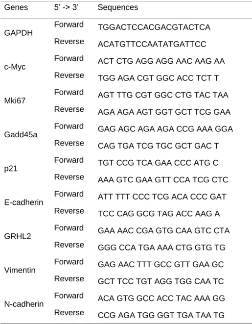
Then, cDNA was amplified by a SYBR green mix (TAKARA, Shinga, Japan) using T100TM Thermalcycler (BIO-RAD, CA, USA) and analyzed. Primers used for real-time PCR were designed as the listed sequences (Table 1).
Fluorescence activated cell sorter (FACS)
A375P, A375SM and HDFn were seeded at a density of 5.0×10⁴ cells per well in 6-well plates in the medium as described above. After 1 day, the medium was replaced with 50, 100, 150, 200, and 300 µg/mL of the LGG extract. 3 days after the treatment of the extract, both cell lines were harvested and stained with Annexin V (Cayman Chemical, MI, USA) and 7- Aminoactinomycin D (7-AAD; Life technologies) dyes. These stained cells were analyzed using FACSVerseTM (BDBiosciences, CA, USA).
Cell migration assay
Migration of A375P and A375SM were assessed by a wound-healing assay.
Both cell lines were seeded at a density of 1.5×10⁵ cells per well in 12-well
6
plates in the medium as described above until they reached 80% confluence.
The monolayers of both melanoma cells were scratched by the pipette tip, and the medium was replaced with 50 and 100 µg/mL of the LGG extract.
The cell migration capabilities were analyzed using the microscope at ×100 magnification.
Statistics
Data is presented with mean + standard deviation (S.D). Statistical analysis was determined using ANOVA. Significance was defined as *p <
0.05, **p < 0.01, and ***p < 0.001.
7
Results
LGG extract affects A375P and A375SM on morphology and growth
To investigate how the extract affects HDFn, A375P, and A375SM, these cell lines were seeded in 12-well plates. After 1 day, the medium was replaced and treated with the various range of 50, 75 and 100 µg/mL of the LGG extract. After 3 days, the morphologies of HDFn, A375P, and A375SM were observed and photographed by microscope. It is observed that the morphologies of both melanoma cell lines was changed by the LGG extract.
Both melanoma cells expanded and the large proportion of the cells was dead. Therefore, the result showed that the LGG extract could inhibit the both melanoma cell lines in a dose-dependent manner (Fig. 1).
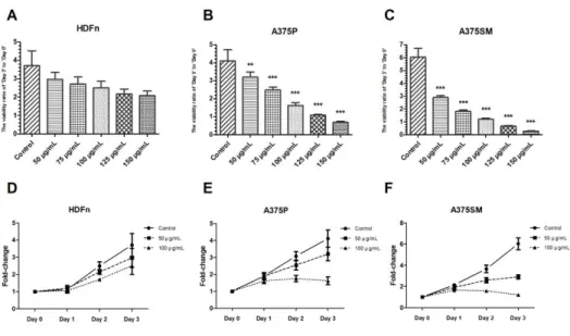
LGG extract inhibits cell viabilities of A375P and A375SM significantly
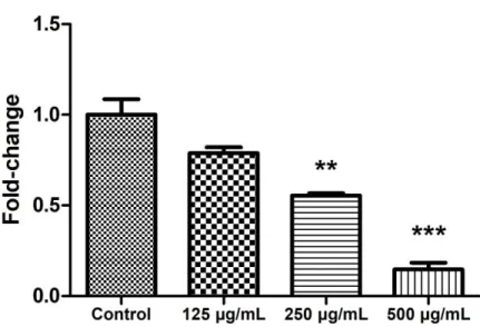
For quantitative analysis, HDFn, A375P, and A375SM were seeded in 96- well plates. In our previous study, the ED50 value of the LGG extract on the HDFn was 250-300 µg/mL (Fig. 2). After 1 day, thus, the medium was
8
replaced with the various concentration ranges of 50, 75, 100, 125 and 150 µg/mL of the LGG extract, which were under the ED50 value of HDFn. The viabilities over time were determined by the WST-1 cell viability assay. The cell viabilities of melanoma cell lines cultured with the extract was significantly decreased in a dose-dependent manner compared to control group (Fig. 3A and B), while HDFn was less influenced by the extract (Fig.
3C). Interestingly, although the A375SM is known for having more aggressive and metastatic properties, it was more susceptible to the LGG extract than A375P (Fig. 1 and 3). When A375P and A375SM were treated with 50 and 100 µg/mL of the LGG extract for 72 h, the proliferation of A375SM was inhibited by 52% and 80% and that of A375P was inhibited by 22% and 61%, respectively. On the other hand, the HDFn was less vulnerable to the LGG extract. To investigate the anti-cancer effect of the LGG extract as time passed, the viabilities at the specific concentrations of the LGG extract were determined each day (Fig. 3E and F). The results showed that the viabilities of A375P and A375SM over time were decreased significantly compared to the Control groups at the 100 µg/mL of the LGG extract, while HDFn maintained the increasing trend.
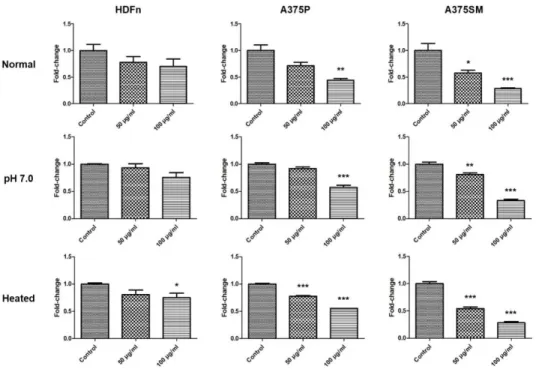
Basically, the LGG extract was acidic because the MRS broth where the LGG grew was acidic, even if it was washed three times. Therefore, to confirm whether the anti-cancer effect of the LGG extract resulted from the acidity of the LGG extract or not, HDFn, A375P, and A375SM were cultured with the LGG extract which had been neutralized to pH 7.0 for 3 days as the
9
same method explained previously. After 3 days, the cell viabilities were analyzed by the WST-1 cell viability assay (Fig. 4). The cell viabilities of melanoma cell lines cultured with the pH 7.0 LGG extract were decreased significantly. Interestingly, the cell viability of the HDFn cultured with the pH 7.0 extract was decreased less than the normal group. These results suggested that the anti-cancer effect of the LGG extract did not result from the acidity and the reason why the LGG extract affected the HDFn a little might be its acidic property. Furthermore, to investigate the molecule which induced the anti-cancer effect on the melanoma cell lines was a protein or not, the cell lines were cultured with the LGG extract heated at 80℃ for 30 min. The cell viabilities of melanoma cell lines cultured with the heated extract were also decreased in the same way as the normal group. These results showed that the molecule which induced the anti-cancer effect was not the protein but the stable molecule which was not denatured in the acidic and high temperature conditions.
LGG extract regulates gene expressions related with cell cycle, proliferation, and apoptosis
To investigate how the LGG extract affects A375P and A375SM, real-time PCR was performed. The mRNAs isolated from A375P and A375SM cultured with the LGG extract for 3 days were reverse-transcribed to cDNA and the
10
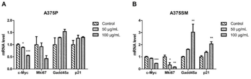
mRNA levels were analyzed. The expressions of c-Myc, which was known as a cancer-associated gene and proliferation marker gene, was significantly decreased in both A375P and A375SM in a dose-dependent manner (Fig. 5).
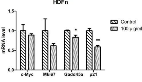
Although the expression of proliferation marker gene, Mki67 was down- regulated by the LGG extract, there was the statistical significant difference only in the A375SM. Furthermore, the expressions of Gadd45a known as cell cycle arrest marker and cyclin-dependent kinase inhibitor, p21 were increased greatly in A375SM. To investigate whether the LGG extract had a negative effect on HDFn or not, the gene expression levels of HDFn cultured with 100 µg/mL of the LGG extract for 3 days were also analyzed by the real- time PCR. The expression level of Mki67 was down-regulated without the statistical significance and the expression of c-Myc was barely changed.
However, the expression levels of Gadd45a and p21 were significantly down- regulated by the LGG extract (Fig. 6).
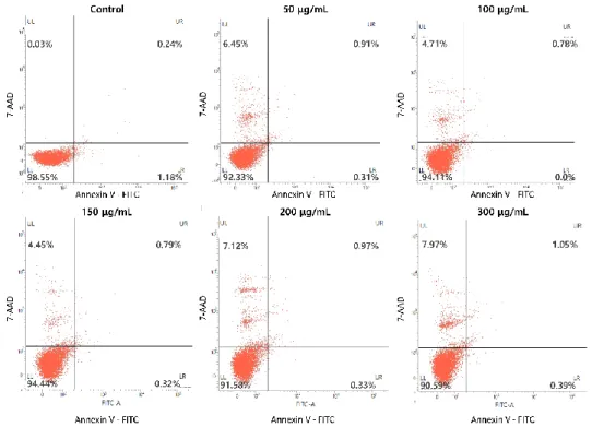
To determine whether the LGG extract induced the anti-cancer effect through apoptosis, the FACS was performed. The staining dye, Annexin V was used to detect cells which have been in process of apoptosis and as a result, express phosphatidylserine on the cell surfaces. The data showed that the control groups were almost not stained for both Annexin V and 7-AAD (Fig. 7). As the concentration of the LGG went up, the cells with higher Annexin V staining intensity were observed, which were considered apoptosis in accordance with real-time PCR data. However, at concentrations above 150 µg/mL, the LGG extract also induced necrosis as
11
well as the apoptosis, especially about the A375P. These results showed the LGG extract regulated the gene expressions related with cell cycle and consequently, induced cell cycle arrest, apoptosis, and necrosis. To investigate the effect of the LGG extract on the HDFn, the HDFn cell cultured with the LGG extract was also analyzed by the FACS. Unlike the melanoma cell lines, the LGG extract could barely induce the apoptosis and the necrosis (Fig. 8).
LGG extract inhibits cell migration by reducing epithelial-mesenchymal transition
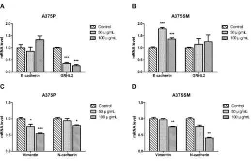
To determine the effect of the LGG extract on EMT of melanomas, the mRNA expression levels of E-cadherin, Grainyhead-like protein 2 homolog (GRHL-2), Vimentin and N-cadherin, which are known as EMT markers, were quantified using real-time PCR. The expression levels of epithelial marker genes, both E-cadherin and GRHL2 were increased in the A375SM, even if the expression level of GRHL2 in the A375P was decreased. Furthermore, the expression levels of both mesenchymal marker genes, Vimentin and N- cadherin were significantly decreased in both cell lines (Fig. 9). This results suggested that the extract made the migration and invasion capabilities of both melanoma cell lines down-regulated. To confirm this hypothesis, the migration assay was performed. Both serum-starved cells were used as
12
negative control, inducing the lack of cell proliferation and cell migration, while the positive control and experiment groups were cultured with 10% FBS and the extract. At 72 h post-wounding, almost full migrations of the A375P and A375SM were observed in the control group, while the extract-treated groups showed delayed migration capabilities in a dose-dependent manner (Fig. 10). These results supported that the LGG extract could suppress the EMT by regulating genes.
13
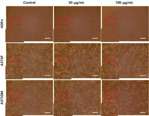
Figure 1. In vitro anti-cancer effect of LGG extract on A375P and A375SM.
HDFn, A375P, and A375SM were cultured with the LGG extract for 3 days and observed at ×40 magnification. The LGG extract had anti-cancer effects on A375P and A375SM in a dose-dependent manner. However, HDFn was less damaged by the LGG extract than both melanoma cell lines, scale bar
= 500 μm.
14
Figure 2. The ED50 value of the LGG extract on HDFn. HDFn was cultured with the LGG extract for 3 days and the viability was analyzed by the WST-1 cell viability assay. The result showed that the ED50 value of the LGG extract on HDFn was about 250-300 µg/mL. Results are the means of three independent experiments (mean + S.D). *p < 0.05, **p < 0.01, and ***p <
0.001 versus the control group.
15
Figure 3. The effect of LGG extract on cell viabilities of HDFn, A375P, and A375SM. HDFn, A375P, and A375SM were cultured with the LGG extract for 3 days and these viabilities were determined by the WST-1 cell viability assay.
The results showed that the effect of the LGG extract on HDFn (A), A375P (B), and A375SM (C), respectively. The cell viability of A375P and A375SM cultured with the extract was significantly decreased in a dose-dependent manner compared to control group. (D-F) The viabilities of those cell lines were also measured each day. The viabilities of A375P and A375SM over time were decreased significantly compared to the Control groups at the 100 µg/mL of the LGG extract, while HDFn maintained the increasing trend.
Results are the means of three independent experiments (mean + S.D). *p <
0.05, **p < 0.01, and ***p < 0.001 versus the control group.
16
Figure 4. Anti-cancer effect of LGG extract on both melanoma cell lines, A375P and A375SM. HDFn, A375P, and A375SM were cultured with the LGG extract which had been neutralized to pH 7.0 or heated for 3 days. The cell viabilities of melanoma cell lines cultured with the pH 7.0 or heated LGG extract were decreased significantly in the same way as the normal group.
Results are the means of three independent experiments (mean + S.D). *p <
0.05, **p < 0.01, and ***p < 0.001 versus the control group.
17
Figure 5. Gene regulation effects of LGG extract on A375P and A375SM.
(A-B) The mRNAs isolated from A375P and A375SM cultured with the LGG extract for 3 days were reverse-transcribed to cDNA and the expressions of several genes related with cell cycle, proliferation, and apoptosis levels were analyzed. Results are the means of three independent experiments (mean + S.D). *p < 0.05, **p < 0.01, and ***p < 0.001 versus the control group.
18
Figure 6. Gene regulation effects of LGG extract on HDFn. The mRNA isolated from HDFn cultured with the LGG extract for 3 days was reverse- transcribed to cDNA and the expressions of several genes related with cell cycle, proliferation, and apoptosis levels were analyzed. Results are the means of three independent experiments (mean + S.D). *p < 0.05, **p < 0.01, and ***p < 0.001 versus the control group.
19
20
Figure 7. Apoptosis and necrosis induced by the LGG extract. A375P and A375SM cultured with the LGG extract for 3 days were harvested. These cells were stained with Annexin V and 7-AAD, respectively, and analyzed by FACS. Control groups of A375P (A) and A375SM (B) were rarely stained for both Annexin V and 7-AAD. However, the higher concentration the LGG extract was treated at, the more apoptosis was induced in both cell lines.
21
Figure 8. The effect of the LGG extract on apoptosis and necrosis of HDFn.
HDFn cultured with the LGG extract for 3 days were harvested. The cells were stained with Annexin V and 7-AAD, respectively, and analyzed by FACS. All groups were rarely stained for both Annexin V and 7-AAD.
22
Figure 9. Modulation of epithelial-mesenchymal transition (EMT) markers by LGG extract. (A-B) The mRNAs isolated from A375P and A375SM cultured with the LGG extract for 3 days were reverse-transcribed to cDNA and the expressions of EMT marker genes were analyzed. Results are the means of three independent experiments (mean + S.D). *p < 0.05, **p < 0.01, and ***p
< 0.001 versus the control group.
23
24
Figure 10. Anti-migration effect of the LGG extract on A375P and A375SM.
The migration capabilities of A375P and A375SM were assessed by the wound-healing assay. When the cells reached to 80% confluence, the monolayers of both melanoma cells were scratched by the pipette tip and the medium was replaced with 50 µg/mL and 100 µg/mL of the LGG extract. The cell migration capabilities were analyzed using the microscope at ×100 magnification. At 72 h post-wounding, almost full migrations of the A375P (A) and A375SM (B) cells were observed in the control group, while the extract- treated groups show delayed migration capabilities in a dose-dependent manner, scale bar = 200 μm.
25
Table 1. Primer sequences used for Real-time PCR Genes 5’ -> 3’ Sequences
GAPDH
Forward TGGACTCCACGACGTACTCA Reverse ACATGTTCCAATATGATTCC c-Myc
Forward ACT CTG AGG AGG AAC AAG AA Reverse TGG AGA CGT GGC ACC TCT T Mki67
Forward AGT TTG CGT GGC CTG TAC TAA Reverse AGA AGA AGT GGT GCT TCG GAA Gadd45a
Forward GAG AGC AGA AGA CCG AAA GGA Reverse CAG TGA TCG TGC GCT GAC T p21
Forward TGT CCG TCA GAA CCC ATG C Reverse AAA GTC GAA GTT CCA TCG CTC E-cadherin
Forward ATT TTT CCC TCG ACA CCC GAT Reverse TCC CAG GCG TAG ACC AAG A GRHL2
Forward GAA AAC CGA GTG CAA GTC CTA Reverse GGG CCA TGA AAA CTG GTG TG Vimentin
Forward GAG AAC TTT GCC GTT GAA GC Reverse GCT TCC TGT AGG TGG CAA TC N-cadherin
Forward ACA GTG GCC ACC TAC AAA GG Reverse CCG AGA TGG GGT TGA TAA TG
26
Discussion
Malignant melanoma is pointed out as the most aggressive cancer and having extremely unfavorable prognosis according to the Centers for Disease Control and Prevention [19], and its incidence is also consistently growing [20]. The fact remains that out of the various skin cancers, malignant melanoma remains the highest cause of death [21,22]. Although various therapeutic methods have been developed so far, its high metastasis capabilities and frequency of intrinsic and obtained resistance to therapies in patients imply the serious need for new therapeutic methods [23].
Lactobacillus has been used as probiotics due to the several enzymes and metabolites it produces [14]. It also has anti-pathogenic bacteria effects, which could decrease the incidence of gastrointestinal diseases [24].
Recently, several strains of Lactobacillus have been reported to have anti- cancer activities, affecting cancer cell growth and metastasis directly or indirectly [15]. Lactobacillus has low or no pathogenic activities, and therefore it is regarded as a potential therapeutic source [25].
Our study showed that the LGG extract induced the dose-dependent anti- cancer effect on human melanoma cells, A375P and A375SM. Both cell lines are generally known for their aggressive growth and high metastasis capabilities. On the other hand, the HDFn was less vulnerable to the LGG
27
extract. These results showed that the LGG extract could be used for cancer therapy with lesser side-effects. However, in the high-dose treatment of the LGG extract, the proliferation of the HDFn also decreased, which might result from the inflammation responses induced by LGG. The several fractions with different molecular weight were separated from the LGG extract, and it was reported that the use of the fraction with specific molecular weight induced less inflammation significantly [26]. It was reported that the probiotics could improve the skin natural defense and benefit immune responses [27]. Thus, the LGG extract could be used as a new therapeutic method such as an ointment or a drug for the malignant melanoma.
To understand the mechanisms underlying the effect of the LGG extract, several genes related to cell cycles were evaluated by real-time PCR. The data showed the expressions of c-Myc and Mki67 were down-regulated in a dose-dependent manner. The oncogene, c-Myc is the master regulator, which is related to many aspects of the cell growth process [28] and the MKi- 67, also known as Ki-67, represents a mitotic index, which could be used for tumor grading due to being observed in proliferating cells [29,30]. Moreover, the expressions of Gadd45a and p21 were up-regulated in a dose-dependent manner. The Gadd45a and p21 were related in negatively regulating the cell cycle and inducing cell cycle arrest [31]. These changes of gene expressions suggested that LGG extract could induce apoptosis and, consequently, down-regulate the growth of the malignant melanoma cell lines. Our Annexin V and 7-AAD double staining assay using the FACS also showed that the
28
apoptosis could be induced by the LGG extract. However, the LGG extract also induced necrosis as well as apoptosis at concentrations above 150 µg/mL. In our study, it was elucidated that the effect molecules were not proteins by using the autoclaved and boiled LGG extract, but it still remained unclear what exactly the effect molecules were. Therefore, further studies are required to investigate the effect molecule to maximize the anti-cancer effect of the LGG extract and reduce the unexpected side-effect [26].
One of the reasons why malignant melanomas have the worst prognosis is due to its aggressive metastasis capability, and therefore, the development of therapies to inhibit the metastasis of malignant melanomas is urgently required. It was reported that the extract of Lactobacillus casei had the anti- metastatic capability [16,32,33]. Our gene expression analysis and cell migration assay indicated that the EMT could be inhibited by the LGG extract [34,35], which suggested that the LGG extract could be used as the agent to inhibit the metastasis in vivo. However, the LGG extract also had cell cytotoxicity effect on the melanoma cell lines, further study should be required to elucidate the accurate effect of the LGG extract on the cell migration of melanoma cell lines.
Surprisingly, A375SM was more susceptible to the LGG extract than A375P overall, although A375SM had the higher proliferation and metastasis rate than A375P. There are several differences in gene expression patterns between the A375P and A375SM, and these differences would be the key
29
factors for the anti-cancer mechanism of the LGG extract. For example, the chemokine CXCL-8, also known as Interleukin-8, is the key regulator of human malignant melanomas and regulates the proliferation, metastasis, and angiogenesis by autocrine and paracrine. The gene expression pattern of CXCL-8 is totally different between A375P and A375SM and as a result, it makes different properties on the proliferation and metastasis [36]. The capabilities of CXCL-8 on human malignant melanomas are the results of the interaction with the CXCL-8 receptors, CXCR-1 and CXCR-2. The CXCR-1 and CXCR-2 are up-regulated in A375SM and it also corresponds with the higher proliferation and metastasis rate of A375SM than A375P [37].
Therefore, the mRNA and the protein levels of CXCL-1, CXCR-1, and CXCR- 2 should be evaluated in further studies.
In conclusion, our study showed the anti-cancer effect of the LGG extract on human malignant melanoma cells by modulating the several genes related to cell cycle, proliferation, and apoptosis. EMT-related genes were also decreased after the treatment of the LGG extract significantly. Thus, this data suggested that the LGG extract could be used as a new therapeutic method with lesser side-effects. To investigate its anti-cancer and –metastasis effects in vivo and accurate mechanism, further studies will be required.
30
References
1. Lee JE. Factors associated with melanoma incidence and prognosis.
Semin Surg Oncol. 1996;12:379–385.
2. Parker SL, Tong T, Bolden S, Wingo PA. Cancer statistics. CA Cancer J Clin. 1996;6:5–27.
3. Jemal A, Murray T, Samuels A, Ghafoor A, Ward E, Thun MJ.
Cancer statistics. CA Cancer J Clin. 2003;53:5–26.
4. Rigel, DS, J Russak, R Friedman. The evolution of melanoma diagnosis: 25 years beyond the ABCDs. CA Cancer J Clin.
2010;60:301–316. doi:10.3322/caac.20074.
5. Faião-Flores F, Coelho P, Arruda-Neto J, Maria DA. Boron neutron capture therapy induces cell cycle arrest and DNA fragmentation in murine melanoma cells. Appl Radiat Isot. 2011;69:1741-1744.
doi:10.1016/j.apradiso.2011.03.005.
6. Dimitrakopoulos I, Lazaridis N, Skordalaki A. Primary malignant melanoma of the oral cavity. Report of an unusual case. Aust Dent J. 1998;43:379-381.
31
7. Pliskin ME, Mastrangelo MJ, Brown AM, Custer RP. Metastatic melanoma of the maxilla presenting as a gingival swelling. Oral Surg Oral Med Oral Pathol. 1976;41:101-104.
8. Chiu NT, Weinstock MA. Melanoma of oronasal mucosa.
Population-based analysis of occurrence and mortality. Arch Otolaryngol Head Neck Surg. 1996;122:985-988.
9. González-García R, Naval-Gías L, Martos PL, Nam-Cha SH, Rodríguez-Campo FJ, Muñoz-Guerra MF, Sastre-Pérez J.
Melanoma of the oral mucosa. Clinical cases and review of the literature. Med Oral Patol Oral Cir Bucal. 2005;10:264-271.
10. Lee B, Mukhi N, Liu D. Current management and novel agents for malignant melanoma. J Hematol Oncol.2012; 5:3.
doi:10.1186/1756-8722-5-3.
11. Kirjavainen PV, El-Nezami HS, Salminen SJ, Ahokas JT, Wright PF.
The effect of orally administered viable probiotic and dairy lactobacilli on mouse lymphocyte proliferation. FEMS Immunol Med Microbiol.
1999;26:131-135.
12. Pessi T, Sütas Y, Hurme M, Isolauri E. Interleukin-10 generation in atopic children following oral Lactobacillus rhamnosus GG. Clinical and experimental allergy. J Br Soc Allergy Clin Immunol.
2000;30:1804-1808.
32
13. Hemarajata P, Versalovic J. Effects of probiotics on gut microbiota:
mechanisms of intestinal immunomodulation and neuromodulation.
Therap Adv Gastroenterol. 2013;6:39–51.
doi:10.1177/1756283X12459294.
14. Hwang JW, Baek YM, Yang KE, Yoo HS, Cho CK, Lee YW, Park J, Eom CY, Lee ZW, Choi JS, Jang IS. Lactobacillus casei extract induces apoptosis in gastric cancer by inhibiting NF-κB and mTOR- mediated signaling. Integr Cancer Ther. 2013;12:165-173.
doi:10.1177/1534735412442380.
15. Khan I, Kang SC. Apoptotic activity of Lactobacillus plantarum DGK- 17-fermented soybean seed extract in human colon cancer cells via ROS-JNK signaling pathway. J Food Sci. 2017;82:1475-1483.
doi:10.1111/1750-3841.13732.
16. Matsuzaki T, Yokokura T, Azuma I. Antimetastatic effect of Lactobacillus casei YIT9018 (LC 9018) on a highly metastatic variant of B16 melanoma in C57BL/6J mice. Cancer Immunol Immunother.
1987;24:99-105.
17. Gamallat Y, Meyiah A, Kuugbee ED, Hago AM, Chiwala G, Awadasseid A, Bamba D, Zhang X, Shang X, Luo F, Xin Y.
Lactobacillus rhamnosus induced epithelial cell apoptosis, ameliorates inflammation and prevents colon cancer development
33
in an animal model. Biomed Pharmacother. 2016;83:536-541.
doi:10.1016/j.biopha.2016.07.001.
18. Riedl S, Rinner B, Schaider H, Liegl-Atzwanger B, Meditz K, Preishuber-Pflügl J, Grissenberger S, Lohner K, Zweytick D. In vitro and in vivo cytotoxic activity of human lactoferricin derived antitumor peptide R-DIM-P-LF11-334 on human malignant melanoma.
Oncotarget. 2017. doi:10.18632/oncotarget.17823.
19. Peterson M, Albertini MR, Remington P. Incidence, survival, and mortality of malignant cutaneous melanoma in wisconsin, 1995-2011.
W M J. 2015;114:196-201.
20. Zehavi L, Schayek H, Jacob-Hirsch J, Sidi Y, Leibowitz-Amit R, Avni D. MiR-377 targets E2F3 and alters the NF-κB signaling pathway through MAP3K7 in malignant melanoma. Mol Cancer. 2015;14:68.
doi:10.1186/s12943-015-0338-9.
21. Balch CM, Gershenwald JE, Soong S-j, Thompson JF, Atkins MB, Byrd DR, Buzaid AC, Cochran AJ, Coit DG, Ding S, Eggermont AM, Flaherty KT, Gimotty PA, Kirkwood JM, McMasters KM, Mihm MC Jr, Morton DL, Ross MI, Sober AJ, Sondak VK. Final version of 2009 AJCC melanoma staging and classification. J Clin Oncol.
2009;27:6199–6206. doi:10.1200/JCO.2009.23.4799.
22. Howlader N, Noone A, Krapcho M, Miller D, Bishop K, Kosary C, Yu
34
M, Ruhl J, Tatalovich Z, Mariotto A, Lewis D, Chen H, Feuer E.
SEER Cancer Statistics Review, 1975-2014. National Cancer Institute. https://seer.cancer. gov/csr/1975_2014/, based on November 2016 SEER data submission, posted to the SEER web site.
23. Hegedüs L, Padányi R, Molnár J, Pászty K, Varga K3, Kenessey I, Sárközy E, Wolf M, Grusch M, Hegyi Z, Homolya L, Aigner C, Garay T, Hegedüs B, Tímár J, Kállay E, Enyedi Á . Histone Deacetylase Inhibitor treatment increases the expression of the plasma membrane Ca2+ pump PMCA4b and inhibits the migration of melanoma cells independent of ERK. Front Oncol. 2017;7:95.
doi:10.3389/fonc.2017.00095.
24. Forestier C, Champs CD, Vatoux C, Joly B. Probiotic activities of Lactobacillus casei rhamnosus: in vitro adherence to intestinal cells and antimicrobial properties. Res Microbiol. 2001;152:167-173.
25. Lilly DM, Stillwell RH. Probiotics: growth-promoting factors produced by microorganisms. Science. 1965;147:747-748.
26. Zhang Z, Zhou Z, Li Y, Zhou L, Ding Q, Xu L. Isolated exopolysaccharides from Lactobacillus rhamnosus GG alleviated adipogenesis mediated by TLR2 in mice. Sci Rep. 2016;6:36083.
doi:10.1038/srep36083.
35
27. Al-Ghazzewi FH, Tester RF. Impact of prebiotics and probiotics on skin health. Benef Microbes. 2014;5:99-107.
doi:10.3920/BM2013.0040.
28. Miller DM, Thomas SD, Islam A, Muench D, Sedoris K. c-Myc and cancer metabolism. Clin Cancer Res. 2012;18:5546–5553.
doi:10.1158/1078-0432.CCR-12-0977.
29. Yalcin SE, Ocal I, Yalcin Y, Selim HS, Caltekin MD, Aydogmus H, Kelekci S. Evaluation of the Ki-67 proliferation index and urocortin expression in women with ovarian endometriomas. Eurasian J Med.
2017;49:107-112. doi:10.5152/eurasianjmed.2017.17070.
30. Huisman MA, De Heer E, Grote JJ. Cholesteatoma epithelium is characterized by increased expression of Ki-67, p53 and p21, with minimal apoptosis. Acta Otolaryngol. 2003;123:377-382.
31. Da Silva GN, Filoni LT, Salvadori MC, Salvadori DMF.
Gemcitabine/Cisplatin treatment induces concomitant SERTAD1, CDKN2B and GADD45A modulation and cellular changes in bladder cancer cells regardless of the site of TP53 mutation. Pathol Oncol Res. 2017. doi:10.1007/s12253-017-0255-x.
32. Matsuzaki T, Yokokura T, Azuma I. Antitumor activity of Lactobacillus casei on Lewis lung carcinoma and Line-10 hepatoma in syngeneic mice and guinea pigs. Cancer Immunol Immunother. 1985;20:18-22.
36
33. Matsuzaki T, Yokokura T. Inhibition of tumor metastasis of Lewis lung carcinoma in C57BL/6 mice by intrapleural administration of Lactobacillus casei. Cancer Immunol Immunother. 1987;25:100-104.
34. Cho IH, Jang EH, Hong D, Jung B, Park MJ, Kim JH. Suppression of LPS-induced epithelial-mesenchymal transition by aqueous extracts of Prunella vulgaris through inhibition of the NF-κB/Snail signaling pathway and regulation of EMT-related protein expression. Oncol Rep. 2015; 34:2445-2450. doi: 10.3892/or.2015.4218.
35. Hsieh CC, Huang YS. Aspirin Breaks the Crosstalk between 3T3-L1 Adipocytes and 4T1 Breast Cancer Cells by Regulating Cytokine Production. PLoS One. 2016; 11:e0147161. doi:
10.1371/journal.pone.0147161
36. Wu S, Singh S, Varney ML, Kindle S, Singh RK. Modulation of CXCL- 8 expression in human melanoma cells regulates tumor growth, angiogenesis, invasion, and metastasis. Cancer Med. 2012;1:306- 317. doi:10.1002/cam4.28.
37. Varney ML, Li A, Dave BJ, Bucana CD, Johansson SL, Singh RK.
Expression of CXCR1 and CXCR2 receptors in malignant melanoma with different metastatic potential and their role in interleukin-8 (CXCL-8)-mediated modulation of metastatic phenotype. Clin Exp Metastasis. 2003;20:723-731.
37
국문초록
인간 악성 흑색종 세포주에 항암 효과를 가지는 Lactobacillus rhamnosus GG 추출물
이재훈 종양 및 발달생물학 전공 치의과학과 서울대학교 대학원 (지도교수: 노상호, D.V.M., Ph.D.)
인간 악성 흑색종은 다른 암에 비해 그 발병률이 급격하게 높아지고 있는 매우 공격적인 피부암 중에 하나이다. 비록 다양한 치료 방법들이 개발되어왔지만, 여전히 이것의 높은 증식 및 전이 속도는 치료에 있어 좋지 않은 예후를 야기하는 장애물로 남아있다. In vitro 및 in vivo 상에서 특정 유산균 추출물이 항암 및 전이를 억제시키는 효과가 있다는 것은 이미 보고가 되어 있으나 Lactobacillus rhamnosus GG (LGG) 추출물이 인간 악성 흑색종에서 어떤 영향을 끼치는지는 아직까지 보고된 것이 없다.
38
따라서 본 연구에서는 LGG 추출물이 인간 악성 흑색종 세포주인 A375P와 A375SM에서 항암 및 전이를 억제시키는 효과를 가지는 지를 알아보고자 하였다. 먼저, WST-1 세포증식속도 분석을 통해 LGG 추출물은 신생아의 보통 피부 섬유아세포에는 거의 영향을 끼치지 않았지만, A375P와 A375SM에는 추출물 농도에 비례하는 항암 효과가 있는 것을 알 수 있었다. 실시간 연쇄중합반응 분석 결과에 따르면 LGG 추출물이 처리된 두 악성 흑색종 세포주에서 세포주기, 증식, 세포자멸사(apoptosis)와 관련된 유전자 발현 패턴이 암세포의 성장을 억제하는 방향으로 조절된다는 것을 확인할 수 있었다. 게다가 상피간엽이행(epithelial-mesenchymal transition) 관련 유전자들의 발현이 LGG 추출물에 의해 암의 전이를 억제시키는 방향으로 조절되었고, 세포 이동 분석을 통해 세포의 이동이 유의적으로 억제되는 것을 확인하였다. 본 연구를 통해 LGG 추출물이 인간 악성 흑색종의 잠재적인 치료 재료가 될 수 있을 것으로 생각된다.
주요어: 유산균, 항암, 전이, 인간 악성 흑색종, 세포 증식,
세포자멸사(apoptosis), 상피간엽이행(epithelial-mesenchymal transition)
학번: 2016-22037