저작자표시
-
비영리-
변경금지2.0
대한민국 이용자는 아래의 조건을 따르는 경우에 한하여 자유롭게l
이 저작물을 복제,
배포,
전송,
전시,
공연 및 방송할 수 있습니다.
다음과 같은 조건을 따라야 합니다:
l
귀하는,
이 저작물의 재이용이나 배포의 경우,
이 저작물에 적용된 이용허락조건 을 명확하게 나타내어야 합니다.
l
저작권자로부터 별도의 허가를 받으면 이러한 조건들은 적용되지 않습니다.
저작권법에 따른 이용자의 권리는 위의 내용에 의하여 영향을 받지 않습니다
.
이것은 이용허락규약(Legal Code)
을 이해하기 쉽게 요약한 것입니다.
Disclaimer
저작자표시
.
귀하는 원저작자를 표시하여야 합니다.
비영리
.
귀하는 이 저작물을 영리 목적으로 이용할 수 없습니다.
변경금지
.
귀하는 이 저작물을 개작,
변형 또는 가공할 수 없습니다.
이학박사학위논문
진핵생물의 전령 RNA 꼬리에 관한 전사체 수준의 연구
Transcriptome-wide studies on eukaryotic mRNA tail
2016 년 8 월
서울대학교 대학원 생명과학부
임재철
Abstract
Transcriptome-wide studies on eukaryotic mRNA tail
Jaechul Lim
School of Biological Sciences The Graduate School Seoul National University
Eukaryotic mRNA is subject to intensive post-transcriptional modifications, which critically influences mRNA stability and translatability. Newly synthesized mRNA acquires a 7-methlyguanosine cap at the 5′ end and polyadenosine tail at the 3′ end. In addition to such canonical modifications, recent studies have revealed untemplated nucleotide additions such as U-tail and G-tail, base modifications including N6-methyladenosine and pseudouridylation, and A-to-I editing, as epitranscriptomic signatures of mRNA.
To investigate RNA tailing at the genomic scale, I recently developed a method called TAIL-seq which accurately measures poly(A) tail length and 3′ end modifications. In- terestingly, I discovered that mammalian cells carried median 50–100 nt of poly(A) tail and widespread uridylation and guanylation at the downstream of poly(A) tail. Moreover, U-tails were mainly found on short poly(A) tails (<~25 nt), implicating its role in mRNA turnover.
Uridylation has been observed on mRNAs in various species, yet its mechanism
and significance remained unknown. By applying TAIL-seq, I identify TUT4 and TUT7
(TUT4/7), also known as ZCCHC11 and ZCCHC6, respectively, as mRNA uridylation
enzymes in mammals. Uridylation readily occurs on deadenylated mRNAs in cells. Con-
sistently, purified TUT4/7 selectively recognize and uridylate RNAs with short A tails
(<~25 nt) in vitro. Moreover, PABPC1 antagonizes uridylation of polyadenylated mRNAs,
contributing to the specificity for short A. In cells depleted of TUT4/7, the vast majority of
mRNAs lose the oligo-U tails, and their half-lives are extended. Suppression of mRNA decay factors leads to the accumulation of oligo-uridylated mRNAs. In line with this, microRNA induces uridylation of its targets, and TUT4/7 are required for enhanced decay of microRNA targets. My study explains the mechanism underlying selective uridylation of deadenylated mRNAs, and demonstrates a fundamental role of oligo-U-tail as a molecular mark for global mRNA decay.
Next, I report a new version of TAIL-seq (mRNA TAIL-seq or mTAIL-seq), which increases sequencing depth for mRNAs by ~1,000 fold compared to the previous version.
Original version of TAIL-seq provided a various information, but its low sensitivity pre- cluded its application to minute amounts of biological materials. With the improved method, I investigate the regulation of poly(A) tail in Drosophila oocytes and embryos.
I find that maternal mRNAs are polyadenylated mainly during late oogenesis, prior to fertilization, and further modulated upon egg activation. Wispy, a noncanonical poly(A) polymerase, adenylates most maternal mRNAs with a few intriguing exceptions such as ribosomal protein transcripts. By comparing mTAIL-seq data to ribosome profiling data, I further find a strong coupling between poly(A) tail length and translational efficiency during egg activation. My data suggest that regulation of poly(A) tail in oocytes shapes the translatomic landscape of embryos, thereby directing the onset of animal development.
By virtue of the high sensitivity, low cost, technical robustness, and broad accessibility, mTAIL-seq will be a potent tool to improve our understanding of mRNA tailing.
Taken together, I investigated mRNA tailings in eukaryotes by using sequencing meth- ods that I develped, TAIL-seq and mTAIL-seq, and found their roles in the control of mRNA stability and translation.
Keywords: Post-transcriptional modification; High-throughput sequencing;
TAIL-seq; mRNA tailing; Poly(A) tail; Uridylation; Cytoplasmic polyadenylation
Student ID: 2011-20349
Contents
Abstract . . . . i
Contents . . . . iii
List of Figures . . . . v
List of Tables . . . . viii
List of Abbreviations . . . . ix
1 Introduction 1 1.1 Life cycle of mRNA . . . . 1
1.2 mRNA tailing in eukaryotes . . . . 1
1.3 TAIL-seq: Transcriptome-wide determination of poly(A) tail length and 3′ end modification . . . . 4
2 Uridylation marks mRNA for degradation 9 2.1 Background . . . . 9
2.2 Results . . . . 12
2.2.1 TUT4 and TUT7 catalyze mRNA uridylation . . . . 12
2.2.2 TUT4/7 selectively oligo-uridylate mRNAs with short A tails in vivo and in vitro . . . . 21
2.2.3 PABP suppresses uridylation of poly(A) + mRNA . . . . 29
2.2.4 Uridylation facilitates global mRNA decay . . . . 29
2.2.5 Uridylation is involved in miRNA-induced gene silencing . . . . 34
2.2.6 mRNA decay factors remove uridylated mRNAs . . . . 39
2.3 Discussion . . . . 44
2.4 Methods and Materials . . . . 48
3 In-depth profiling of poly(A) tail length by mTAIL-seq 99 3.1 Background . . . . 99
3.2 Results . . . . 101
3.2.1 mTAIL-seq: a solution for limited materials . . . . 101 3.2.2 Global poly(A) tail length measurement in Drosophila . . . . 111 3.2.3 Distinct patterns of poly(A) tail regulation . . . . 121 3.2.4 Correlation between poly(A) tail length and translational effi-
ciency . . . . 131 3.3 Discussion . . . . 136 3.4 Methods and Materials . . . . 139
4 Conclusion 157
Summary (in Korean) 159
Bibliography 161
List of Figures
1.1 Life cycle of mRNA. . . . 2
1.2 Experimental procedure of TAIL-seq. . . . 6
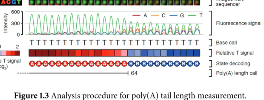
1.3 Analysis procedure for poly(A) tail length measurement. . . . 7
2.1 Domain architecture of human TUTases. . . . 13
2.2 The level of seven TUTases after RNAi of TUT1/3/5/6 and TUT2/4/7. . . 13
2.3 Uridylation frequency measured by small-scale TAIL-seq. . . . 14
2.4 Validation of each knockout cell line by western blotting. . . . 14
2.5 Uridylation frequency measured by small-scale TAIL-seq. . . . 15
2.6 Summary of TUT4/7 double knockout cell generation. . . . 16
2.7 Western blotting after TUT4 and TUT7 depletion. . . . 17
2.8 Cell proliferation rates shown in growth curve. . . . 17
2.9 FACS analysis. . . . 18
2.10 Uridylation frequency measured by small-scale TAIL-seq. . . . 18
2.11 Changes in uridylation of individual mRNA species upon TUT4/7 knock- down. . . . 19
2.12 Examples of gene-level uridylation changes. . . . 19
2.13 Changes in uridylation at transcript level upon TUT4/7 knockdown. . . 20
2.14 Distribution of mono- and oligo-uridylation according to the length of poly(A) tails. . . . 21
2.15 In vitro uridylation assay with immunopurified TUT4. . . . 23
2.16 In vitro uridylation assay using immunopurified TUT7. . . . 24
2.17 In vitro uridylation assay using immunopurified TUT7 (A25 and A25R). 25 2.18 Coomassie staining of TUT4 and TUT7 used in this study. . . . 26
2.19 In vitro uridylation assay with recombinant TUT7. . . . 27
2.20 In vitro uridylation assay with recombinant TUT7. . . . 28
2.21 PABP inhibits uridylation of polyadenylated mRNA. . . . 30
2.22 Western blotting after subcellular fractionation. . . . 31
2.23 Measurement of mRNA half-life by RNA-seq. . . . 32
2.24 Measurement of mRNA half-life by qRT-PCR. . . . 33
2.25 In vitro tethring assay. . . . 35
2.26 Western blotting showing the expression of transfected proteins. . . . 36
2.27 Changes in uridylation after miR-1 transfection. . . . 37
2.28 Measurement of half-life of miR-1 targets by qRT-PCR. . . . 38
2.29 Validation of knockdown by qRT-PCR or sequencing. . . . 40
2.30 The 5′ and 3′ mRNA decay factors degrade uridylated mRNAs. . . . 41
2.31 Uridylation frequency change upon poly(A) tail. . . . 42
2.32 Poly(A) tail length distribution. . . . 43
2.33 Model for uridylation-dependent mRNA decay in humans. . . . 45
3.1 Comparison of TAIL-seq protocols. . . . 102
3.2 Ligation efficiency test. . . . 103
3.3 Design of the 3′ hairpin adaptor. . . . 104
3.4 Schematic description of experimental procedure. . . . 105
3.5 Accuracy assessment using poly(A) spike-ins. . . . 105
3.6 Comparison of efficiency between TAIL-seq and mTAIL-seq. . . . 106
3.7 Position of the 5′ end of read 1 relative to the annotated 3′ end. . . . 106
3.8 Comparison of sensitivity between TAIL-seq and mTAIL-seq. . . . 107
3.9 Global distributions of poly(A) tails. . . . 107
3.10 Reproducibility between four different amounts of input RNA. . . . 108
3.11 Comparison of poly(A) tail lengths measured by TAIL-seq and mTAIL-seq. 109 3.12 Detection of U-tails by mTAIL-seq. . . . 110
3.13 3′ uridylation frequency detected by TAIL-seq. . . . 111
3.14 Global poly(A) tail distribution of Drosophila early embryos. . . . 112
3.15 Schematic view of late oogenesis and egg activation in Drosophila. . . . . 113
3.16 Reproducibility of mTAIL-seq libraries. . . . 115
3.17 Global measurement of poly(A) tail length. . . . 116
3.18 Global measurement of poly(A) tail length in replicates . . . . 116
3.19 Intragenic poly(A) tail length change. . . . 117
3.20 Changes of mRNA abundance. . . . 117
3.21 Comparison of poly(A) length changes to mRNA abundance changes. . 118
3.22 Examples of individual genes. . . . 119
3.23 Results of Hire-PAT assay. . . . 120
3.24 Poly(A) tail length profiles. . . . 122
3.25 Intragenic poly(A) tail length distributions in two replicates. . . . 123
3.26 Poly(A) tail distribution of representative genes. . . . 124
3.27 Gene ontology groups. . . . 125
3.28 Reproducibility between two biological replicates of wisp mutant. . . . . 128
3.29 Comparison of poly(A) tail lengths between wild type and wisp mutant. 129 3.30 mRNA abundance change in wisp mutant. . . . 130
3.31 Wispy-dependent groups in mature oocytes and activated eggs. . . . 130
3.32 Comparison of poly(A) tail lengths between wild type and wisp mutant. 132 3.33 Poly(A) tail length change during late oogenesis in wisp mutants. . . . . 133
3.34 Relationship between poly(A) tail length and translational efficiency. . . 133
3.35 Relationship between poly(A) tail length and translational efficiency (0–1 hr). . . . 134
3.36 Correlation between poly(A) tail length change and TE change upon egg activation. . . . 135
3.37 Correlation between poly(A) tail length change and TE change upon egg activation (0–1 hr). . . . 135
3.38 Translational controls on poly(A) tail length profiles. . . . 136
3.39 Ribosome occupancy changes over poly(A) tail length profiles. . . . 137
List of Tables
1.1 mRNA uridylating enzymes in eukaryotes. . . . 3
2.1 TAIL-seq data from TUT4/7-depleted cells. . . . 68
2.2 List of mRNA half-life following TUT4/7 knockdown. . . . 96
2.3 Lists of oligonucleotides used in this study. . . . 98
3.1 The list of genes belong to 8 groups. . . . 155
List of Abbreviations
AEL After Egg Laying cDNA complementary DNA CDS Coding Sequence
CPE Cytoplasmic Polyadenylation Element CPEB CPE Binding protein
CRISPR Clustered Regularly Interspaced Short Palindromic Repeats DNA Deoxyribonucleic Acid
GO Gene Ontology
mRNA messenger RNA
NMD Nonsense-Mediated Decay PABP Poly(A) Bnding Protein PAP Poly(A) Polymerase PAT Poly(A) Tail
PCR Polymerase Chain Reaction RACE Rapid Amplification of cDNA Ends RNA Ribonucleic Acid
rRNA ribosomal RNA
RT Reverse Transcription snoRNA small nucleolar RNA snRNA small nuclear RNA
TALEN Transcription Activator-like Effector Nuclease TE Translational Efficiency
tRNA transfer RNA
TUT Terminal Uridylyl Transferase UTP Uridine Triphosphate
UTR Untranslated Region
1. Introduction
1.1 Life cycle of mRNA
The fate of messenger RNAs (mRNAs) is exquisitely modulated by multiple layers of post-transcriptional regulation. In the nucleus, pre-mRNAs undergo three processing steps for their maturation: 5′ capping, splicing, and 3′ processing and polyadenylation (Figure 1.1). Once mRNAs are exported to the cytoplasm, mRNAs are associated with cytoplasmic poly(A) binding proteins (PABPCs) and translational machinery to produce proteins (Figure 1.1) (Norbury, 2013; Weill et al., 2012). During the process, transcripts failed to become functionally competent are targeted to surveillance system (Houseley &
Tollervey, 2009). For instance, as a quality control mechanism, nonsense-mediated decay (NMD) eliminates prematurely terminated mRNAs to prevent an unproductive translation (Schoenberg & Maquat, 2012). After translation is finished, the bulk of mRNAs are subject to the first step of decay initiated by poly(A) tail shortening, called deadenylation (Figure 1.1) (Garneau et al., 2007). If the length of poly(A) tails decreases to a certain threshold, either of two decay pathways – the 5′-3′ or the 3′-5′ pathway which are not mutually exclusive – is activated to degrade transcripts (Figure 1.1) (Garneau et al., 2007).
1.2 mRNA tailing in eukaryotes
The 3′ termini of nascent mRNAs are generated by endonucleolytic cleavage (Norbury,
2013). Soon afterwards, the poly(A) tails are processively added by canonical poly(A)
polymerase (PAP) and associated with poly(A) binding proteins (PABPs), which is known
to stabilize mRNA and facilitate translation (Figure 1.1) (Weill et al., 2012). In addition
to the canonical PAP, many noncanonical PAPs have been reported to carry out nontem-
plated nucleotide additions (uridylation or adenylation) which affect mRNA turnover and
translation (Table 1.1) (Martin & Keller, 2007; Norbury, 2013).
Nucleus
Cytoplasm RNA pol II
5´ Capping Transcription
Splicing
Nuclear pore
3´ End processing and poyladenylation
Deadenylation
Translation
Decay
3´-5´ decay 5´-3´ decay
PABP
Deadenylases
Decay factors
Ribosome AAAAAAAAA
m
7G
AAAAA
AAAAAAAAA m
7G
AAAAA
m
7G AAAA
m G
7AA AAAAAAA
m G
7AA AAAAAAA
Export
Figure 1.1 Life cycle of mRNA.
Sp ecies Enzyme Ref er en ce P o ly(A) + mRN A s
S. po m b e Cid1 (R is sl an de ta l.,2 0 0 7; R is sl an d&N o rb u ry ,2 0 0 9 ) A. th al ia n a UR T1 (S emen t et al ., 2013) A. n id u la ns C u tA an d C u tB * (M o ro zo v et al ., 2010, 2012) ma mmals unkno w n P o ly(A) - mRN A s (hist o n e mRN A) H. sa pi ens TUT4 (TUT1/3) (M ullen & M arzl uff, 20 08; S chmid t et al ., 2011; Su et al ., 2013) miRN A -dir ec te d cle av ag e p ro d u ct s A. th al ia n a HESO1 (S hen & G o o d ma n, 20 0 4 ;R en et al ., 201 4 ) M. m u scul us unkno w n (S hen & G o o d ma n, 20 0 4 ) T abl e 1.1 mRN A u ri d yla tin g enzymes in euka ry o tes. * M ix edt ai li n gw it hCa n d U .
In fission yeast, Cid1 has been described as the first terminal uridylyl transferase (TUT) which attaches U residues to poly(A) tails of several mRNAs (Rissland et al., 2007;
Rissland & Norbury, 2009). Similarly, a homolog of Cid1, URT1 in Arabidopsis was reported to uridylate decapped mRNAs (Sement et al., 2013). In Aspergillus nidulans, CutA and CutB, noncanonical PAPs, catalyze mixed tailing with cytidines and uridines (Morozov et al., 2010, 2012). Uridylation was also found on human replication-dependent histone mRNAs that lack a poly(A) tail (Mullen & Marzluff, 2008). For histone mRNAs, TUT1 (also known as MTPAP/PAPD1) and TUT3 (also known as PAPD5/TRF4-2) were initially reported (Mullen & Marzluff, 2008), but recent studies revealed that TUT4 (also known as ZCCHC11) is a responsible enzyme for uridylation of histone mRNAs (Schmidt et al., 2011; Su et al., 2013). Enzymes that carry out uridylation of poly(A) + mRNAs in mammals have been uncharacterized yet.
Apart from poly(A) tailed mRNAs, miRNA-directed cleavage products (5′ fragments) in plants and mammalian cells were also known to be uridylated (Shen & Goodman, 2004). In plants, HESO1 (HEN1 suppressor 1), which is a miRNA nucleotidyl transferase, uridylates cleaved products to facilitate their degradation (Ren et al., 2014). An enzyme for uridylation of mammalian 5′ fragments remains elusive.
In Xenopus, Drosophila, and mouse, elongation of A-tails, called cytoplasmic polyadeny- lation has been extensively studied(Mendez & Richter, 2001; Richter, 2007; Salles et al., 1994). A noncanonical PAP, TUT2 (also known as GLD-2/PAPD4) adds long adenosine tail to deadenylated mRNAs stored in the cytoplasm (Kwak et al., 2004; Norbury, 2013).
In neurons, oocytes and early embryos where new transcription is limited, deadenylated mRNAs are not degraded, but reused upon stimulation by cytoplasmic polyadenylation (D’Ambrogio et al., 2013; Weill et al., 2012).
1.3 TAIL-seq: Transcriptome-wide determination of poly(A) tail length and 3 ′ end modification
Technically, transcriptome-wide investigation of the 3′ extremity of mRNA (3′-terminome)
has been infeasible because of difficulties associated with sequencing homopolymers. Deep
sequencing technologies have not been able to read homopolymers of longer than ~30 nt, although Illumina sequencing chemistry deals with homopolymeric sequences relatively well (Bragg et al., 2013). Even in Illumina sequencer, however, fluorescent signals from T stretches (which corresponds to the poly(A) tail in reverse orientation) tend to accumulate over cycle because of incomplete cleavage of T fluorophore (Ledergerber & Dessimoz, 2011). So, it was difficult to discern non-poly(T) sequences following poly(T), according to standard base calling algorithm. In addition, another global approach, microarray combined with differential elution from oligo(dT) column, has been often used to estimate the ratio of long and short poly(A) tails, however the resolution and accuracy were highly limited (Cui et al., 2013; Du & Richter, 2005; Meijer et al., 2007; Novoa et al., 2010).
Therefore, current knowledge is restricted to studies from individual genes which were carried by low-throughput methodologies such as northern blotting and RT-PCR (Norbury, 2013; Salles et al., 1999).
Moreover, in practice, detection of 3′ termini of mRNA was hampered by relative scarcity of mRNA. Highly abundant non-coding RNAs such as rRNAs and snRNAs over- whelm cDNA library unless poly(A) + mRNAs are selectively enriched. Although the enrichment of mRNAs using poly(A) tail is a conventional way, it is not suitable for detect- ing 3′-terminome because oligo(dT) pull-down introduces bias towards long poly(A) tail and splint ligation can preclude the non-A sequences at the very 3′ end of poly(A) tail. So, despite the importance, the information of actual sequences of 3′ end remains obscure.
Overcoming the challenges, I recently developed a sequencing technique named TAIL-seq to determine the 3 ′ end sequences of transcriptome in collaboration with Dr.
Hyeshik Chang (Chang et al., 2014b). TAIL-seq has several main features compared to the common RNA-seq. First, TAIL-seq removes rRNAs and small non-coding RNAs (<200 nt) by rRNA depletion and size fractionation, respectively (Figure 1.2 1 ). Enrichment of mRNAs by oligo(dT) is not used in TAIL-seq. Second, the 3′ adaptor is directly ligated to RNAs before RNA fragmentation to preserve the sequence information of 3′ end. Third, 3′ adaptor ligated RNAs are partially digested by RNase T1 which cleaves selectively after G residues. Fourth, two biotin residues in the 3 ′ adaptor enable a streptavidin purification
1
Chang, H., Lim, J., Ha, M., & Kim, V. N. (2014b). Tail-seq: Genome-wide determination of poly(a) tail
length and 3’ end modifications. Mol Cell, 53(6), 1044–52.
Figure 1.2 Experimental procedure of TAIL-seq.
of the 3 ′ -most fragments. Fifth, the 3 ′ adaptor contains 15 nt of random sequences which improve sequencing performance by diversifying sequence composition in initial cycles of read 2. The degenerate sequences also serve as a duplication filter to eliminate PCR amplification artifacts. Sixth, TAIL-seq library is sequenced on Illumina platform in paired-end to yield 51 nt from the 5′ end (read 1), which is used to identify the transcript and 231 nt from the 3′ end (read 2), used for 3′ end sequence determination. Seventh, Dr.
Hyeshik Chang developed a new algorithm that is specialized in detecting signal transition between poly(T) and non-T sequences (Figure 1.3 2 ). Looking at the raw images from the sequencer, he noticed that the signal from T homopolymers decreases while non-T signals increase once the cycle reaches non-T region. He adopted a machine learning algorithm to detect the position of transition from T stretches (poly[A] tail) to heterogeneous sequences (3′ UTR) and precisely measured poly(A) tail length.
TAIL-seq is the first method that provides global measurement of poly(A) length and 3 ′ end modification of mRNAs (Chang et al., 2014b). In designing the TAIL-seq, I intended to be as comprehensive as possible, which allowed us to cover the complex 3′
2
Chang, H., Lim, J., Ha, M., & Kim, V. N. (2014b). Tail-seq: Genome-wide determination of poly(a) tail
length and 3’ end modifications. Mol Cell, 53(6), 1044–52.
Figure 1.3 Analysis procedure for poly(A) tail length measurement.
terminome including uridylation, guanylation, and endonucleolytic cleavage sites. In
future studies, current version of TAIL-seq can be modified further in order to focus
on particular types of 3′ termini in greater depth with lower cost. In addition, it will be
exciting to identify the enzymes involved in mRNA tailing and endonucleolytic cleavage
event, and to understand underlying mechanisms and biological significance of them. To
this end, TAIL-seq would be a valuable tool to tackle various questions regarding mRNA
life cycle: the dynamics of mRNA deadenylation, cytoplasmic polyadenylation, translation,
and decay. Especially, in specialized biological contexts, such as in neuronal synapse,
late oogenesis, early embryogenesis, and circadian rhythm will be of great subjects to
study where cytoplasmic polyadenylation is known to play a crucial role in translational
activation (D’Ambrogio et al., 2013; Weill et al., 2012).
2. Uridylation marks mRNA for degradation
2.1 Background
RNA tailing (non-templated nucleotide addition to the 3′ end of RNA) is one of the most frequent types of RNA modification, with a deep evolutionary root and diverse molecular functions. In bacteria, adenylation of mRNA triggers RNA degradation whereas polyadeny- lation in eukaryotes increases the stability and translatability of mRNA (Dreyfus & Regnier, 2002). Tailing is catalyzed by a group of template-independent ribonucleotidyl transferases that contain DNA polymerase β-like nucleotidyl transferase domain (Aravind & Koonin, 1999). Apart from canonical poly(A) polymerases (PAPs) that generate poly(A) tail of mRNA, many noncanonical PAPs have been described from fission yeast to human (Mar- tin & Keller, 2007; Norbury, 2013). Because some noncanonical PAPs catalyze uridylation instead of adenylation, noncanonical PAPs are also called terminal uridylyl transferases (TUTases or TUTs). Some PAPs/TUTs have more relaxed nucleotide specificity and carry out both uridylation and adenylation. Humans have seven noncanonical PAPs/TUTs with distinct substrate specificity and subcellular localization.
Uridylation of mRNA was initially noticed at the 3′ ends of miRNA-directed cleavage products in Arabidopsis and mammalian cells (Shen & Goodman, 2004). U tails were also detected on human replication-dependent histone mRNAs that lack a poly(A) tail (Mullen & Marzluff, 2008). Histone mRNAs are uridylated and degraded at the end of S phase or upon inhibition of DNA replication (Mullen & Marzluff, 2008). TUT4 (ZCCHC11) was reported to catalyze histone mRNA uridylation (Schmidt et al., 2011; Su et al., 2013), although two other TUTs (TUT1/MTPAP/PAPD1 and TUT3/PAPD5/TRF4-2) were proposed in an earlier study (Mullen & Marzluff, 2008). Uridylation induces rapid decay of histone mRNA through both the 5′–3′ degradation by XRN1, DCP2, and LSM1 and the 3′–5′ degradation by exosome and ERI1 (3′hExo) (Hoefig et al., 2013; Mullen &
Marzluff, 2008; Slevin et al., 2014).
Interestingly, uridylation occurs not only on poly(A)-lacking mRNAs but also on poly(A)+ mRNAs, as shown first with the actin (act1) mRNA in fission yeast S. pombe (Rissland et al., 2007). When six mRNAs were examined by circularized rapid amplification of cDNA ends (cRACE) technique, all of them were found to bear short U-tails (usually one or two uridines) at the end of poly(A) tails albeit at varying frequencies, indicating that mRNA uridylation may be widespread in fission yeast (Rissland & Norbury, 2009).
The stability of the urg1 mRNA increased in a mutant lacking Cid1 which is one of the TUTs in fission yeast (Rissland et al., 2007; Rissland & Norbury, 2009). The uridylation frequency was enhanced in mutants defective of deadenylase and decapping enzyme (ccr4Δ and dcp1-ts). Based on these results, it was proposed that uridylation and deadenylation may act redundantly to induce decapping. A more recent study showed that Arabidopsis mRNAs are also subject to uridylation (Sement et al., 2013). Short uridyl residues (1–2 uridines) were detected on deadenylated, decapped mRNAs. The Cid1 homolog URT1 is required for uridylation. But, curiously, URT1 mutation did not have a major impact on mRNA turnover, and instead inhibited trimming of mRNA from the 3′ end (Sement et al., 2013), implying that uridylation may be necessary to establish the directionality (5′–3′) rather than to control the rate of mRNA decay. Therefore, although these observations are intriguing, it was unclear if uridylation has a conserved function across species and whether animal poly(A) + mRNAs are also uridylated. In addition, because previous studies examined a few individual mRNAs by RACE and small-scale cloning, it remained to be tested whether or not uridylation occurs globally and if the observed changes in uridylation and poly(A) length are statistically significant.
So as to investigate tail structures at the genomic scale, I recently developed a method called TAIL-seq which deep-sequences the 3′ most fragments of RNAs in collaboration with Dr. Hyeshik Chang (Chang et al., 2014b). The TAIL-seq protocol begins with removal of abundant noncoding RNAs such as rRNA, tRNA, snRNA, and snoRNA by affinity- based depletion and size fractionation. To avoid any bias against unconventional tails, TAIL-seq does not use splint ligation or oligo(dT) enrichment. The resulting RNA sample enriched with mRNA is subsequently ligated to the 3′ adaptor that contains biotin residues.
Following partial fragmentation, the 3′ most fragments are purified using streptavidin
beads, and ligated to the 5 ′ adaptor. Paired-end sequencing of the cDNA library yields 51
nt from the 5 ′ terminus of the fragment (to identify the transcript) and 231 nt from the 3 ′ terminus (to examine the tail sequences).
TAIL-seq provided us with a unique opportunity to investigate poly(A) tail length and additional 3 ′ modifications simultaneously at the genomic scale. Surprisingly, the vast majority of mRNAs are found to be subject to uridylation in mammals. Over 85 % of mRNAs are terminally uridylated at a frequency of higher than 1 % in both NIH 3T3 and HeLa cells(Chang et al., 2014b). Interestingly, U tails are found mainly on mRNAs with short A tails (<~25 nt), indicating that uridylation may occur following deadenylation.
Furthermore, a negative correlation between uridylation frequency and mRNA half-life was detected, suggesting a role of uridylation in general mRNA decay.
Current model for eukaryotic mRNA decay pathway is mainly based on the pioneering genetic and biochemical studies in S. cerevisiae (Garneau et al., 2007; Houseley & Tollervey, 2009; Norbury, 2013; Parker & Song, 2004). Decay is generally initiated by deadenylation that is mediated by multiple deadenylases such as the Pan2-Pan3 complex and the Ccr4- Not complex. Subsequently, deadenylated mRNAs are subject to either of two major decay pathways. In the 5′–3′ decay pathway, the Lsm1–7 complex binds to the 3′ end of deadenylated mRNA and recruits the decapping complex (Dcp1/2) that removes 5 ′ cap structure. Subsequently, 5′ monophosphate-dependent exoribonuclease, Xrn1, digests mRNA processively. From the opposite orientation, a multi-subunit exosome complex degrades deadenylated mRNAs from the 3′ end. This model seems to apply generally to most, if not all, eukaryotic species. However, S. cerevisiae is unusual among eukaryotes in that it does not have any known TUT homolog with uridylation activity and that mRNAs in S. cerevisiae do not carry terminal U tails (Norbury, 2013). Thus, the current model for mRNA decay, particularly in mammals, may need to be revised to incorporate the recent findings of pervasive uridylation (Lee et al., 2014b).
In this study, I aimed to identify enzyme(s) that catalyze mRNA uridylation in mam-
mals and understand the significance of uridylation in the mRNA decay pathway. I discover
TUT4 and TUT7 as uridylyl transferases for poly(A) + mRNAs in humans and delineate in
detail the action mechanism and molecular function of uridylation in the mRNA decay
pathway. Based on these results, I propose a revised model for general mRNA decay in
mammals.
2.2 Results
2.2.1 TUT4 and TUT7 catalyze mRNA uridylation
In order to identify enzyme(s) responsible for mRNA uridylation, I took a candidate approach by depleting seven human TUTases (Figure 2.1). Because TUT2 (also known as GLD2 and PAPD4), TUT4 (ZCCHC11), and TUT7 (ZCCHC6) act redundantly in mono-uridylation of precursor of let-7 (pre-let-7) (Heo et al., 2012), I knocked down TUTases in two subgroups (TUT1/3/5/6 and TUT2/4/7) by transfecting siRNA mixtures into HeLa cells (Figure 2.2), and carried out TAIL-seq (Figure 2.3) 1 . Overall frequency of uridylation was quantified by dividing the read number of terminally uridylated mRNAs by that of total mRNAs. Because short A tails are preferentially uridylated (Chang et al., 2014b), uridylation frequency in short A tail range (5–25 nt) is shown. Interestingly, when TUT2/4/7 were depleted, terminal uridylation was significantly reduced while RNAi of TUT1/3/5/6 did not affect uridylation. To narrow down on individual TUTases, I generated knockout HeLa cell lines using TALENs (transcription activator-like effector nucleases) against the genes coding TUT2, TUT4, or TUT7 proteins (Figure 2.4). I observed a modest decrease of uridylation in both TUT4 and TUT7 knockout cells, but not in TUT2 knockout cells (Figure 2.5). Repeated attempts to generate double knockout of TUT4 and TUT7 by utilizing the TALEN and CRISPR/Cas9 (clustered regularly interspaced short palindromic repeats/CRISPR associated protein 9) systems have failed (Figure 2.6). Although genomic deletion was effectively introduced by the nucleases, mutant clones disappeared during clonal selection processes (Figure 2.6), which indicates that the combined activity of TUT4 and TUT7 is essential for cell viability. Of note, previous studies have shown that TUT4 and TUT7 are highly similar in their domain organization and activity in pre-miRNA uridylation (Heo et al., 2012; Liu et al., 2014; Thornton et al., 2012). Thus, TUT4 and TUT7 (TUT4/7) may act redundantly in mRNA uridylation as well as in pre-miRNA uridylation.
To confirm this notion, I carried out simultaneous transient RNAi against TUT4/7 by
1
In collaboration with Dr. Minju Ha and Dr. Hyeshik Chang
TUTase4 (ZCCHC11) 1644 a.a
TUTase7 (ZCCHC6) 1495 a.a
TUTase1 (MTPAP/PAPD1) 582 a.a
TUTase2 (GLD2/PAPD4) 484 a.a
TUTase3 (PAPD5/TRF4-2) 572 a.a
TUTase5 (PAPD7/TRF4-1/POLS) 542 a.a
TUTase6 (TUT1/Star-PAP/RBM21) 874 a.a
Catalytic motif
Figure 2.1 Domain organization of human TUTases. Yellow, nucleotidyl transferase do- main; green, PAP-associated domain; light yellow, inactive nucleotidyl transferase domain due to sequence variations; blue, C2H2 zinc finger domain; red, CCHC zinc finger domain;
pink, RNA recognition motif.
siLuc siTUT1/3/5/6
0 0.4 0.8 1.2
0.2 0.6 1.0
TUT1 TUT2 TUT3 TUT4 TUT5 TUT6 TUT7 mRNA level (normalized to GAPDH)
qRT-PCR Western blotting
TUT7 GAPDH TUT2 TUT4
siTUT2/4/7 siLuc
1 2
Figure 2.2 The level of seven TUTases after RNAi of TUT1/3/5/6 and TUT2/4/7, measured
by qRT-PCR (left) and western blotting (right). GAPDH was used as a negative control in
both qRT-PCR and western blotting.
0 5 10 15 20 25
Frequency ( % )
siLuc
siTUT1/3/5/6 siTUT2/4/7 U UU U 3
Figure 2.3 Uridylation frequency measured by small-scale TAIL-seq (with Illumina MiSeq) following RNAi of the indicated genes. Frequency (y-axis) is the fraction of uridylated reads among the total number of mRNA reads with short poly(A) tail (5–25 nt). Light blue refers to mono-uridylation (U), blue indicates di-uridylation (UU), and dark blue represents
≥ 3 uridines (U ≥ 3). Uridylation frequency significantly decreased in siTUT2/4/7 (P = 0.0378 for U; 0.0388 for UU; 0.0201 for U ≥ 3 by one-tailed t test). Error bar represents the standard error of the mean from two biologically independent replicates (n=2).
TUT7 GAPDH
TUT7 KO Mock
5 6
TUT4 GAPDH
TUT4 KO Mock
3 4
TUT2 GAPDH
TUT2 KO Mock
*
1 2
Figure 2.4 Validation of each knockout cell line by western blotting. An asterisk in TUT2
refers to a non-specific band. Dashed lines indicate discontinuous lanes from the same gel.
Mock TUT2
KO TUT4
KO TUT7
KO 0
5 10 15 20 30 25
Frequency ( % )
U UU U 3
Figure 2.5 Uridylation frequency of mRNAs with short poly(A) tails (5–25 nt) measured by small-scale TAIL-seq in knockout HeLa cell lines. Uridylation frequency was reduced modestly in TUT4 and TUT7 knockout cells (P = 0.109 for U, 0.0273 for UU, 0.142 for U ≥ 3 of TUT4 KO; P = 0.150 for U, 0.00685 for UU, 0.0713 for U ≥ 3 of TUT7 KO by one-tailed t test). Error bar indicates the standard error of the mean from two replicates.
transfecting siRNAs (Figures 2.10 and 2.7). The TUT4/7 knockdown cells looked largely normal and proliferated at a modestly reduced rate with a slight increase of apoptosis after 4 days of siRNA treatment (Figures 2.8 and 2.9) 2 . Under this condition, uridylation of mRNA was significantly reduced when both TUT4 and TUT7 are depleted (Figure 2.10). Oligo-uridylation (≥ 2 U) was more sensitive to TUT4/7 knockdown than mono- uridylation was (3.71 and 1.36 fold decrease, respectively), suggesting that a relatively high level of TUT4/7 may be required to generate oligo-U tails on mRNA.
Gene-level analyses revealed that the majority of mRNA species (638 out of 746 genes, 85.5 %) are decreased in uridylation following TUT4/7 knockdown (P = 7.69 × 10 -100 , one- tailed Mann-Whitney U test) (Figure 2.11 and Table 2.1) 3 . This result strongly indicates that TUT4/7 uridylate most, if not all, mRNAs. Figure 2.12 presents 21 most abundant mRNAs as examples, the majority of which are reduced in uridylation upon TUT4/7 knockdown.
Two biological replicate experiments showed a comparable decrease of uridylation (Figure
2
These experiments were carried out by Dr. Minju Ha.
3
All analyses in this section are carried out by Dr. Hyeshik Chang.
TUT7 KO cells 48 hr
T7E1 assay and Sequencing
96-well seeding 2 weeks
3-5 weeks MACS
(c)
: 20/24 (wild type), 4/24 (heterozygote)
M
a b c d
-T7E1
+T7E1
*
*
*
*
1 2 3 4 5
Transfection of
TUT4 CRISPR T7E1 assay
(d) (b) (a)
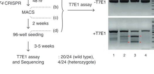
Figure 2.6 One of the unsuccessful attempts to generate TUT4/7 double knockout cell.
TUT7 KO HeLa cell was used as a parental cell line, and transfected with TUT4 CRISPR- Cas9 to delete the TUT4 gene. To detect mutation in genomic DNA, target region was amplified by PCR and T7E1 (T7 Endonuclease I) assay was carried out. T7E1 recognizes and cleaves heteroduplex formed between wild type and mutated target sequence. “a to d”
indicate the steps at which genomic DNA was extracted. (a) The parental TUT7 KO cells, (b) before MACS (magnetic-activated cell sorting for enrichment of mutated clones), (c) right after MACS, and (d) 2 weeks after MACS. Red asterisks denote cleaved DNA frag- ments detected by T7E1 assay, which indicate that genomic deletions had been effectively introduced. 3–5 weeks after 96-well seeding, ToolGen analyzed 24 individual colonies.
T7E1 negative clones were thought to be ‘wild type’ clones. T7E1 positive clones were sequenced but all of them contained at least one wild type allele for TUT4 (heterozygote).
Because none of the clones carried homozygous deletions for TUT4, our efforts to generate double knockout has failed despite the fact that the initial genomic deletion was successful.
This indicates that double knockout of TUT4 and TUT7 may be lethal.
siCont siTUT4/7
TUT7 TUT4
GAPDH
1 2
Figure 2.7 RNAi to deplete TUT4 and TUT7 simultaneously by transfecting siRNAs into HeLa cells. The TUT4 and TUT7 protein levels were monitored by western blotting.
0 1 2 3 4 5 6 7
0 2 4
Day Cell number (x10
6)
siTUT4/7 siCont
Figure 2.8 Cell proliferation rates shown by cell counting. Cell numbers are presented as
mean ± standard error of the mean (SEM) (n=3).
siControl
siTUT4/7 2n 4n
2n 4n
Counts Counts
0 20
40 65.4
±0.7 18.0
±0.5 16.3
±1.2
0.3
±0.0
16.3
±0.3 14.2
±0.3
66.1
±0.3
60 80 100
siCont siTUT4/7
Sub G1 G2-M S G1
Proportion (%)
3.4
±0.4
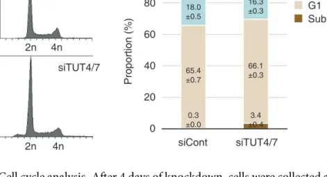
Figure 2.9 Cell cycle analysis. After 4 days of knockdown, cells were collected and profiled using BD FACSCanto and analyzed by BD FACSDiva software. (left) Cell cycle profiles.
(right) The proportion of each cell cycle phase is displayed in stacked bar graph. The average percentages ± standard error of the mean (SEM) from three independent experiments are shown.
siCont siTUT4/7 0
5 10 15 20 25
Frequency ( % )
U UU U 3
Figure 2.10 Uridylation frequency of mRNAs with short poly(A) tails (5–25 nt) measured
by TAIL-seq following simultaneous TUT4 and TUT7 knockdown (siTUT4/7). Uridyla-
tion was reduced when both TUT4 and TUT7 were depleted (P = 0.0941 for U, 0.00922
for UU, 0.0105 for U≥3; one-tailed t test). Error bar represents the standard error of the
mean from three biological replicates (n=3).
0.0 0.2 0.4 0.6 0.8 1.0 siCont
(Average U length per tail (nt)) 0.0
0.2 0.4 0.6 0.8 1.0
siTUT4/7 (Average U length per tail (nt))
n=746
638 108
Figure 2.11 Changes in uridylation of individual mRNA species upon TUT4/7 knock- down. “Average U length per tail” (y-axis) is the sum of the number of all uridines on short A tails (5–25 nt) divided by the total number of reads with short A tails. Note that unlike “uridylation frequency”, average U length per tail weighs every uridine in oligo- U tails. Each dot represents a transcript with ≥ 15 reads in both samples. Uridylation was significantly decreased following TUT4/7 knockdown (P = 7.69 × 10 -100 , one-tailed Mann-Whitney U test). The full list is shown in Table 2.1.
0.5
0.0 0.5
0.0 0.5
0.0
Average U length per tail (nt)
siCont siTUT4/7
1.17
1.04
HSPA8 TM4SF1 GNB2L1 LDHB CCT5 CCT2 GAPDH
PPIB TMSB10
HSP90AB1 SNRPD2 LDHA PABPC1 ATP5B
SERBP1 MYL6 PTP4A1 EIF4G2 PSMB3 ACTB CPS1
Figure 2.12 Examples of gene-level uridylation changes. Twenty-one most abundant
mRNAs (not including ribosomal protein mRNAs and histone mRNAs) are shown in the
order of mRNA abundance.
0.0 0.2 0.4 0.6 0.8 1.0 siCont
(Average U length per tail (nt)) 0.0
0.2 0.4 0.6 0.8 1.0
siTUT4/7 (Average U length per tail (nt))
n=48
43 5
0.0 0.2 0.4 0.6 0.8 1.0 siCont
(Average U length per tail (nt))
Replicate #2 Replicate #3 (Small-scale TAIL-seq using MiSeq)
0.0 0.2 0.4 0.6 0.8 1.0
siTUT4/7 (Average U length per tail (nt))
n=477
324 153
Figure 2.13 Changes in uridylation at transcript level upon TUT4/7 knockdown. Average U length per tail (y-axis) is the number of uridines on short A tails (5–25 nt) divided by the total number of reads with short A tails. Each dot represents a transcript with ≥ 15 reads in both samples. Replicate #3 was from “small-scale” TAIL-seq using Illumina MiSeq. Uridylation was significantly reduced when both TUT4 and TUT7 were knocked down (P = 1.39 × 10⁻¹⁵ for replicate #2, and P = 1.38 × 10 -9 for replicate #3 by one-tailed Mann-Whitney U test).
2.13).
Histone mRNAs that lack poly(A) tails are also uridylated and their uridylation is
dependent modestly on TUT4/7, but not on TUT1/2/3/5/6 (data not shown). However,
poly(A) histone mRNAs were excluded from my current data analyses because I used
non-synchronous cell population for my experiments, and it is known that uridylation of
histone mRNA occurs specifically at the end of S phase (Mullen & Marzluff, 2008; Schmidt
et al., 2011; Su et al., 2013). It would be more appropriate to investigate histone mRNAs
using synchronous cells in future studies.
10 20 30 40 50 100 150 200 0
10 20 30
M odi fi cat ion f requency ( % )
Mono-U siCont siTUT4/7
10 20 30 40 50 100 150 200
Poly(A) tail length (nt) 0
10 20 30
Oligo-U (2)
Figure 2.14 Distribution of mono-uridylation (top) and oligo-uridylation (bottom) ac- cording to the length of poly(A) tails. Poly(A) tail lengths from 5 nt to 231 nt are pooled into equal-width bins in the logarithmic scale (base 2) (x-axis). The left sides (inclusive) of bins are 5, 7, 9, 12, 15, 21, 28, 38, 50, 67, 89, 119, 159, and 212 nt. Uridylation frequency (y-axis) indicates the percentage of uridylated reads within each poly(A) tail size range.
Error bar represents the standard error of the mean (n=3).
2.2.2 TUT4/7 selectively oligo-uridylate mRNAs with short A tails in vivo and in vitro
It is intriguing that uridylation occurs preferentially on shortened A tails in plants and
animals (Chang et al., 2014b; Sement et al., 2013). Figure 2.14 shows the distribution of U
tails over different lengths of A tails in HeLa cells. The frequency of uridylation on the
transcripts with a short A tails (5–25 nt) is higher than that on the rest (A tails of >25 nt),
especially when only oligo-U (≥ 2 U) is counted. Note that mRNAs with A tails of shorter
than 5 nt were excluded from this analysis as it is sometimes difficult to distinguish them
from genomic A-rich sequences in 3′ UTR. When TUT4/7 were depleted, uridylation on
short A tails was selectively reduced (especially for oligo-U), indicating that TUT4/7 are responsible for the specific uridylation of short A tails (Figure 2.14).
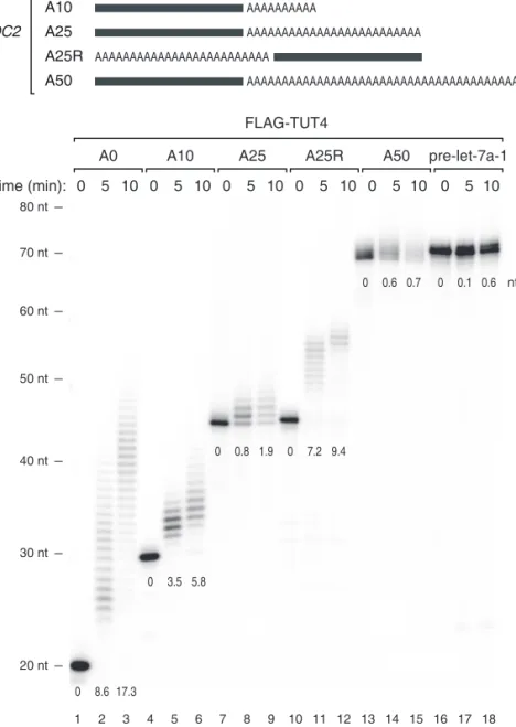
To understand the mechanism underlying such strong association with A tail length, I performed in vitro uridylation assays using immunopurified full-length TUTases (Figures 2.15, 2.16, 2.17, and 2.18) 4 . Substrate RNAs were chemically synthesized to contain het- erogenous sequences (the last 20 nt from the SHOC2 3′ UTR) linked to A tails of various lengths (0, 10, 25, and 50 nt) at the 3′ end (Figure 2.15). I also used a “swapped” control (A25R) that has a 25 nt A segment at the 5′ side of the SHOC2 3′ UTR such that the RNA is identical to SHOC2-A25 (A25) in the overall length and base composition, but lacks an A tail at the 3′ end (Figure 2.15).
Interestingly, RNAs with no tail (A0) or a short A tail (A10) were oligo-uridylated efficiently by TUT4 under the condition where pre-let-7a-1 is mono-uridylated weakly (Figure 2.15). A25 and A50 were less efficiently uridylated than A0 and A10 were. The A25R RNA was a much better substrate than the A25 was, indicating that it is the 3′ A tail length (not the overall RNA length) that is measured by TUT4 (Figure 2.15). Comparable results were obtained with full-length TUT7 protein (Figures 2.16 and 2.17), again demonstrating that these two related enzymes are functional paralogs. The U-tail length in Figures S2A–B was overall shorter than those in Figure 2.15 because the amount of immunoprecipitated TUT7 was smaller than that of TUT4 in Figure 2.15 (data not shown).
I also prepared recombinant TUT7 protein (951–1,495 aa) from E. coli and used the fragment for in vitro uridylation assay (Figure 2.18). Apart from the SHOC2 RNAs (Figure 2.19), I synthesized and tested another series of RNAs based on the CALM1 3′
UTR sequences (Figure 2.20) 5 . The purified protein fragment was fully capable of carrying out uridylation in an A tail length-dependent manner with both RNAs (Figures 2.20 and 2.19, see below). Thus, the C-terminal half of TUT7 is sufficient to recognize and uridylate single-stranded RNAs with a short A tails (<~25 nt), in a 3′ UTR sequence-independent manner. These results suggest that TUT4/7 possess an intrinsic ability to measure the 3′
terminal A length and avoid uridylation of long A tails.
4
These experiments were carried out by Dr. Minju Ha.
5
These experiments were carried out by Dr. Minju Ha.
20 nt 30 nt 40 nt 50 nt 60 nt 70 nt 80 nt
1 2 3 4 5 6 7 8 9 10 11 12 13 14 15 16 17 18 0 8.6 17.3
0 3.5 5.8
0 0.8 1.9 0 7.2 9.4
0 0.6 0.7 0 0.1 0.6
0 5 10 0 5 10 0 5 10 0 5 10
A0 A10 A25
0 5 10
A25R A50
FLAG-TUT4
Time (min):
pre-let-7a-1 0 5 10 SHOC2 3´ UTR (20 nt)
A0 SHOC2
AAAAAAAAAA A10
AAAAAAAAAAAAAAAAAAAAAAAAA A25
AAAAAAAAAAAAAAAAAAAAAAAAAAAAAAAAAAAAAAAAAAAAAAAAAA A50
AAAAAAAAAAAAAAAAAAAAAAAAA A25R
UUUAUUACAGCUCUACCUAG
nt
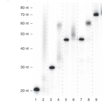
Figure 2.15 (top) Illustration of chemically synthesized RNA substrates. Grey bars repre-
sent the last 20 nt of SHOC2 3′ UTR and “A” indicates an adenosine. (bottom) In vitro
uridylation assay using immunopurified FLAG-TUT4. 0.45 nM of RNA was used in each
reaction. The products were resolved on 6 % polyacrylamide sequencing gel contain-
ing 7 M urea. The average length of uridylation is shown below each band. See 2.4 for
quantification method.
1 2 3 4 5 6 7 8 9 10 11 12 13 14 15 0 5 10 0 5 10 0 5 10 0 5 10
A0 A10 A25 A50
FLAG-TUT7
Time (min):
SHOC2: pre-let-7a-1
0 5 10
20 nt 30 nt 40 nt 50 nt 60 nt 70 nt 80 nt
Figure 2.16 In vitro uridylation assay using immunopurified full-length TUT7 and SHOC2
RNAs (the same RNA substrates and reaction conditions as shown in Figure 2.15). Dashed
lines indicate discontinuous lanes from the same gel, which applies to all the dashed lines
in the figure.
1 2 3 4 5 6 7 8 SHOC2:
Time (min):
FLAG-TUT7
0 5 10
A25
0 2.5
2.5 5 10
A25R
20 nt 30 nt 40 nt 50 nt 60 nt 70 nt 80 nt
Figure 2.17 In vitro uridylation assay using immunopurified full-length TUT7 and SHOC2
RNAs (A25 and A25R). Uridylation efficiency for A25R was higher than A25. Reaction
conditions are the same as in Figure 2.16, except that the amount of TUT7 was smaller in
Figure 2.17 than in Figure 2.16.
Mock M M M FLAG-TUT4 Mock FLAG-TUT7 Recombinant TUT7
1ug 2ug 4ug
130
70
55
45
35
25 100 170 kDa
1 2 3 4 5 6 7 8 9 10
Figure 2.18 Coomassie staining of immunopurified FLAG-TUT4 (full length), FLAG- TUT7 (full length), and recombinant TUT7 (951–1,495 aa) resolved on 10 % SDS-PAGE
gel. Each protein is indicated by arrowheads. M, size marker.
SHOC2:
- + - + - + - + A0 A10 A25 A25R
- + A50 rTUT7 (951-1495):
1 2 3 4 5 6 7 8 9 10
20 nt 30 nt 40 nt 50 nt 60 nt 70 nt 80 nt
Figure 2.19 In vitro uridylation assay using recombinant TUT7 (951–1,495 aa) and SHOC2
RNAs. 0.45 nM of RNA and 14 nM of recombinant TUT7 (rTUT7) were used in the
reaction. Extension products were resolved on 12.5 % polyacrylamide gel with 7 M urea.
20 nt 30 nt 40 nt 50 nt 60 nt 70 nt 80 nt
1 2 3 4 5 6 7 8 9 10 11 12 13 14 15
0 6.4 11.4
0 7.2 13.0
0 2.8 4.7 0 8.5 9.1
0 2.4 2.9
0 5 10 0 5 10 0 5 10 0 5 10
A0 A10 A25
0 5 10
A25R A50
Recombinant TUT7
Time (min):
CALM1 3´ UTR (20 nt)
A0 CALM1
AAAAAAAAAA A10
AAAAAAAAAAAAAAAAAAAAAAAAA A25
AAAAAAAAAAAAAAAAAAAAAAAAAAAAAAAAAAAAAAAAAAAAAAAAAA A50
AAAAAAAAAAAAAAAAAAAAAAAAA A25R
GCCUUUCAUCUCUAACUGCG
nt
Figure 2.20 (top) Illustration of chemically synthesized RNA substrates. Green bars
represent the last 20 nt of CALM1 3′ UTR and “A” indicates an adenosine. (bottom)
In vitro uridylation assay using recombinant TUT7 C-terminal fragment (951-1,495 aa)
purified from E. coli. 0.45 nM of RNA and 14 nM of recombinant TUT7 were used in
each reaction. Extension products were resolved on 6 % polyacrylamide sequencing gel
containing 7 M urea. The average length of uridylation was quantified as in Figure 2.15.
2.2.3 PABP suppresses uridylation of poly(A) + mRNA
As poly(A) + mRNAs are associated with poly(A) binding protein (PABP) in cells, I asked if PABP has an influence on mRNA uridylation. It was previously shown that PABP preferentially interacts with poly(A) or A-rich sequences (Eliseeva et al., 2013). The binding affinity increases as the A stretch gets longer (Eliseeva et al., 2013; Khanam et al., 2006;
Kuhn & Pieler, 1996; Sachs et al., 1987). Full length PABP occupies a ~25 nt A tail as determined by nuclease digestion assay (Baer & Kornberg, 1983; Eliseeva et al., 2013).
In order to test an effect of PABP on uridylation, in vitro uridylation assays was carried out in the presence of recombinant PABPC1 (Figure 2.21) 6 . When PABPC1 was added to RNA, uridylation of RNAs with long poly(A) tail (A25 and A50) was suppressed even at a low concentration of PABPC1 (10 nM) while those with no or short A-tail (A0, A10 and A25R) remained largely unaffected (Figure 2.21). This result suggests that PABPC1 binds preferentially to long poly(A) tails and protects them from TUT4/7, and thereby enhances the selectivity of uridylation according to poly(A) tail length.
Taken together, my results suggest that the strict dependence on the A tail length observed in vivo may be determined by the combination of two factors: (1) the intrinsic ability of TUT4/7 to measure poly(A) stretch (Figures 2.14, 2.15, and 2.20) and (2) the protective activity of PABP (Figure 2.21). As deadenylation is thought to occur mainly in the cytoplasm, I examined the localization of TUT4/7 by western blotting. The TUT4 and TUT7 proteins are mainly localized in the cytoplasm (Figure 2.22). Thus, TUT4/7 may function mainly in the metabolism of cytoplasmic, deadenylated mRNAs.
2.2.4 Uridylation facilitates global mRNA decay
To understand the functional consequences of uridylation, I measured mRNA half-life in HeLa cells with or without TUT4/7 knockdown (Figure 2.23) 7 . mRNA levels were determined by RNA-seq at 0, 1, 2, and 4 hr after actinomycin D treatment that blocks transcription. To avoid any bias from tail length variation, I omitted the oligo-dT enrich- ment step and instead used Ribo-Zero to remove abundant rRNAs prior to cDNA library
6
These experiments were carried out by Dr. Minju Ha.
7
All analyses in this section are carried out by Dr. Hyeshik Chang.
1 2 3 4 5 6 7 8 9 10 11 12 13 14 15 16
A0 A10 A25 A25R
rPABPC1 (nM):
- + + + - + + + - + + + - + + + rTUT7 (951–1495):
A50 - + + + 0 0 10 40 0 0 10 40 0 0 10 40 0 0 10 40 0 0 10 40
17 18 19 20 0 16.7 14.4 13.1
0 8.1 7.6 4.0
0 3.1 1.3 0.1 0 7.9 8.2 8.2
0 2.1 0.7 0 nt
20 nt 30 nt 40 nt 50 nt 60 nt 70 nt 80 nt SHOC2:
Figure 2.21 In vitro uridylation assay by using recombinant TUT7 (951–1,495 aa) with a
varying concentration of recombinant PABPC1 (0, 10, or 40 nM). 0.45 nM of RNA and
160 nM of recombinant TUT7 (rTUT7) were used in the reaction. Extension products
were resolved on 6 % polyacrylamide sequencing gel containing 7 M urea. The average
length of uridylation was quantified as described in Methods and Materials and shown
below each band.
Nuc Cyto HeLa
TUT7
GAPDH TUT4
Tubulin Lamin
1 2
Figure 2.22 Relative amount of TUT4 and TUT7 proteins in the nucleus and cytoplasm was measured by western blotting. Lamin was used as a nuclear marker while Tubulin and GAPDH were used as cytoplasmic markers.
construction. I could measure turnover rates of 1,829 mRNAs. In TUT4/7-depleted cells, the majority of mRNAs (1,426 out of 1,829 [78.0 %]) showed increase stability (Figure 2.23, left panel, and Table 2.2). Half-lives were increased by ~30 % on average, and median half-life was extended from 9.45 hr to 11.2 hr (Figure 2.23, right panel).
Of note, although TUT4/7 contribute to let-7 biogenesis, double knockdown of TUT4/7 (without simultaneous knockdown of TUT2) did not substantially affect the let-7 level (Heo et al., 2012). In fact, the transcriptome analyses show that mRNAs are globally upregulated, indicating that the changes in mRNA half-life observed in this study cannot be attributed to specific regulation of let-7 biogenesis.
For validation of the impact of TUT4/7 deletion on mRNA stability, five mRNAs (SHOC2, TRIM24, RB1CC1, MET, and XRN1) were measured by quantitative RT-PCR after actinomycin D treatment (Figure 2.24) 8 . None of these mRNAs contains a let-7 binding site with seed match in their 3′ UTR, yet all of them showed increased stability when TUT4/7 were depleted. Therefore, my results demonstrate that TUT4/7 play an important role in bulk mRNA degradation in a let-7 independent manner.
8
In collaboration with Dr. Minju Ha.
RNA-seq
48 hr
1st transfection of siRNA 2nd transfection of siRNA Actinomycin D treatment
44 hr 0 hr 1 hr 2 hr 4 hr si Cont si TUT4/7 0 5 10 15 20 25 30 si Cont (hal f- lif e ( hr) )
0
5 10
15
20
25
30
siTUT4/7 (half- lif e (hr) )
2
4
8 16
32
64 Ha lf-life (h r)
333
*** 1,496 N=1,829 Fi g u re 2 .2 3 T ra n scr ip to me-wide ch an ge o f mRN A h alf-lif e det er mined b y RN A -s eq . (left) Exp er imen tal sc heme . H eL a cells w er e tra n sf ec te d twice an d h ar ve st ed at 0, 1, 2, an d 4 h r fo llo win g ac tino m ycin D tr ea tmen t. (cen ter) Cha n ges o f av erag e mRN A h alf-lif e u p o n TUT4/7 kno ck do w n fr o m tw o b io logical rep lica tes. The ra n ge o f disp la y is limi te d to b etw een 0 and 30 hr fo r the b et ter visual re cogni tio n (2 32 o u t o f 1,8 29 mRN A s ar e o u tside o f th e vie w). The full list is av ai la b le in T ab le 2.2. (rig h t) D istr ib u tio n o f mRN A half-li ves in co n tr o lo r TUT4/7 kno ck do w n cells. A b o x rep res en ts the fir st an d thir d q ua rtiles an d an in ter nal b ar indica tes media n . W h isk er s spa n b etw een the 9t h and th e 91st p er cen tiles. H alf-li ve s o f mRN A s ar e significa n tl y ext ended b y TUT4/7 kno ck d o w n (*** P=4 .0 6×1 0 -15 5 ,o ne-t ai led p ai re d M ann- W h it ne y U test). S ee M et ho ds an d M at er ials fo r the det ai led des cr ip tio n o f p ro ced ur e.
Relative quantity (%)
Relative quantity (%)
qRT-PCR
48 hr
1st transfection of siRNA 2nd transfection of siRNA Actinomycin D treatment
44 hr 0 hr 2 hr 4 hr Time (hr) Time (hr)
Half-life (hr)
0
5 10
15
20
25
ME T
SH C O 2
RB1CC1 TR 24 IM
XRN 1
siTUT4/7
siCont
0 2 4 100 90 80 70 60 50 XRN1
0 2 4 100 90 80 70 60 50 ME T
0 2 4 100 90 80 70 60 50 RB1CC1
0 2 4 100 90 80 70 60 50 SHOC2
0 2 4 100 90 80 70 60 50 TRI M 24 F igur e 2.24 M easur emen t o f mRN A h alf-lif e b y q R T -PCR . (left) The exp er imen tal sc heme . (r ig h t) F o llo w in g 0 , 2, and 4 h r o f ac tino m ycin D tr ea tmen t, rela ti ve ab unda nce (y-axis) o f fi ve selec te d genes w er e me asur ed . F o r no rm aliza tio n , GAP D H mRN A was u se d b eca u se it was hig hl y st ab le (half-lif e > 24 hr ,d at a n o t sho wn) and did no t cha n ge no tice ab ly b y TUT4/7 dep letio n. Er ro r ba r rep re se n ts the st an da rd er ro r o f the me an (n=3). H alf-li ve s ar e calc ula ted b y line ar fi tt in g o f the log-tra n sf o rm ed exp o n en tial deca y func tio n.
Next, to examine the effect of overexpressed TUTase on mRNA expression, I carried out tethering experiments in HeLa cells (Figure 2.25, left panel) 9 . A related experiment was reported recently in Xenopus oocytes: when Xenopus TUT7 homolog was tethered to the 3′ UTR of luciferase reporter mRNA, luciferase activity was reduced without signifi- cant changes in mRNA, implicating translational repression (Lapointe & Wickens, 2013).
However, since mRNA decay activity is generally suppressed in oocytes (Barckmann &
Simonelig, 2013), it was unclear if the observation from frog oocytes can be generalized.
For tethering experiments, I generated constructs that express proteins tagged with the λN peptide that interacts with its specific binding sites (BoxB sites) in the 3′ UTR of luciferase mRNA (Figures 2.25 and 2.26) 10 . Expression of λN protein modestly increased luciferase expression non-specifically for an unknown reason (Figure 2.25, middle panel).
Nevertheless, tethering of AGO2 repressed luciferase reporter expression (Figure 2.25), as previously shown (Pillai et al., 2004), indicating that this is a valid system to test the effect of RNA silencing factors. Neither the negative control TUT2 nor its mutant repressed luciferase reporter expression. But when wild type TUT4 was tethered to the reporter mRNA, luciferase activity was decreased to ~60 % while such reduction was not observed with the catalytically dead point mutant (D1011A) of TUT4 (Figure 2.25, middle panel), indicating that TUT4 suppressed gene expression via uridylation. Quantitative RT-PCR further showed that tethering of TUT4 induced a reduction of mRNA (Figure 2.25, right panel). Thus, these results collectively indicate that TUT4/7 function as suppressors of gene expression through mRNA destabilization.
2.2.5 Uridylation is involved in miRNA-induced gene silencing
My model predicts that if a gene-specific inducer of deadenylation is introduced into cells, uridylation of the given transcript will take place, which in turn will facilitate RNA decay.
To test the model, I examined the effect of miRNA as an example, which is well established to induce specific deadenylation of its complementary targets (Ameres & Zamore, 2013;
Djuranovic et al., 2011; Huntzinger & Izaurralde, 2011; Krol et al., 2010).
9
These experiments were carried out by Dr. S.Chul Kwon.
10
This experiment was carried out by Dr. S.Chul Kwon.
Firefly luciferase
or A
(n)m
7G 5X BoxB sites
O N-HA-Protein HA-Protein
qRT-PCR 0
0. 4
0.8
1. 2 0. 2
0.6
1. 0
RNA level (FL/RL)
O N-tag: - + - +
HA-TUT4 D1011A HA-TUT4
**
Luciferase assay * 0
0. 4
0.8 1. 2
1.6 0. 2
0.6 1. 0
1. 4
Luciferase activity (FL/RL)
O N-tag:
HA-AGO2 HA-TUT2 HA-TUT2 D215A HA-TUT4 D1011A HA-TUT4
-+ -+ - + -+-+
-+ HA
** * Fi g u re 2 .2 5 (left) S chema tic rep re se n ta tio n o f rep o rt er ass ay syst em w it h the λ N tet her in g. (cen ter) Rep o rt er (fir efl y) lucif eras e ac ti vi ty was m easur ed and no rmalized to renilla lucif eras e ac ti vi ty (n=3). (r ig h t) R ep o rt er mRN A le vels w er e det er mined b y qR T -PCR (n=4). Er ro r b ar s rep re se n t th e st anda rd er ro r o f th e m ea n. L ucif eras e ac ti vi ty o r RN A le vel w er e significa n tl y red uced w h en A GO2 o r TUT4 w er e te th er ed (* P <0.01, ** P <0.0 01; tw o-t ai led t test).
HA GAPDH
1 2
HA-AGO2 O N-HA-AGO2
HA-TUT2 O N-HA-TUT2
HA-TUT2-D215A O N-HA-TUT2-D215A HA-TUT4 O N-HA-TUT4
HA-TUT4-D1011A O N-HA-TUT4-D1011A
3 4 5 6 7 8 9 10
Figure 2.26 Western blotting showing the expression of transfected proteins in Figure 2.25. GAPDH was used as a loading control.
I first analyzed the TAIL-seq data from my previous experiment where miR-1 mimic was transfected into HeLa cells (Chang et al., 2014b). As expected, miR-1 targets undergo deadenylation and subsequent downregulation following miR-1 mimic transfection (Figure 2.27, middle and lower panels, respectively) 11 . Importantly, I detected a specific increase of uridylation on miR-1 targets whereas the rest of genes stayed largely unaffected (Figure 2.27, upper panel). This result is consistent with my model that deadenylation leads to uridylation.
I next measured turnover rates of miR-1 targets with or without TUT4/7 knockdown.
The mRNAs tested (PTMA, ADAR, and PGM2) are normally stable (half-lives >24 hr) in cells that do not contain miR-1 (Figure 2.28, black line) 12 . When miR-1 was introduced, their half-lives were shortened to 6.5, 10.0, and 9.4 hr, respectively (Figure 2.28, blue line).
Upon TUT4/7 depletion, the miR-1 target mRNAs were stabilized (with extended half- lives of 14.0, 36.1, and 18.1 hr, respectively) (Figure 2.28, red line). Therefore, TUT4/7 are necessary for the facilitated decay of miRNA targets. I propose that other factors that cause deadenylation may also induce uridylation and decay, as shown here with an example of miR-1.
11
Analysis was done by Dr. Hyeshik Chang.
12
In collaboration with Dr. Minju Ha.
TAIL-seq 0 hr 3 hr 6 hr 9 hr miR-1 transfection
0 1 2 3
1.0 0.5 0.0 0.5
0 3 6 9
Time after transfection (hr) 1.0
0.5 0.0 0.5 Average U length change (ratio) Poly(A) tail length change (log
2ratio) mRNA abundance change (log
2fold)
miR-1 targets The rest
* ** ***
Figure 2.27 Changes in uridylation after miR-1 transfection. (left) Experimental scheme.
miR-1 was transfected into HeLa cells and the cells were harvested after the indicated time for TAIL-seq. Targets are the transcripts with ≥1 miR-1 3′ UTR site and down-regulated by ≥30 % on 12 hr post-transfection of miR-1 (Guo et al., 2010). (right top) Average U length change relative to 0 hr is shown in each time point. Average U length per tail is the number of uridines on short A tails (5–25nt) divided by the total number of reads with short A tails. Box represents the interval between the first and third quartiles, and the internal bar indicates the median. Whiskers span between the 9th and 91st percentiles.
Average U length of miR-1 target is significantly extended after miR-1 transfection (* P = 0.0152, ** P = 0.00318, *** P = 5.79 × 10 -4 ; one-tailed Mann-Whitney U test). (right middle) Poly(A) tail length change relative to 0 hr. The length change is represented by log 2 odds ratio between long tails (>25 nt) and short tails (≤25 nt) in one among 3, 6, or 9 hr and 0 hr. A negative value (<0) indicates increase of the fraction of short tails compared to 0 hr.
Error bars indicate standard deviation among mRNAs. The portion of short poly(A) tails
expanded more for miR-1 targets than the others (P=1.80×10 -6 for 3 hr, P=8.47×10 -13 for 6 hr,
P=1.48×10 -11 for 9 hr; one-tailed Mann-Whitney U test). (right bottom) mRNA abundance
(poly(A) + tag counts) change relative to 0 hr. Error bars indicate standard deviation among
mRNAs. Expression levels of miR-1 targets were decreased more than the rest transcripts
(P=2.09×10 -4 for 3 hr, P=2.65×10 -14 for 6 hr, P=5.46×10 -18 for 9 hr; one-tailed Mann-Whitney
U test).
qRT-PCR 62 hr Mock or miR-1 transfection
Actinomycin D treatment 4 hr
0 hr 2 hr 4 hr Transfection of siRNA
Relative quantity (%)
Time (hr)
Half-life (hr)
0 10 20 30 40
PTMA ADAR PGM2
Relative quantity (%) Relative quantity (%) siTUT4/7 siCont
0 2 4
100 90 80 70 60
50 ADAR
0 2 4
100 90 80 70 60
50 PGM2
0 2 4
100 90 80 70 60
50 PTMA
siCont (Mock transfection) siCont (miR-1 transfection) siTUT4/7 (miR-1 transfection)
Figure 2.28 Measurement of half-life of miR-1 targets by qRT-PCR. (left) The experimental scheme. Following siRNA transfection for 62 hr, HeLa cells were transfected with miR-1 or mock transfected. After 4 hr, actinomycin D was treated and cells were harvested at 0, 1, 2, and 4 hr. (right) Relative abundance (y-axis) of miR-1 target mRNAs were measured.
For the normalization, highly stable GAPDH mRNA was used because it did not change
significantly by siTUT4/7 or miR-1 transfection. Error bar represents the standard error
of the mean (n=3). Half-lives are determined by linear fitting of the log-transformed
exponential decay function.
2.2.6 mRNA decay factors remove uridylated mRNAs
To understand downstream events of uridylation, I disrupted 5 ′ –3 ′ or 3 ′ –5 ′ exonucleolytic decay factors and examined the mRNA terminome (Figure 2.29) 13 . The popsicle-shaped bars in Figure 2.30 display the relative quantity of reads with a U tail (thick stem) or without a U tail (thin stem) 14 . As U-tail frequencies vary depending on poly(A) tail length, different A-tail ranges are shown separately along the horizontal axis. For more information, the overall uridylation frequency and poly(A) length distribution are presented in Figures 2.31 and 2.32, respectively.