This is an Open Access article distributed under the terms of the Creative Commons Attribution Non-Commercial License (http: //creativecommons.org/licenses/by- nc/4.0/) which permits unrestricted non-commercial use, distribution, and reproduction in any medium, provided the original work is properly cited.
© 2019 THE KOREAN SOCIETY OF MYCOLOGY.
RESEARCH ARTICLE
A new record of Trichocladium griseum in Korea: morphological and molecular characterization
Setu Bazie Tagele
1,†, Thuong T.T. Nguyen
2,†, Sang Woo Kim
1, Mahesh Adhikari
1, Sun Kumar Gurung
1, Hyun Goo Lee
1, Byeong Heon Gwon
1, Han Jun Ju
1, San Kosol
1, Hyang Burm Lee
2,*, and Youn Su Lee
1,*
1
Division of Biological Resources Sciences, Kangwon National University, Chuncheon 24341, Korea
2
Division of Food Technology, Biotechnology & Agrochemistry, College of Agriculture and Life Sciences, Chonnam National University, Gwangju 61186, Korea
*Corresponding author: [email protected], [email protected]
†
This authors contributed equally to this work.
ABSTRACT
A unrecorded species of Trichocladium, Trichocladium griseum, was isolated in 2017 during a survey of fungal diversity in Ulsan province, South Korea. This species was identified based on morphological characteristics and phylogenetic analysis of the internal transcribed spacer (ITS) rDNA and β-tubulin gene sequences. T. griseum has not yet been reported in South Korea. Thus, we report for the first time a new record of Trichocladium griseum in Korea, and we include the descriptions and morphological illustrations of this fungus.
Keywords: β-tubulin, ITS rDNA, Morphology, Taxonomy, Trichocladium griseum
Introduction
The genus Trichocladium, which belongs to the subphylum Pezizomycotina, class Sordariomycetes, order Sordariales, and family Chaetomiaceae, was first described by Harz in 1871 with Trichocladium asperum as the type species [1]. Members of this genus are characterized by the production of darkly pigmented, septate conidia. Species belonging to the genus Trichocladium have been frequently isolated from soil, estuarine sediment, leaf, dung, grass, meal, and Usnea cf. aurantio-atra [2]. Recently, Wang et al. [2]
redefined Humicola sensu stricto and related genera in the family Chaetomiaceae based on the phylogenetic analyses of multi-gene sequences combined with morphological observations, and they transferred certain species of Beniowskia, Chaetomium, Chaetomidium, Gilmaniella, Humicola, Monodictys, Thielavia, and Monodictys to the new genus Trichocladium. To date, 12 species belonging to this genus are known [2].
Prior to its renaming, Trichocladium griseum was classified under the name Humicola grisea.
Knowledge of the taxonomy of certain fungi within the genera Trichocladium in Korea is limited. A recent study reported the presence of Trichocladium asperum in soil [3]. A survey of Korean indigenous
Accepted: June 17, 2019 Revised: June 12, 2019 Received: April 16, 2019 https://doi.org/10.4489/KJM.20190013 Kor. J. Mycol. 2019 June, 47(2): 105-12
OPEN ACCESS pISSN : 0253-651X eISSN : 2383-5249
Mahesh Adhikari
https://orcid.org/0000-0001-9646-8633
fungal diversity was conducted in 2017 in Ulsan, South Korea. Fungal isolates possessing various morphologies were isolated from soil samples obtained from paddy fields. Consequently, an unrecorded species belonging to the genus Trichocladium (T. griseum) was discovered. This fungal species has not previously been reported in Korea. In this report, macro-morphological and micro-morphological characteristics of this newly recorded species are presented.
Materials and methods
Isolation of the fungal isolate
Soil samples were collected in 2017 from a paddy field located in Ulsan (35°31'45.36''N, 129°06'23.12'' E), South Korea. Crop debris were removed and soil samples were collected at a depth of 10~15 cm. The soil samples were dried and then stored at 4°C in a sterile polythene bag prior to use. Morphologically different isolates of fungal species were isolated on potato dextrose agar (PDA, Difco Laboratories, Detroit, Michigan, USA) supplemented with 100 μg L
-1chloramphenicol using conventional soil dilution techniques [4]. The diluted soil suspensions were streaked onto the petri plates and then incubated for 5 days at 25°C. The developing morphologically different colonies were then aseptically transferred to fresh PDA plates to obtain pure cultures. The pure fungal isolates were finally reserved on PDA slants at 4 °C for further use.
Table 1. The ITS and β-tubulin gene sequences obtained from Trichocladium griseum used in this study, but also the reference strains and their GenBank accession numbers
Taxon name Collection no.
(strain no.) GenBank accession numbers
ITS tub2
Trichocladium griseum CBS 119.14 (NT) LT993639 LT993720
T. griseum EML-KNU17-5 MK785135 MK782052
T. griseum CGMCC 3.13888 LT993641 LT993722
T. asperum CBS 903.85 (eT) LT993632 LT993713
T. gilmaniellae CBS 388.75 (T) LT993638 LT993719
Trichocladium sp. CBS 351.77 LT993647 LT993728
T. crispatum CBS 149.58 LT993636 LT993717
T. antarcticum CBS 123565 (T) LT993629 LT993710
T. arxii CBS 104.79 (T) LT993631 LT993712
T. seminis-citr ulli CBS 143.58 (T) LT993645 LT993726
T. amorphum CBS 127763 (T) LT993628 LT993709
T. acropullum CBS 114580 (T) LT993626 LT993707
Melanocarpus albomyces CBS 638.94 (T) KX976679 KX977021
Ovatospora medusarum CBS 148.67 (T) KX976684 KX977032
O. senegalensis CBS 728.84 (T) KX976687 KX977035
O. mollicella CBS 583.83 (T) KX976685 KX977033
Microascus trigonosporus CBS 218.31 (T) LM652443 LM652655
Bold letters indicate isolates and accession numbers determined by our study. CBS: Centraalbureau voor
Schimmelcultures (Utrecht, The Netherlands); CGMCC: China General Microbiological Culture Collection
Morphological examination
For detailed morphological studies, the EML-KNU17-5 strain was cultured on malt extract agar (MEA), oatmeal agar (OA; 30 g of oatmeal and 20 g of agar in 1 L of deionized water), and PDA at 25°C for 7 days.
The colonies grown on OA were observed microscopically. Fragments of mycelia were removed from the cultures and placed on microscope slides with lactic acid. An Olympus BX51 microscope possessing differential interference contrast optics (Olympus, Tokyo, Japan) was used to obtain digital images.
Genomic DNA extraction, PCR, sequencing, and phylogenetic analysis
The genomic fungal DNA was isolated from mycelia of the fungal isolate using the DNeasy Plant Mini Kit (Qiagen, Hilden, Germany). The β-tubulin (tub2) gene and the ITS region from EML-KNU17-5 isolates were amplified using the primer pairs Bt2a and Bt2b [5] and ITS1 and ITS4 [6], respectively. The PCR products were sequenced at Macrogen (Seoul, Korea) using an ABI PRISM 3730XL Analyzer (Life Technologies, Gaithersburg, MD, USA). The DNA sequences were subjected to BLAST analysis using the GenBank database. The construction of phylogenetic trees was accomplished by neighbor-joining (NJ) method using Kimura �s 2-parameter model [7] implemented in the program MEGA version 6 [8] with 1,000 bootstrap replicates. The EML-KNU17-5 isolate used in this study was deposited at the National Institute of Biological Resources (NIBR, Incheon, Korea), as NIBRFG000499999. The sequences of EML-KNU17-5 were deposited into the NCBI database under the accession numbers indicated in Table 1.
Results
Phylogenetic analysis
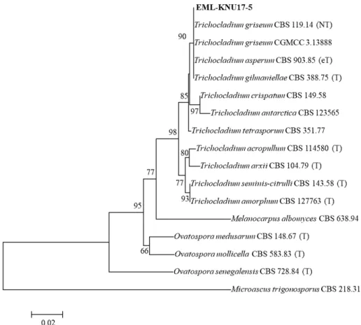
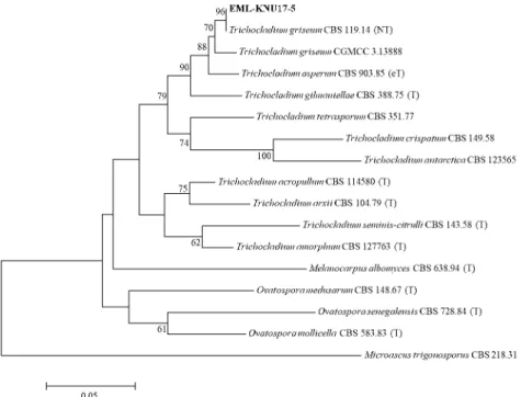
For the phylogenetic analysis, ITS and β-tubulin gene sequences were used to analyze the phylogenetic relationships between EML-KNU17-5 and related species. BLASTn search of ITS rDNA indicated that the strain EML-KNU17-5 was most closely related to H. grisea CGMCC 3.13888 (current name: T. griseum) (GenBank accession no. LT993641) with 100% identity. BLASTn analysis of tub2 of EML-KNU17-5 revealed a similarity of 99.6% to H. grisea CBS 119.14 (current name: T. griseum) (Genbank accession no. LT993720). The phylogenetic relationships among the strain EML-KNU17-5 and related species were analyzed using single genes and the combined datasets for ITS and tub2 (Fig. 1 -3). The strain EML- KNU17-5 was grouped within the same clade as T. griseum with 90%, 96%, and 99% bootstrap support according to ITS, tub2, and a combination of ITS and tub2 sequences, respectively.
Morphological characterization
Taxonomic descriptions of the morphological structures of the T. griseum EML-KNU17-5 are
detailed below.
Colony characteristics
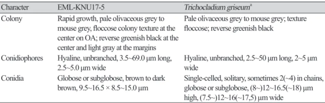
Trichocladium griseum (Traaen) X. Wei Wang & Houbraken, Studies in Mycology 93: 141 (2018) [MB#824469] (Table 2, Fig. 4).
Colony characteristics of the fungal isolate EML-KNU17-5 on various agar media are shown in Figure 4. Colonies on PDA exhibited greenish-black centers and grey-white margins, and they were reverse olivaceous black with moderate sporulation and reached 37~40 mm in diameter after 7 days at 25 °C.
Colonies on MEA were olivaceous grey with a velvety to floccose texture, and they were also reverse greenish black and reached 35~38 mm in diameter after 7 days at 25 °C. On OA, colonies were pale olivaceous grey to mouse-grey throughout with greenish-black centers and light grey margins, and they possessed floccose colony texture and reached 40~43 mm in diameter after 7 days at 25 °C.
Micromorphology
Conidiophores were hyaline, unbranched, and occasionally exhibited swelling at the tops, and these structures were 3.5~69.0 µm long and 2.5~5.0 µm wide. Conidia were globose or subglobose, brown to dark brown, smooth, and measured 9.5~16.5 × 8.5~15.0 µm. Acremonium-like conidia formed in chains that were obovoid to ellipsoidal.
Fig. 1. Phylogenetic tree based on neighbor-joining analysis of the internal transcribed rDNA sequence obtained from EML-KNU17-5. Microascus trigonosporus was used as outgroup. Bootstrap scores of
> 60% are indicated at the nodes. The scale bar represents the number of substitutions per site. The new
Fig. 2. Phylogenetic tree based on neighbor-joining analysis of β-tubulin gene sequence obtained from EML-KNU17-5. Microascus trigonosporus was used as outgroup. Bootstrap scores of > 60% are indicated at the nodes. The scale bar represents the number of substitutions per site. The new isolate described in this study is shown in bold.
Fig. 3. Phylogenetic tree based on neighbor-joining analysis of the combined datasets for ITS and β-tubulin sequences obtained from EML-KNU17-5. Microascus trigonosporus was used as outgroup.
Bootstrap scores of ≥ 60% are indicated at the nodes. The scale bar represents the number of substitutions
per site. The new isolate described in this study is shown in bold.
Table 2. Morphological characteristics of EML-KNU17-5 and the reference species Trichocladium griseum cultured on different agar mediums at 25℃
Character EML-KNU17-5 Trichocladium griseum
aColony Rapid growth, pale olivaceous grey to mouse grey, floccose colony texture at the center on OA; reverse greenish black at the center and light gray at the margins
Pale olivaceous grey to mouse grey; texture floccose; reverse greenish black
Conidiophores Hyaline, unbranched, 3.5~69.0 μm long,
2.5~5.0 μm wide Hyaline, unbranched, 2.5~50 μm long, 2~5 μm wide
Conidia Globose or subglobose, brown to dark
brown, 9.5~16.5 × 8.5~15.0 µm Single-celled, solitary, sometimes 2(~4) in chains, globose or subglobose, (8~)12~16.5(~18) µm high, (7.5~)12~16(~17,5) µm wide
a