Received: 17 November, 2017 Revised: 23 November, 2017 Accepted: 23 November, 2017
Ⓒ The Korean Society of Mycology
This is an Open Access article distributed under the terms of the Creative Commons Attrib- ution Non-Commercial License (http://creative- commons.org/licenses/by-nc/4.0/) which permits unrestricted non-commercial use, distribution, and reproduction in any medium, provided the original work is properly cited.
Kor. J. Mycol. 2017 December, 45(4): 345-354 https://doi.org/10.4489/KJM.20170040
pISSN : 0253-651X eISSN : 2383-5249
OPEN ACCESS
RESEARCH NOTE
목본식물의 잎에서 분리된 5종의 미기록 내생균
박혁, 심재성, 김지수, 최항석, 엄안흠*
한국교원대학교 생물교육과
Five Previously Unreported Endophytic Fungi Isolated from the Leaves of Woody Plants in Korea
Hyeok Park, Jae-Sung Shim, Ji-Su Kim, Hang-Seok Choi, Ahn-Heum Eom
*Department of Biology Education, Korea National University of Education, Cheongju 28173, Korea
*Corresponding author: [email protected]
Abstract
The leaves of two woody plant species, Pinus densiflora and Aronia melanocarpa, were collected in Korea, and endophytic fungi were isolated from these surface-sterilized leaves. The fungal isolates were identified based on their morphological characteristics and the results of the phylogenetic analysis involving nucleotide sequences of the internal transcribed spacer region (ITS), including 5.8S rDNA, D1/D2 regions of 28S rDNA, and β-tubulin genes. Pestalotia lawsoniae and Zasmidium fructicola were isolated from Pinus densiflora, and three species, Pestalotiopsis chamaeropis, Pestalotiopsis jesteri, and Stagonosporopsis cucurbitacearum were isolated from Aronia melanocarpa. To the best of our knowledge, these species have not been previously reported in Korea.
Keywords: Endophytic fungi, Pestalotia lawsoniae, Pestalotiopsis chamaeropis, Pestalotiopsis jesteri, Stagonosporopsis cucurbitacearum, Zasmidium fructicola
내생균(endophytic fungi)은 살아 있는 식물의 식물체에서 병증을 유발하지 않으며 공생하는
균류를 의미하는데[1], 식물체의 잎과 뿌리, 줄기 등 모든 조직 내에 서식하고 있다. 특히 식물
의 잎에 서식하는 내생균은 병원체로 작용하는 미생물의 잎 조직으로의 침투를 제한하는 역
할을 하기도 하고[2], 식물의 포식자에게 독소가 될 수 있는 alkaloid 형태의 2차 대사산물을
분비하기도 한다[3]. 최근에는 내생균들이 잎 조직 내에서 분비하는 이러한 물질들이 다른 미
생물 병원체에 대한 항생 효과 또한 낼 수 있다는 사례들이 연구되면서[4, 5], 식물의 잎에서
분리된 내생균에 대한 연구와 균주 확보의 중요성이 점차 높아지고 있다. 본 연구에서는 우리
나라의 다양한 식물의 잎에서 내생균을 분리하여 다양성을 조사하는 연구를 진행하던 과정
에서 충북 단양의 아로니아(Aronia melanocarpa)와 전북 부안의 소나무(Pinus densiflora)의
잎에서 5종의 국내 미기록 내생균을 분리하여 그 균의 형태적 특징과 계통 분석 결과를 보고
하고자 한다.
아로니아는 충청북도 단양군 적성면 현곡리의 아로니아 재배 농경지(N36°57'05.5", E128°18'01.3") 에서 채집되었고, 소나무는 전북 부안군 주산리 사산면의 산림(N35°39' 23.3", E126°40'59.2") 에서 채집되었다. 채집된 시료는 지퍼백에 담아 24시간 내에 실험실로 운반하여 외관상 병증이 없는 잎 시료를 선별하여 증류수로 세척 후 30% H 2 O 2 로 30초~1분 간 처리하여 표면을 살균하였다. 표면 살균 시간은 잎의 면적에 따라 다르게 수행하였다. 표 면살균 후 잎을 potato dextrose agar (PDA) 배지에 4조각씩 치상하였고 25°C 암소에서 배양 한 후 균사가 뻗어 나오면 새로운 PDA 배지에 계대배양하였다. 순수 분리된 균주는 PDA 배 지와 malt extract agar (MEA) 배지에서 7일간 배양하여 형태학적 특징을 관찰하였다(Fig. 1).
Fig. 1. Colonies of strain 16B102 (Pestalotia lawsoniae) grown on PDA (A) and MEA (F);
Colonies of strain 16C083 (Pestalotiopsis chamaeropis) grown on PDA (B) and MEA (G), conidia (K); Colonies of strain 16C094 (Pestalotiopsis jesteri) grown on PDA (C) and MEA (H), conidia (L); Colonies of strain 16C067 (Stagonosporopsis cucurbitacearum) grown on PDA (D) and MEA (I), conidia (M, type 1; N, type 2); Colonies of strain 16B021 (Zasmidium fructicola) grown on PDA (E) and MEA (J), conidia (O); PDA, potato dextrose agar; MEA, malt extract agar (scale bars = 10 µm).
DNeasy Plant mini kit (Qiagen, Germantown, MD, USA) 을 사용하여 Genomic DNA를
추출하였으며, 이 DNA를 주형으로 하여 PCR 반응을 수행하였다. 모든 균주의 DNA는 균 특
이적 primer인 ITS1F와 ITS4를 이용하여 internal transcribed spacer (ITS) 지역을 증폭하였
고[6], primer LR0R과 LR16을 이용하여 ribosomal DNA의 large subunit (LSU) 지역을[7],
그리고 primer Bt2a과 Bt2b를 이용하여 β-tubulin (TUB) 영역을 증폭하였다[8]. PCR 조건
중 annealing 온도는 ITS 지역은 50°C, LSU rDNA 지역은 44°C, β-tubulin 영역은 58°C로
설정하여 수행하였으며, PCR 최종 산물은 1.5% agarose gel에서 20분 간 전기영동을 실시하
였고, 예상되는 크기의 DNA band를 확인한 후 염기서열 분석을 의뢰하였다(SolGent,
joining 계통수를 작성하였다(Table 1, Figs. 2~5). 분리된 균주는 국립생물자원관(NIBR)에 기탁하였으며, 염기서열은 GenBank에 제출하였다(Table 2).
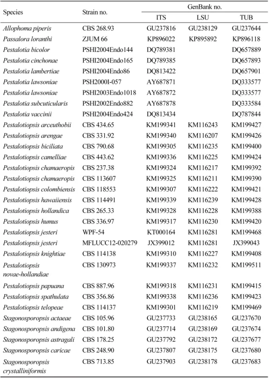
Table 1. Fungal strains used in phylogenetic analysis, including outgroups
Species Strain no. GenBank no.
ITS LSU TUB
Allophoma piperis CBS 268.93 GU237816 GU238129 GU237644 Passalora loranthi ZJUM 66 KP896022 KP895892 KP896118 Pestalotia bicolor PSHI2004Endo144 DQ789381 DQ657889 Pestalotia cinchonae PSHI2004Endo165 DQ789385 DQ657893 Pestalotia lambertiae PSHI2004Endo86 DQ813422 DQ657901
Pestalotia lawsoniae PSH2000I-057 AY687871 DQ333577
Pestalotia lawsoniae PSHI2003Endo1018 AY687872 DQ333577 Pestalotia subcuticularis PSHI2002Endo882 AY687878 DQ333584 Pestalotia vaccinii PSHI2004Endo424 DQ813434 DQ787844 Pestalotiopsis arceuthobii CBS 434.65 KM199341 KM116243 KM199427 Pestalotiopsis arengae CBS 331.92 KM199340 KM116207 KM199426 Pestalotiopsis biciliata CBS 790.68 KM199305 KM116235 KM199400 Pestalotiopsis camelliae CBS 443.62 KM199336 KM116225 KM199424 Pestalotiopsis chamaeropis CBS 237.38 KM199324 KM116217 KM199392 Pestalotiopsis chamaeropis CBS 113607 KM199325 KM116211 KM199390 Pestalotiopsis colombiensis CBS 118553 KM199307 KM116222 KM199421 Pestalotiopsis hawaiiensis CBS 114491 KM199339 KM116239 KM199428 Pestalotiopsis hollandica CBS 265.33 KM199328 KM116228 KM199388 Pestalotiopsis humus CBS 336.97 KM199317 KM116230 KM199420 Pestalotiopsis jesteri WPF-54 KT000164 KM116281 KM199468 Pestalotiopsis jesteri MFLUCC12-020279 JX399012 KM116281 JX399043 Pestalotiopsis knightiae CBS 114138 KM199310 KM116227 KM199408 Pestalotiopsis
novae-hollandiae
CBS 130973 KM199337 KM116232 KM199511
Pestalotiopsis papuana CBS 887.96 KM199318 KM116231 KM199415 Pestalotiopsis spathulata CBS 356.86 KM199338 KM116236 KM199423 Pestalotiopsis telopeae CBS 114137 KM199301 KM116219 KM199469 Stagonosporopsis actaeae CBS 105.96 GU237733 GU238165 GU237670 Stagonosporopsis andigena CBS 101.80 GU237714 GU238169 GU237674 Stagonosporopsis astragali CBS 178.25 GU237792 GU238172 GU237677 Stagonosporopsis caricae CBS 248.90 GU237807 GU238175 GU237680 Stagonosporopsis
crystalliniformis
CBS 713.85 GU237903 GU238178 GU237683
ITS, internal transcribed spacer; LSU, large subunit; TUB, β-tubulin.
Table 1. (Continued)
Species Strain no. GenBank no.
ITS LSU TUB
Stagonosporopsis cucurbitacearum
CBS 133.96 GU237780 GU238181 GU237686
Stagonosporopsis cucurbitacearum
PD 91/310 GU237922 GU238180 GU237685
Stagonosporopsis dennisii CBS 135.96 GU237782 GU238183 GU237688 Stagonosporopsis hortensis CBS 104.42 GU237730 GU238198 GU237703 Stagonosporopsis loticola CBS 562.81 GU237890 GU238192 GU237697 Stagonosporopsis
oculi-hominis
CBS 634.92 GU237901 GU238196 GU237701
Stagonosporopsis trachelii CBS 379.91 GU237850 GU238173 GU237678 Stagonosporopsis
valerianellae
CBS 273.92 GU237819 GU238200 GU237705
Xylaria hypoxylon CBS 122620 KY610407 KY610495 KX271279 Zasmidium anthuriicola CBS 118742 FJ839626 FJ839662 KF252763 Zasmidium citri-griseum ZJUM 103 KP896039 KP895909 KP896134 Zasmidium fructicola ZJUM 9 KP896043 KP895913 KP896138 Zasmidium fructicola ZJUM 68 KP896048 KP895918 KP896142 Zasmidium fructigenum ZJUM 100 KP896061 KP895931 KP896154 Zasmidium lonicericola CBS 125008 KF251283 KF251787 KF252765 Zasmidium nocoxi CBS 125009 KF251284 KF251788 KF252766 Zasmidium podocarpi CBS 142529 KY979766 KY979821 KY979930 Zasmidium pseudoparkii CBS 111049 DQ303025 KF901976 KF902976 Zasmidium scaevolicola CBS 127009 KF251285 KF251789 KF252767 Zasmidium xenoparkii CBS 111185 DQ303028 JF700966 KF902978
ITS, internal transcribed spacer; LSU, large subunit; TUB, β-tubulin.
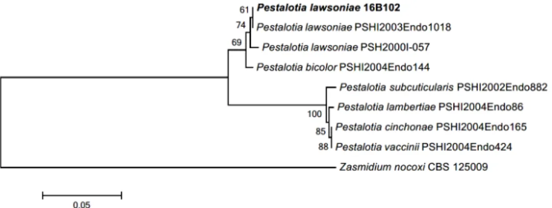
Fig. 2. Neighbor-joining phylogenetic tree based on a combined alignment of both internal
transcribed spacer (ITS) and β-tubulin (TUB) sequences. Zasmidium nocoxi was used as an
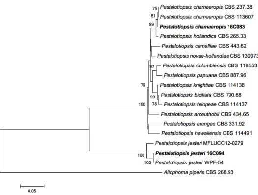
Fig. 3. Neighbor-joining phylogenetic tree based on a combined alignment of both internal transcribed spacer (ITS), large subunit (LSU) and β-tubulin (TUB) sequences. Xylaria longipes was used as an outgroup. Numbers on branches indicate bootstrap values (1,000 replicates).
Fungal strain isolated in this study are in bold.
Fig. 4. Neighbor-joining phylogenetic tree based on a combined alignment of both internal
transcribed spacer (ITS), large subunit (LSU) and β-tubulin (TUB) sequences. Allophoma
piperis was used as an outgroup. Numbers on branches indicate bootstrap values (1,000
replicates). Fungal strain isolated in this study are in bold.
Pestalotia lawsoniae Mundk. & Khesw. Mycologia 34: 315 (1942)
PDA 배지에서 7일간 배양된 균총의 직경은 42~45 mm 정도로 매우 빠르게 자라며, 균총의 색은 앞뒷면 모두 전체적으로 흰색이다. 고도는 중앙부에서 볼록 융기되어 있고 중앙부에서 부터 가장자리로 공중균사가 형성되어 방사형으로 뻗어 나간다(Fig. 1A). MEA 배지에서 배 양된 균총의 직경은 44~45 mm 정도이고 형태적 특성은 PDA 배지에서 배양된 균주와 대부 분 일치하나 색이 전체적으로 연한 갈색을 띤다(Fig. 1F). NCBI 상에서 ITS 지역의 분석결과 P. lawsoniae AY687872 와 100.0%의 일치도를 확인하였고, LSU 지역의 분석결과 HM535714 와 98.0%의 유사도를, TUB 지역의 분석결과 DQ333577과 97.0%의 유사도를 확인하였다. Pestalotia속은 1841년 De Notaris에 의해 명명되었으며[10], Pestalotiopsis와 같이 Amphisphaeriaceae에 속한다. Pestalotiopsis속과 비슷한 형태적 특성을 공유하고 실제
Fig. 5. Neighbor-joining phylogenetic tree based on a combined alignment of both internal transcribed spacer (ITS), large subunit (LSU) and β-tubulin (TUB) sequences. Passalora loranthi was used as an outgroup. Numbers on branches indicate bootstrap values (1,000 replicates). Fungal strain isolated in this study are in bold.
Table 2. GenBank accession numbers and NIBR numbers for fungal strains isolated from this this study
Isolates NIBR specimen no. GenBank no. Closest taxon Host plants 16B102 NIBRFG0000499923 MG436878, Pestalotia lawsoniae Pinus densiflora 16C083 NIBRFG0000499919 MG436879 Pestalotiopsis
chamaeropis
Aronia melanocarpa
16C094 NIBRFG0000499921 MG436876 Pestalotiopsis jesteri Aronia melanocarpa 16C067 NIBRFG0000499920 MG462717 Stagonosporopsis
cucurbitacearum
Aronia melanocarpa
16B021 NIBRFG0000499920 MG462717 Zasmidium fructicola Pinus densiflora
에서는 숙주식물의 잎에 반점(leaf spot)을 발생시키는 병원성 균류로 기록되었다[12]. 본 연 구에서는 소나무 잎에서 분리한 내생균이며, Pinus속의 침엽수 잎에서 내생균으로 분리된 기 록도 존재한다[13].
Pestalotiopsis chamaeropis Maharachch., K. D. Hyde & Crous, Stud Mycol 79: 158 (2014)
PDA 배지에서 7일간 배양된 균총의 직경은 42~45 mm 정도이고, 앞면은 중앙부는 올리브색 이고 주변부는 흰색의 공중 균사로 덮여 있다. 뒷면의 색은 중앙부는 베이지색이고 주변부는 흰색이며, 균총의 고도는 융기되어 있으며, 가장자리는 불규칙한 형태이다(Fig. 1B). MEA 배지에서 7일간 배양한 균총의 직경은 42~45 mm 정도이고, PDA 배지에서 배양한 균주와 형태적 특성이 일치하나 뒷면 균총의 중앙부에서 방사형으로 주름이 있다(Fig. 1G). 분지된 균사 끝에서 분생자를 형성하는데, 분생자는 격벽이 있으며 검은색의 세포층으로 나뉘어 있 고, 바깥층은 투명한 무색의 유리질 막이 감싸고 있다. 분생자의 크기는 13.9~23.2 × 9.0~10.9 µm 이다(Fig. 1K). ITS 지역의 염기서열은 KR259104와 99.0%, LSU 지역은 KM116217 과 99.0%, 그리고 TUB 지역은 KR259103과 98.0%의 유사도를 나타냈다.
Pestalotiopsis 속은 1949년 Steyaert [11]에 의해 최초로 명명된 속으로, 검은 색의 분생포자 층(acervulus)이 불규칙하게 혹은 균열이 있는 상태로 종방향으로 길게 뻗어 나가며, 유리질 의 격막으로 분리된 갈색의 분생자를 형성하는 것이 특징이다. 본 연구에서 이 균주는 아로니 아의 잎에서 분리되었으며, 이탈리아에서는 야자나무과(Arecaceae)에 속하는 식물인 Chamaerops humilis 의 잎에서 분리된 내생균으로 보고되었다[14, 15].
Pestalotiopsis jesteri Strobel, J.Yi Li, E. J. Ford & W. M. Hess. Mycotaxon 76: 260 (2000)
PDA 배지에서 7일간 배양된 균총의 직경은 31~33 mm 이며, 균총의 앞뒷면은 중앙부는 청 록색을 띠는 흰색이며 가장자리에 흰색의 띠가 생긴다. 가장자리의 띠는 7일 이상 배양하면 파도형으로 여러 층을 형성하여 점차 나이테와 같은 형태로 자란다. 균사는 중앙부에서 성기 고 가장자리로 갈수록 조밀해져 전체적으로 방사형을 이룬다. 균총의 고도는 배지에 납작 붙 어 있는 형태이다(Fig. 1C). MEA 배지에서 7일간 배양된 균총의 직경은 35~40 mm 정도로 PDA 배지에 배양할 때보다는 약간 크게 자라고, 균총의 앞면 중앙부는 아이보리색이며 가장 자리는 흰색을 띤다. 뒷면은 중앙부에서 연한 갈색을 띠며 가장자리는 흰색이다. 가장자리에 는 방사형의 공중균사가 발달하고, 고도는 볼록 융기된 형태이다(Fig. 1H). 분생자는 3중의 격벽에 의해 2개의 세포로 분리된 방추형 혹은 타원형이고, 색깔은 진한 갈색이며 크기는 11.8~14.4 × 5.5~6.7 µm 정도이다(Fig. 1L). ITS 지역의 염기서열은 KM199380과 98.0%, LSU 지역은 KM116281와 99.0%, TUB 지역은 KM199468과 98.0%의 유사도를 나타냈다.
이 종은 파푸아뉴기니의 용담과(Gentianaceae) 식물의 줄기에서 처음으로 분리되었다[14].
Pestalotiopsis 에 속하는 내생균들 중 일부는 항생 작용을 하는 alkaloid를 분비하는 것으로
알려져 있는데, Pestalotiopsis microspora와 Pestalotiopsis guepinii의 경우 Taxus와
Wollemia 속의 침엽수 잎에서 항암성분인 taxol을 생산하는 것으로 보고되어 있으며[16, 17],
이 종 역시 침엽수의 잎에서 항진균성 물질인 jesterone을 분비하는 것으로 보고되었다[18].
Stagonosporopsis cucurbitacearum (Fr.) Aveskamp, Gruyter & Verkley, Stud Mycol 65: 45.
PDA 배지에서 7일간 배양된 균총의 직경은 44~45 mm 정도로 매우 빠르게 자라며, 앞뒷면 이 전체적으로 무색에 가까운 흰색을 띠며 가장자리는 undulate 형태이다. 균총의 표면에는 전체적으로 작은 점 형태의 사마귀(wart)들이 밀집해 있으며, 균총의 고도는 배지에 납작하 게 붙은 형태이다(Fig. 1D). MEA 배지에서 7일간 배양된 균총의 직경은 31~35 mm 정도로 PDA 배지에서 보다 느리게 자라며, 대부분의 형태적 특성은 PDA배지에서 배양된 균총과 일치하나 균총의 앞뒷면 색깔이 베이지색에 가깝고 균사가 가장자리로 가면서 방사형으로 뻗어 나가는 점이 다르다(Fig. 1I). 첫 번째 형태의 분생자(type 1)는 원통형 혹은 타원형으로 길쭉한 형태이며 크기는 10.0~10.1 × 2.7~4.2 µm 정도이고(Fig. 1M), 두 번째 형태의 분생 자(type 2)는 구형에 가까운 모양으로 직경은 4.2~5.4 µm 정도였다(Fig. 1N). ITS 지역의 염 기서열은 KM489071과 98.0%, LSU 지역은 EU167563과 100%, TUB 지역은 KY930337 과 97.0%의 일치도를 확인하였다. 본 균주는 아로니아의 잎에서 분리되었으며, 원 기재문 내 에 Stagonosporopsis에 해당하는 종들이 타원형 혹은 구형의 두 가지 형태의 분생자를 형성 한다고 기록되어 있는데[19], 본 연구에서도 두 가지 형태의 분생자를 확인할 수 있었다. 인간 에게 급성 칸디다증을 유발하는 원인균인 Candida albicans에 대해 항생 효과를 보이는 pyridone 계열의 alkaloid 물질을 분비하는 것으로 연구된 바 있다[20]. Stagonosporopsis속 은 1912년 Diedicke에 의해 최초로 명명된 속으로[21], Stagonospora속과 비슷하게 multi-septate 형의 분생자를 생성하는 것이 특징이고[21], Stagonosporopsis oculihominis는 난초과(Orchidaceae)에 속하는 식물인 Dendrobium huoshanense의 줄기에서 내생균으로 작용하며, alkaloid를 분비하여 병원성 균류에 대한 내성을 제공하는 것으로 알려져 있다[22].
Zasmidium fructicola Crous, F. Huang & Hong Y. Li, Mycologia 107: 1165 (2015)
PDA 배지에서 7일간 배양된 균총의 직경은 13~15 mm로 중앙부에 균사가 밀집하여 느리게
자라고, 앞면은 중앙부는 회색빛이며 가장자리에 약간의 어두운 녹색(dark green)을 띤다. 뒷
면은 전체적으로 검은색이며, 균총의 고도는 낮아, 납작하게 붙어있으며 배지 속으로 들어 가
는 형태이다(Fig. 1E). MEA 배지에서 7일간 배양된 균총의 직경은 15~20 mm 정도로 PDA
에서보다 크게 자란다. 형태적 특성은 PDA 배지에서 배양된 균주와 일치한다(Fig. 1J). 분생
자의 형태는 장타원형 혹은 원통형의 분생자경(conidiophore) 끝에서 분지되어 형성되는 것
을 관찰할 수 있다[23]. 분생자의 색은 연하거나 진한 갈색이며, 크기는 22.6~27.9 ×
12.4~15.8 µm 이다(Fig. 1O). ITS 지역의 염기서열은 KP896053와 99.0%, LSU 지역은
KP895915 와 99.0%, TUB 지역은 KP896138과 99.0%의 일치도를 확인하였다. Zasmidium
속은 Mycosphaerellaceae에 속하는 균류로, conidium은 격벽이 구분되지 않으며 표면에 치
아 형태의 중심축이 자리잡고 있는 것이 특징이다[24]. 원 기재문에서는 감귤류에 속하는 과
에 서식하는 침엽수 잎에서 내생균으로 분리된 결과가 있다[25].
적 요
본 연구에서는 소나무(Pinus densiflora)와 아로니아(Aronia melanocarpa)의 잎을 채취하여 표면살균한 후 내생균을 분리하였다. 분리한 균주는 형태적 특징과 internal transcribed spacer (ITS) rDNA 지역과 28S rDNA 지역 그리고 β-tubulin 유전자의 염기서열을 이용하 여 계통분석을 통해 동정하였다. 소나무에서 분리한 두 종의 균주인 Pestalotia lawsoniae와 Zasmidium fructicola, 그리고 아로니아에서 분리한 세 종의 균주인 Pestalotiopsis chamaeropis, Pestalotiopsis jesteri, Stagonosporopsis cucurbitacearum 는 국내 미기록 진 균으로 보고하고자 한다.
Acknowledgements
This work was supported by the Project on Survey and Discovery of Indigenous Species of Korea funded by NIBR of the Ministry of Environment (MOE), Republic of Korea.
REFERENCES