http://dx.doi.org/10.11620/IJOB.2013.38.1.029
Molecular Discrimination of Mitis Group Streptococci Isolated from Koreans using RpoB Nucleotide Sequences
Soon-Nang Park and Joong-Ki Kook*
Korean Collection for Oral Microbiology, Department of Oral Biochemistry, and Oral Biology Research Institute, School of Dentistry, Chosun University, 375 Seosuk-Dong, Dong-Gu, Gwangju 501-759, Korea
(received Feburary 13, 2013 ; revised March 7, 2013 ; accepted March 8, 2013)
*Correspondence to: Joong-Ki Kook, Department of Oral Bio- chemistry, School of Dentistry, Chosun University, 375 Seosuk- Dong, Dong-Gu, Gwangju 501-759, Korea. Tel: +82-62-230- 6877, Fax: +82-62-224-3706, E-mail: [email protected] This is an Open-Access article distributed under the terms of the Creative Commons Attribution Non-Commercial License(http://creati- vecommons.org/licenses/by-nc/3.0) which permits unrestricted non- commercialuse, distribution, and reproduction in any medium, pro- vided the original work is properly cited.
Mitis group streptococci (MGS) were classified based on the nucleotide sequences 16S rRNA gene (16S rDNA) and comprised 13 Streptococcus species. However, 16S rDNA homogeneity among MGS was too high to discriminate bet- ween clinical strains at the species level, notably between Streptococcus mitis, Streptococcus oralis, Streptococcus pneu- moniae, and Streptococcus pseudopneumoniae. The pur- pose of this study was to discriminate between 37 strains of MGS isolated from Korean oral cavities using phylogenetic ana- lysis of the DNA-dependant RNA polymerase beta‐subunit gene (rpoB). 16S rDNA and rpoB from clinical strains of MGS were sequenced using the dideoxy chain termination method and analyzed using MEGA version 5 software. The resul- ting phylogenetic data showed that the rpoB sequences could delineate clinical strains of MGS at the species level. Phylo- genetic analysis of rpoB is therefore a useful approach for iden- tifying MGS at the species level.
Key words: Discrimination, mitis group streptococci, 16S rDNA, rpoB
서 론
구강 내에 존재하는 mitis group streptococci (MGS)는 주 로 치아표면에 집락을 이루며 구강질환의 원인균들이 치아 에 부착하는 것을 촉진하거나 저해 한다. 예를 들어 Strepto- coccus gordonii는 획득피막의 타액 단백질에 결합하여 초 기 치면세균막 형성에 있어서 일종의 선구역할을 한다[1,2].
또한 치주질환의 주요한 원인균종 중 하나인 Porphyromo- nas gingivalis와 결합할 수 있는 부위를 제공한다[3]. 반 면에 이들 MGS는 치아우식증의 주요한 원인균종인 Strep- tococcus mutans 및 Streptococcus sobrinus들과 경쟁적으로 치아 표면에 부착하는 것으로 보고되었다[4].
MGS에 속하는 균종들은 구강질환뿐만 아니라 아급성 세균성 심내막염[5,6], 호중구감소성 암환자에게 두드러지 게 나타나는 패혈증[7-9], 뇌수막염[10] 및 안구감염[11]의 중요 병인체로도 알려져 있다.
MGS들을 종 수준으로 정확하게 동정하는 것은 기회감 염의 발병기전과 균주들의 증가하는 항생제 내성에 관한 분 자역학 등의 연구를 수행하는 데 매우 중요하다. 현재 임 상에서 연쇄구균 균주들을 동정하기 위해, Rapid ID 32 STREP system (BioMéieux, La Balme les Grottes, France)과 STREPTOGRAM (Wako Pure Chemicals, Osaka, Japan) 같 은 세균의 특정 효소활성 여부를 검사하는 일종의 표현형분 석 실험 키트를 일반적으로 사용하고 있다. 하지만, 이들의 종 수준에서의 동정률은 30-50%인 것으로 보고되었다[12,13].
세균의 종 수준에서의 동정에 있어서 일종의 황금기준 은 DNA-DNA 혼성화법과 16S rRNA 유전자(16S rDNA)
29
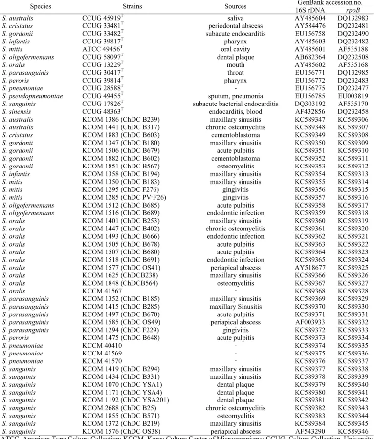
Table 1. The strains used in this study and accession numbers for nucleotide sequences of 16S rRNA (16S rDNA) and DNA‐
dependant RNA polymerase beta subunit gene (rpoB)
Species Strains Sources GenBank accession no.
16S rDNA rpoB
S. australis CCUG 45919T saliva AY485604 DQ132983
S. cristatus CCUG 33481T periodontal abscess AY584476 DQ232481
S. gordonii CCUG 33482T subacute endocarditis EU156758 DQ232490
S. infantis CCUG 39817T pharynx AY485603 DQ232482
S. mitis ATCC 49456T oral cavity AY485601 AF535188
S. oligofermentans CCUG 58097T dental plaque AB682364 DQ232508
S. oralis CCUG 13229T mouth AY485602 AF535168
S. parasanguinis CCUG 30417T throat EU156771 DQ132985
S. peroris CCUG 39814T pharynx EU156772 DQ232483
S. pneumoniae CCUG 28588T - EU156775 DQ232477
S. pseudopneumoniae CCUG 49455T sputum, pneumonia EU156785 EU003819
S. sanguinis CCUG 17826T subacute bacterial endocarditis DQ303192 AF535170
S. sinensis CCUG 48363T endocarditis, blood AF432856 DQ232458
S. australis KCOM 1386 (ChDC B239) maxillary sinusitis KC589347 KC589306
S. australis KCOM 1441 (ChDC B317) chronic osteomyelitis KC589348 KC589307
S. cristatus KCOM 1883 (ChDC B603) cementoblastoma KC589349 KC589308
S. gordonii KCOM 1347 (ChDC B180) maxillary sinusitis KC589350 KC589309
S. gordonii KCOM 1506 (ChDC B679) acute pulpitis KC589351 KC589310
S. gordonii KCOM 1882 (ChDC B602) cementoblastoma KC589352 KC589311
S. gordonii KCOM 1851 (ChDC B567) osteomyelitis KC589353 KC589312
S. infantis KCOM 1358 (ChDC B194) maxillary sinusitis KC589354 KC589313
S. mitis KCOM 1350 (ChDC B183) maxillary sinusitis KC589355 KC589314
S. mitis KCOM 1295 (ChDC F276) gingivitis KC589356 KC589315
S. mitis KCOM 1285 (ChDC PV‐F26) gingivitis KC589357 KC589316
S. oligofermentans KCOM 1512 (ChDC B685) acute pulpitis KC589358 KC589317
S. oligofermentans KCOM 1516 (ChDC B689) endodontic infection KC589359 KC589318
S. oralis KCOM 1401 (ChDC B253) maxillary sinusitis KC589360 KC589319
S. oralis KCOM 1447 (ChDC B402) chronic osteomyelitis KC589361 KC589320
S. oralis KCOM 1493 (ChDC B666) endodontic infection KC589362 KC589321
S. oralis KCOM 1505 (ChDC B678) acute pulpitis KC589363 KC589322
S. oralis KCOM 1507 (ChDC B680) acute pulpitis KC589364 KC589323
S. oralis KCOM 1518 (ChDC B691) endodontic infection KC589365 KC589324
S. oralis KCOM 1577 (ChDC OS41) periapical abscess AY518677 KC589325
S. oralis KCOM 1625 (ChDCB238) maxillary sinusitis KC589366 KC589326
S. oralis KCOM 1848 (ChDCB564) osteomyelitis KC589367 KC589327
S. oralis KCCM 41567 ‐ KC589368 KC589328
S. parasanguinis KCOM 1352 (ChDC B185) maxillary sinusitis KC589369 KC589329
S. parasanguinis KCOM 1415 (ChDC B285) maxillary Sinusitis KC589370 KC589330
S. parasanguinis KCOM 1497 (ChDC B670) acute pulpitis KC589371 KC589331
S. parasanguinis KCOM 1585 (ChDC OS49) periapical abscess AF003933 KC589332
S. parasanguinis KCOM 1294 (ChDC F229) gingivitis KC589372 KC589333
S. peroris KCOM 1475 (ChDC B648) acute pulpitis KC589373 KC589334
S. pneumoniae KCCM 40410 ‐ KC589374 KC589335
S. pneumoniae KCCM 41569 ‐ KC589375 KC589336
S. pneumoniae KCCM 41570 ‐ KC589376 KC589337
S. sanguinis KCOM 1419 (ChDC B294) maxillary sinusitis KC589377 KC589338
S. sanguinis KCOM 1434 (ChDC B331) maxillary sinusitis KC589378 KC589339
S. sanguinis KCOM 1070 (ChDC YSA1) dental plaque KC589379 KC589340
S. sanguinis KCOM 1171 (ChDC YSA4) dental plaque KC589380 KC589341
S. sanguinis KCOM 1192 (ChDC YSA201) dental plaque KC589381 KC589342
S. sanguinis KCOM 2688 (ChDC B25) chronic osteomyelitis KC589382 KC589343
S. sanguinis KCOM 1855 (ChDC B571) osteomyelitis KC589383 KC589344
S. sanguinis KCOM 1372 (ChDC B219) maxillary sinusitis KC589384 KC589345
S. sanguinis KCOM 1576 (ChDC OS38) periapical abscess AF543290 KC589346
ATCC, American Type Culture Collection; KCCM, Korea Culture Center of Microorganisms; CCUG, Culture Collection, University of Gothenburg, Sweden; KCOM, Korean Collection for Oral Microbiology; ChDC, Dental College of Chosun University.
핵산염기서열 비교분석법이다[14]. 특히, 세균 종들의 16S rDNA 핵산염기서열은 진화론적 측면에서 종 간에 잘 보 존되어 있고, 데이터베이스가 잘 구축되어 있으며, 다른 분 류학적 방법들에 비해 수월하기 때문에 가장 널리 사용 되고 있다[14]. 그러나 MGS 종 중의 S. pneumoniae, S. mitis 및 S. oralis의 경우는 16S rDNA 핵산염기서열들은 서로 99%의 상동성을 가지고 있어 이들 세균 종을 동정하는 데 한계가 있다[15]. DNA-dependent RNA polymerase gene beta- subunit (rpoB) 유전자는 약 4-5 kbp로 구성되어 있으며, 16S rDNA처럼 유전학적으로 상동성이 잘 보존된 영역과 그렇지 않는 영역이 반복적으로 존재한다[16]. 이러한 이 유로 현재 rpoB 핵산염기서열 비교 분석법이 세균의 종 수준에서의 분류에 급속히 이용되고 있다[17-20]. 최근 Hoshino 등[15]은 rpoB 핵산염기서열(742 bp)을 바탕으로 MGS에 속하는 13종의 연쇄구균을 종 수준으로 S. sinesis 를 제외하고는 성공적으로 동정할 수 있다고 보고하였다. 그러므로, 본 연구에서는 한국인에서 분리되어 16S rDNA 핵산염기서열 비교분석법으로 MGS임이 확인되었지만, 종 수준에서 동정이 되지 않은 임상균주를 rpoB 핵산염기서 열을 바탕으로 종 수준으로 동정하고자 한다.
재료 및 방법
세균 및 세균 배양
본 연구에서 사용된 균주들은 Table 1과 같다. 이들 균주 들은 ATCC (Type Culture Collection, USA), CCUG (Culture Collection, University of Gothenburg, Sweden), KCCM (Korea Culture Center of Microorganisms, Korea) 및 KCTC (Korean Collection for Type Cultures, Biological Resource Center, Korea)에서 구입하여 사용하였다. 그리고, 한국인에서 분 리된 MGS 균주들은 한국구강미생물자원은행(Korean Collec- tion for Oral Microbiology, Gwangju, Korea)에서 분양 받아 사용하였다. 이들 균주들은 모두 한국인의 구강으로 분리된 것으로 구강외과, 보존과, 예방치과 및 교정과에서 분리된 것 이었다(Table 1). 이들 균주들 중 cementoblastoma 병소에서 분리된 균주들(ChDC B603 및 ChDC B602)은 검체를 채취하 는 과정에서 구강 내 타액 등에서 오염된 것으로 생각된다. 본 연구에 사용된 모든 연쇄구균 균주들은 BHI (Brain heart infusion, Difco Laboratory, Detroit, MI, USA)배지를 사용하여 37oC 세균배양기에서 24시간 동안 배양하여 다 음 실험에 사용하였다
rpoB 클로닝
세균의 지놈 DNA들은 G-spinTM Genomic DNA Extrac-
tion Kit (iNtRON Co., Seoul, Korea)를 이용하여 제조회 사의 지시에 따라 추출하였다.
본 연구에서 분석하고자 한 16S rDNA 및 rpoB 유전자를 PCR법으로 증폭하기 위해 참고 문헌에 보고되었던 프라 이머들[21,22]을 사용하였고, 이들 프라이머는 Bioneer사(Dae- jeon, Korea)에 의뢰하여 제작하였다. 본 연구에서 사용된 각 유전자를 증폭하기 위한 프라이머와 프라이머 핵산염 기서열 그리고 중합효소연쇄반응은 선행 논문에서 제시한 조건을 따랐다[21,22].
PCR 증폭물은 pGEM-T easy vector (Promega Crop., Ma- dison, WI, USA)에 제조회사의 지시에 따라 클로닝하고, E.
coli DH5α에 형질전환 하였다. 재조합 플라스미드는 Accu- PrepTM Plasmid Extraction Kit (Bioneer Corp., Daejeon, Korea)를 이용하여 제조회사의 지시대로 추출하였다.
핵산염기서열 결정
16S rDNA 및 rpoB 핵산염기서열은 Big Dye Termina- tor Cycle Sequencing Kit (Applied Biosystems, Foster City, CA, USA)와 ABI PRISM 310 Genetic Analyzer (Applied Biosystems)를 이용하여 결정하였다. 16S rDNA 핵산염기 서열 결정을 위해 ChDC-GEM-F (5'-TTC CCA GTC ACG ACG TTG TAA AA-3'), ChDC-GEM-R (5'-GTG TGG AAT TGT GAG CGG ATA AC-3')과 Seq-F1 (5'-CCT ACG GGA GGC AGC AG-3') 프라이머들[23]을 핵산염기서열 결정에 사용하였다. 또한 rpoB 핵산염기서열 결정을 위해 ChDC- GEM-F 및 ChDC-GEM-R 프라이머를 사용하였다.
계통발생 분석(Phylogenetic analysis)
본 연구에서 사용한 균주들 16S rDNA 및 rpoB 핵산염 기서열은 MEGA version 5 [24] 프로그램을 이용하여 ClustalW 법으로 정렬하고 neighbor-joining methods 법으 로 계통도(phylogenetic tree)를 작성하였다. 이때 계통도의 신뢰도를 확인할 수 있는 bootstrap 값은 1,000회의 re- sampled data로부터 계산하였다. 16S rDNA 및 rpoB 핵산 염기서열들의 세균 종들 및 세균 종 간 진화적 거리(%
distance로 표현함)는 Maximum Composite Likelihood model [25]을 이용하여 계산하였다.
결 과
본 연구에서 사용한 37개 MGS 임상균주들의 16S rDNA 와 rpoB 유전자들을 PCR 법으로 증폭한 결과 각각 약 1502 bp와 742 bp의 PCR 산물을 얻어 핵산염기서열을 결정하였다. MGS에 속하는 13종 표준균주 16S rDNA와
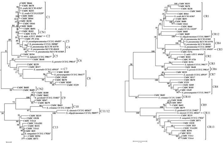
Fig. 1. Phylogenetic trees base on the partial nucleotide sequences of (A) 16S rDNA (about 1.33 kb) and (B) rpoB (678 bp) of type strains and clinical isolates of mitis group streptococci. The resulting tree topology was evaluated by bootstrap analyses of the neighbor‐
joining tree based on 1,000 resamplings. The scale bar (neighbor‐joining distance) represents a 5% difference in nucleotide sequence.
rpoB 핵산염기서열은 GenBank에서 다운받아 사용하였다 (Table 1). MGS 균주들의 16S rDNA와 rpoB 핵산염기서 열의 계통발생 분석(phylogenetic analysis)에는 각각 1,328 bp와 678 bp를 이용하였다. 이는 두 유전자를 증폭할 때 사용되었던 프라이머 염기서열 부분을 모두 삭제하였고, Gen- Bank에 등록된 참고균주들의 16S rDNA 염기서열과 같은 크기를 분석에 이용하기 위함이었다. 이들의 핵산염기서 열을 GenBank에 등록하였다(Table 1).
본 연구에서 사용한 37개 MGS 임상균주들과 4개 참고 균주들을 13종 표준균주들의 16S rDNA 및 rpoB 핵산염 기서열을 기준으로 하여 형성된 계통도를 바탕으로 종 수 준으로 동정하였다(Fig. 1). 표준균주는 단독 혹은 임상 및 참고균주와 같은 클러스터(cluster)를 형성할 때 C1-C13 (16S rDNA) 또는 CR1-CR13 (rpoB)라는 그룹명을 부여하였으 며, 표준균주와 같은 클러스터에 속하는 균주들은 표준균 주와 같은 종으로 동정하였다(Fig. 1). 이때 아라비아 숫 자가 같은 것은 동일한 종을 의미한다. 16S rDNA 핵산 염기서열을 기준으로 형성된 계통도에서 표준균주와 함께 그룹이 안 된 단일 균주에는 그룹 명을 부여하지 않았고 (ChDC PV-F26, ChDC B239 및 ChDC B317), 표준균주
와는 같은 클러스터를 형성하지 않았더라도 임상균주들 간에 하나의 그룹이 형성된 경우 CN1 (ChDC B183과 ChDC F276)과 CN2 (ChDC B685와 ChDC B689)로 그룹 명을 부여하였다(Fig. 1A). 그 결과 16S rDNA를 바탕으로 형 성된 계통도에서 표준균주들과 함께 그룹이 형성된 클러 스터(C1-C13)에 존재하는 임상균주들은 모두 rpoB를 기 준으로 형성된 클러스터(CR1-CR13)와 동일하게 일치하였 다. 즉, C13에 속하는 임상균주들은 모두 CR13에 존재하여 모두 S. sanguinis로 동정되었다(Fig. 1). 16S rDNA를 기 준으로 특정 그룹에 속하지 않거나 CN1과 CN2에 속하는 임상균주들은 rpoB를 기준으로 형성된 계통에 의하면, S.
mitis (ChDC PV-F26, ChDC B183 및 ChDC F276), S. aus- tralis (ChDC B239과 ChDC B317) 및 S. oligofermentans (ChDC B685와 ChDC B698)인 것으로 조사되었다(Fig. 1).
본 연구에서 사용된 MGS들의 16S rDNA 및 rpoB 유 전자들의 핵산염기서열을 바탕으로 세균 종들 및 세균 종 간 진화적 거리(% distance로 표현함)를 Maximum Compo- site Likelihood model [25]을 이용하여 계산하였다(Table 3과 4). 이때 16S rDNA 핵산염기서열을 바탕으로 진화적 거리를 계산할 때, rpoB를 기준으로 형성된 계통도를 통
Table 3. The percent mean distance of 16S rDNA nucleotide sequences between the species
Sgo Smi Spa Sif Ssa Sor Sau Scr Spe Sol Spn Sps Ssi
Sgo -
Smi 2.5 ± 0.4 -
Spa 3.0 ± 0.5 2.7 ± 0.4 -
Sif 2.5 ± 0.4 1.0 ± 0.2 2.7 ± 0.4 -
Ssa 1.9 ± 0.3 2.1 ± 0.4 2.4 ± 0.4 2.2 ± 0.4 -
Sor 2.4 ± 0.4 0.3 ± 0.1 2.6 ± 0.4 0.8 ± 0.2 2.0 ± 0.4 -
Sau 2.5 ± 0.4 1.4 ± 0.3 2.0 ± 0.3 1.5 ± 0.3 2.3 ± 0.4 1.4 ± 0.3 -
Scr 2.1 ± 0.4 3.0 ± 0.5 2.9 ± 0.4 3.1 ± 0.5 2.8 ± 0.4 3.1 ± 0.5 2.4 ± 0.4 -
Spe 2.8 ± 0.4 1.8 ± 0.3 3.3 ± 0.5 2.0 ± 0.3 2.8 ± 0.4 1.7 ± 0.3 2.0 ± 0.3 3.2 ± 0.5 -
Sol 2.6 ± 0.4 2.2 ± 0.3 2.9 ± 0.4 2.3 ± 0.3 2.4 ± 0.3 2.2 ± 0.3 2.6 ± 0.4 2.4 ± 0.3 2.9 ± 0.4 -
Spn 2.9 ± 0.5 0.6 ± 0.2 3.1 ± 0.5 1.3 ± 0.3 2.4 ± 0.4 0.7 ± 0.2 1.8 ± 0.3 3.3 ± 0.5 2.1 ± 0.4 2.4 ± 0.3 -
Sps 2.6 ± 0.4 0.4 ± 0.1 2.8 ± 0.5 1.0 ± 0.3 2.2 ± 0.4 0.4 ± 0.2 1.5 ± 0.3 3.0 ± 0.5 1.9 ± 0.3 2.1 ± 0.3 0.4 ± 0.2 -
Ssi 2.8 ± 0.5 4.0 ± 0.6 3.0 ± 0.5 4.2 ± 0.6 3.1 ± 0.5 3.8 ± 0.6 3.6 ± 0.5 2.2 ± 0.4 3.8 ± 0.5 3.0 ± 0.4 4.4 ± 0.6 4.1 ± 0.6 ‐ Sgo, Streptococcus gordonii; Smi, Streptococcus mitis; Spa, Streptococcus parasanguinis; Sif, Streptococcus infantis; Ssa, Strep- tococcus sanguinis; Sor, Streptococcus oralis; Sau, Streptococcus australis; Scr, Streptococcus cristatus; Spe, Streptococcus peroris; Sol, Streptococcus oligofermentans; Spn, Streptococcus pneumoniae; Sps, Streptococcus pseudopneumoniae; Ssi, Streptococcus sinensis.
Table 2. The percentage distance of 16S rDNA and rpoB nu- cleotide sequences within the species
Species % diversity (Mean ± Standard Error)
16S rDNA rpoB
S. gordonii 0.3 ± 0.1 1.5 ± 0.3
S. mitis 0.3 ± 0.1 2.9 ± 0.5
S. parasanguinis 0.8 ± 0.2 2.5 ± 0.4
S. infantis 0.2 ± 0.1 3.2 ± 0.7
S. sanguinis 0.8 ± 0.2 5.1 ± 0.7
S. oralis 0.4 ± 0.1 3.6 ± 0.5
S. australis 0.2 ± 0.1 4.8 ± 0.8
S. cristatus 0.5 ± 0.2 5.1 ± 0.9
S. peroris 2.1 ± 0.4 5.2 ± 0.9
S. oligofermentans 2.8 ± 0.4 3.5 ± 0.6
S. pneumoniae 0.3 ± 0.1 0.7 ± 0.2
S. pseudopneumoniae n/c n/c
S. sinensis n/c n/c
하여 동정된 임상균주를 같은 종으로 그룹화하였다(Fig.
1B와 Table 3). 그 결과 표준균주만 이용한 S. pseudop- neumoniae와 S. sinensis를 제외하고 11개 MGS 균종 내의 평균 distance는 16S rDNA와 rpoB가 각각 0.8%와 3.5%
로 rpoB 핵산염기서열이 같은 세균 종 내에서 균주 간의 상동성이 16S rDNA 에 비해 distance가 4배 정도 높았다
(Table 2). 13개 MGS 세균 종 간의 16S rDNA와 rpoB의 distance는 각각 0.3%-4.2%및 0.84%-17.7%로 다양하였으 며, 이 역시 rpoB 유전자 핵산염기서열이 16S rDNA의 것보다 상동성이 낮음을 알 수 있었다(Table 3과 4).
고 찰
본 연구결과 한국인의 구강에서 분리되어 16S rDNA 핵산염기서열비교결정법에 의해 MGS로 판정된 37개 임 상균주를 rpoB 핵산염기서열을 바탕으로 계통발생 분석 으로 종 수준으로 동정할 수 있었다(Fig. 1B). 그 결과 이들 37개 MGS 임상균주들은 S. oralis (9 균주), S. sanguinis (9 균주), S. parasanguinis (5 균주), S. mitis (3 균주) S. oli- gofermentans (2 균주), S. australis (2 균주), S. gordonii (4 균주), S. infantis (1 균주), S. peroris (1 균주) 및 S. cris- tatus (1 균주)들로 동정되었다(Fig. 1B). 56개 MGS 균주들 16S rDNA 및 rpoB 핵산염기서열을 바탕으로 세균 종들 및 세균 종 간 진화적 거리를 계산한 결과 rpoB의 것이 16S rDNA의 것보다 상이성이 커서 rpoB를 이용하여 MGS 균 주들을 세균 종 수준으로 동정하는 데 더 효과적임을 알 수 있었다(Table 2-4). Drancourt 등[22]이 rpoB (740 bp)를 이용하여 Streptococcus, Enterococcus, Gemella, Abiotrop- hia 및 Granulicatella 속(Genus)의 종의 동정을 처음으로 시도하였다. 이들의 연구에서 사용한 13종 Streptococcus의
Table 4. The percent mean distance of rpoB nucleotide sequences between the species
Sgo Smi Spa Sif Ssa Sor Sau Scr Spe Sol Spn Sps Ssi
Sgo -
Smi 15.8 ± 2.0 -
Spa 15.2 ± 1.9 13.6 ± 1.7 -
Sif 15.6 ± 1.9 7.5 ± 1.0 14.6 ± 1.8 -
Ssa 9.9 ± 1.3 17.6 ± 2.115.9 ± 1.91.55 ± 0.32 -
Sor 14.6 ± 1.8 6.5 ± 0.9 13.6 ± 1.73.05 ± 0.4916.1 ± 1.9 -
Sau 14.3 ± 1.813.6 ± 1.6 6.6 ± 0.9 2.3 ± 0.3415.1 ± 1.814.0 ± 1.7 -
Scr 11.8± 1.5 11.1 ± 1.413.8 ± 1.70.96 ± 0.2412.6 ± 1.511.3 ± 1.412.4 ± 1.6 -
Spe 15.7 ± 1.9 7.7 ± 1.0 14.6 ± 1.70.84 ± 0.2317.1 ± 2.0 7.5 ± 1.0 15.0 ± 1.713.1 ± 1.6 -
Sol 15.2 ± 1.9 6.6 ± 1.0 14.5 ± 1.81.99 ± 0.3415.4 ± 1.9 5.6 ± 0.8 13.9 ± 1.711.1 ± 1.4 8.4 ± 1.2 -
Spn 16.7 ± 2.1 5.2 ± 0.9 14.3 ± 1.81.26 ± 0.2917.2 ± 2.1 6.9 ± 1.0 14.2 ± 1.810.8 ± 1.4 8.8 ± 1.2 6.5 ± 1.0 -
Sps 14.8 ± 1.9 4.3 ± 0.7 14.4 ± 1.81.04 ± 0.2717.0 ± 2.1 6.2 ± 1.0 14.3 ± 1.810.9 ± 1.5 8.3 ± 1.2 6.3 ± 1.0 5.1 ± 1.0 -
Ssi 10.1 ± 1.416.8 ± 2.014.7 ± 1.94.16 ± 0.6 7.0 ± 0.9 15.5 ± 1.913.6 ± 1.711.5 ± 1.517.0 ± 2.114.9 ± 1.917.1 ± 2.117.1 ± 2.1 - Sgo, Streptococcus gordonii; Smi, Streptococcus mitis; Spa, Streptococcus parasanguinis; Sif, Streptococcus infantis; Ssa, Strep- tococcus sanguinis; Sor, Streptococcus oralis; Sau, Streptococcus australis; Scr, Streptococcus cristatus; Spe, Streptococcus peroris; Sol, Streptococcus oligofermentans; Spn, Streptococcus pneumoniae; Sps, Streptococcus pseudopneumoniae; Ssi, Streptococcus sinensis.
rpoB 핵산염기서열 상동성은 94%로 성공적으로 종 수준 으로 동정하였다. 하지만, 그들의 연구에서는 MGS에 속하 는 균종은 S. mitis, S. oralis 및 S. pneumoniae 만을 사용하 였다. 그 후 Hoshino 등[15]은 S. oligofermentans를 제외한 12종 MGS, 3종 anginosus group streptococci (AGS), 5종 bovis group streptococci (BGS), Streptococcus mutans 및 S. salivarius 148 균주에서 D-alanine:D-alanine ligase (ddl, 563 bp), glutamate dehydrogenase (gdh, 642 bp), rpoB (742 bp), 및 manganese-dependent superoxide dismutase (sodA, 438 bp) 유전자를 이용하여 mutlilocus sequence typing (MLST) 법으로 이들 균주들을 종 수준으로 동정하였다. 현재 MLST 법은 여러 유전자 핵산염기서열을 순차적으로 연결하여 하 나의 핵산염기서열로 만든 다음, 이를 계통발생 분석을 통 하여 세균의 종 수준 및 아종 수준까지도 동정할 수 있는 방법으로 많이 사용되고 있다[26,27]. 또한, MLST 결과와 4가지 유전자 각각만을 이용하여 계통도를 만들어 비교 분 석한 결과 MLST 결과와 sodA만을 이용한 결과가 서로 같 았다고 보고하였다. 하지만, ddl, gdh, sodA의 경우 148 균 주 중 각각 81.1%, 70.9% 및 96% 균주 핵산염기서열을 이 용하여 “pairwise deletion” 조건에서 neighbor-joining algori- thm으로 분석한 것이기 때문에 모든 균주에서 이들 유전 자 핵산염기서열을 이용하여 같은 방법으로 계통발생 분석 을 하면 다른 결과가 나올 수 있을 것으로 생각된다. 이 들의 결과에서 모든 균주에서 100% 클로닝에 성공하여 핵산염기서열을 계통발생 분석한 것은 rpoB 뿐이었다[15].
그 결과 MLST로 분석하여 S. sanguinis로 동정된 하나의
균주(SK36)가 rpoB로는 S. sinesis로 잘못 동정된 것을 제 외하고는 MLST와 rpoB 결과가 모두 일치하였다. 이러한 결과를 바탕으로 본 연구에서도 37개 임상균주를 rpoB 핵 산염기서열을 기준으로 계통발생 분석을 통하여 종 수준 으로 동정하였다. 최근 Glazunova 등[28]은 65개 연쇄구균 (58 종과 9개 아종) 표준균주들의 16S rDNA (1468-1478 bp), rpoB (659-680 bp), sodA (435-462 bp), 60 kDa heat-shock protein (groEL, 757 bp) 및 beta subunit of DNA gyrase (gyrB, 458-461 bp) 핵산염기서열을 계통발생 분석한 결과 종 간의 상이성(divergence)이 그들이 사용한 5가지 유전 자들 중에서 groEL이 가장 크기 때문에 종 수준에서의 동 정에서 groEL을 이용하는 것이 가장 효과적일 것이라고 보고하였다. 하지만, 이들의 연구에서는 표준균주들만을 이 용하였기 때문에 차후 임상균주들을 포함하여야 할 것으 로 생각된다. 또한 이들 5가지 유전자들을 순서적으로 나 열하여 하나의 핵산염기서열을 만들어 MLST 법으로 계통 발생 분석을 한다면 어느 유전자가 가장 효율적으로 이 들 연쇄구균을 종 수준으로 동정할 수 있을지 알 수 있을 것으로 생각된다.
이러한 결과를 종합할 때, 본 연구에서 37개 MGS 임상 균주를 rpoB 핵산염기서열로 계통발생 분석하여 S. oralis (9 균주), S. sanguinis (9 균주), S. parasanguinis (5 균주), S.
mitis (3 균주) S. oligofermentans (2 균주), S. australis (2 균 주), S. gordonii (4 균주), S. infantis (1 균주), S. peroris (1 균주) 및 S. cristatus (1 균주)들로 동정하였다. 하지만, 향 후 연구에서 rpoB와 함께 기존에 보고된 sodA, groEL, gyrB,
gdh 및 ddl 유전자들의 핵산염기서열을 결정하여 MLST 법 으로 계통발생 분석을 시행한다면 더 정확한 결과를 얻을 수 있을 것으로 생각된다.
감사의 글
이 논문의 일부분은 2010년도 정부(교육과학기술부)의 재원으로 한국연구재단의 기초연구사업 지원을 받아 수 행된 것임(2010-0025411)
References
1. Gibbons RJ, Hay DI, Schlesinger DH. Delineation of a segment of adsorbed salivary acidic proline-rich proteins which promotes adhesion of Streptococcus gordonii to apa- titic surfaces. Infect Immun. 1991;59:2948-2954.
2. Scannapieco FA, Torres GI, Levine MJ. Salivary amylase pro- motes adhesion of oral streptococci to hydroxyapatite. J Dent Res. 1995;74:1360-1366.
3. Lamont RJ, Bevan CA, Gil S, Persson RE, Rosan B. Invo- lvement of Porphyromonas gingivalis fimbriae in adhe- rence to Streptococcus gordonii. Oral Microbiol Immunol.
1993;8:272-276.
4. Nyvad B, Kilian M. Comparison of the initial streptococcal microflora on dental enamel in caries-active and in caries- inactive individuals. Caries Res. 1990;24:267-272.
5. Baddour LM. Virulence factors among gram-positive bac- teria in experimental endocarditis. Infect Immun. 1994;62:
2143-2148.
6. Douglas CW, Heath J, Hampton KK, Preston FE. Identity of viridans streptococci isolated from cases of infective endocarditis. J Med Microbiol. 1993;39:179-182
7. Beighton D, Carr AD, Oppenheim BA. Identification of viri- dans streptococci associated with bacteraemia in neutro- penic cancer patients. J Med Microbiol. 1994;40:202-204.
8. Bochud Bochud PY, Eggiman P, Calandra T, Van Melle G, Saghafi L, Francioli P. 1994. Bacteremia due to viridans streptococcus in neutropenic patients with cancer: clinical spectrum and risk factors. Clin Infect Dis. 1994;18:25-31.
9. Jacobs JA, Schouten HC, Stobberingh EE, Soeters PB.
Viridans streptococci isolated from the bloodstream. Rele- vance of species identification. Diagn Microbiol Infect Dis.
1995;22:267-273.
10. Cabellos C, Viladrich PF, Corredoira J, Verdaguer R, Ariza J, Gudiol F. Streptococcal meningitis in adult patients:
current epidemiology and clinical spectrum. Clin Infect Dis. 1999;28:1104-1108.
11. Arbique JC, Poyart C, Trieu-Cuot P, Quesne G, Carvalho Mda G, Steigerwalt AG, Morey RE, Jackson D, Davidson RJ, Facklam RR. Accuracy of phenotype and genotypic testing for identification of Streptococcus pneumoniae and description of Streptococcus pseudopneumoniae sp. nov. J
Clin Microbiol. 2004;42:4686-4696.
12. French GL, Talsania H, Charlton JR, Phillips I. A phy- siological classification of viridans streptococci by use of the API-20STREP system. J Med Microbiol. 1989;28:275- 286.
13. Hinnebusch CJ, Nikolai DM, Bruckner DA. Comparison of API Rapid Strep, Baxter MicroScan Rapid Pos ID Panel, BBL Minitek Differential Identification System, IDS RapID STR System, and Vitek GPI to conventional bioc- hemical tests for identification of viridans streptococci. Am.
J Clin Pathol. 1991;96:459-463.
14. Gillis M, Vandamme P, De Vos P, Swings J, Kersters K.
Polyphasic taxonomy. 2nd ed. In G. Garrity (ed.), Bergey's Manual of Systematic Bacteriology, Springer Verlag, New York, USA. 2001;43-48.
15. Hoshino T, Fujiwara T, Kilian M. Use of phylogenetic and phenotypic analyses to identify nonhemolytic streptococci isolated from bacteremic patients. J Clin Microbiol. 2005;
43:6073-6085.
16. Severinov K, Mustaev A, Kukarin A, Muzzin O, Bass I, Darst SA, Goldfarb A. Structural modules of the large su- bunits of RNA polymerase. Introducing archaebacterial and chloroplast split sites in the beta and beta' subunits of Escherichia coli RNA polymerase. J Biol Chem. 1996;
271:27969-27974.
17. Drancourt M, Raoult D. rpoB gene sequence-based iden- tification of Staphylococcus species. J Clin Microbiol 2002;
40:1333-1338.
18. Khamis A, Raoult D, La Scola B. rpoB gene sequencing for identification of Corynebacterium species. J Clin Micro- biol. 2004;42:3925-3931.
19. Kim HS, Lee DS, Chang YH, Kim MJ, Koh S, Kim J, Seong JH, Song SK, Shin HS, Son JB, Jung MY, Park SN, Yoo SY, Cho KW, Kim DK, Moon S, Kim D, Choi Y, Kim BO, Jang HS, Kim CS, Kim C, Choe SJ, Kook JK. Appli- cation of rpoB and zinc protease gene for use in molecular discrimination of Fusobacterium nucleatum subspecies. J Clin Microbiol 2010;48:545-553.
20. Park SN, Park JY, Kook JK. Development of quantitative real-time PCR primers for the detection of Aggregatibacter actinomycetemcomitans. Int J Oral Biol. 2011;36(1):1-6.
21. Lane DJ, Pace B, Olsen GJ, Stahl DA, Sogin ML, Pace NR. Rapid determination of 16S ribosomal RNA sequen- ces for phylogenetic analyses. Proc Natl Acad Sci USA.
1985;82:6955-6959.
22. Drancourt M, Roux V, Fournier PE, Raoult D. rpoB gene sequence-based identification of aerobic gram-positive cocci of the genera Streptococcus, Enterococcus, Gemella, Abio- trophia, and Granulicatella. J Clin Microbiol. 2004; 42:
497-504.
23. Yoo SY, Kim KJ, Lim SH, Kim KW, Hwang HK, Min BM, Choe SJ, Kook JK. First isolation of Streptococcus downei from human dental plaques. FEMS Microbiol Lett. 2005;
249:323-326.
24. Tamura K, Peterson D, Peterson N, Stecher G, Nei M, Kumar S. MEGA 5: Molecular evolutionary genetics ana- lysis using maximum likelihood, evolutionary distance, and
maximum parsimony method. Mol Biol Evol. 2011;28:
2731-2739.
25. Tamura K, Nei M., and Kumar S. Prospects for inferring very large phylogenies by using the neighbor-joining me- thod. Proc Natl Acad Sci USA. 2004;101:11030-11035.
26. Cesarini S, Bevivino A, Tabacchioni S, Chiarini L, Dal- mastri C. RecA gene sequence and Multilocus Sequence Typing for species-level resolution of Burkholderia cepacia complex isolates. Lett Appl Microbiol. 2009;49:580-588.
27. Kook JK, Park SN, Lim YK, Choi MH, Cho E, Kong SW, Shin Y, Paek J, Chang YH. Fusobacterium nucleatum sub- sp. fusiforme Gharbia and Shah 1992 is a later synonym of Fusobacterium nucleatum subsp. vincentii Dzink et al.
1990. Curr Microbiol. 2013;66:414-417.
28. Glazunova OO, Raoult D, Roux V. Partial sequence com- parison of the rpoB, sodA, groEL and gyrB genes within the genus Streptococcus. Int J Syst Evol Microbiol. 2009;
59:2317-2322.