Characterization of
Miniimonas sp. S16 isolated from activated sludge
Hyeon-Woo Koh1, Hongik Kim1*, and Soo-Je Park2*1R&D Division, VITABIO, Inc., Daejeon 34025, Republic of Korea
2Department of Biology, Jeju National University, Jeju 63243, Republic of Korea
활성슬러지로부터 분리된 Miniimons sp. S16 세균의 특성
고현우1 ・ 김홍익1* ・ 박수제2*1비타바이오 생물자원개발연구소, 2제주대학교 생물학과
(Received July 5, 2019; Revised July 31, 2019; Accepted August 1, 2019)
*For correspondence. (H. Kim) E-mail: [email protected]; Tel.: +82-42-864-4647; Fax: +82-42-864-4645 /
(S.J. Park) E-mail: [email protected]; Tel.: +82-64-753-3524; Fax: +82-64-756-3541
The GenBank/EMBL/DDBJ accession number for the 16S rRNA gene sequence of strain S16 is MF457652.
Biological factors (e.g. microorganism activity) in wastewater treatment plant (WWTP) play essential roles for degradation and/or removal of organic matters. In this study, to understand the microbial functional roles in WWTP, we tried to isolate and characterize a bacterial strain from activated sludge sample. Strain S16 was isolated from the activated sludge of a municipal WWTP in Daejeon metropolitan city, the Republic of Korea. The cells were a Gram-stain-positive, non-motile, facultative anaerobe, and rod-shaped. Strain S16 grew at a temperature of 15~40°C (optimum, 30°C), with 0~9.0% (w/v) NaCl (optimum, 1.0~2.0%), and at pH 5.5~9.0 (optimum, pH 7.0~7.5). Phylogenetic analysis based on 16S rRNA gene sequences indicated that strain S16 was most closely related to the unique species Miniimonas arenae NBRC 106267T (99.79%, 16S rRNA gene
sequence similarity) of the genus Miniimonas. The cell wall contained alanine, glutamic acid, serine, and ornithine. Although the isolation source of the type strain NBRC 106267T which considered as a marine microorganism is sea sand, that of strain S16 is terrestrial environment. It might raise an ecological question for habitat transition. Therefore, comparative genome analysis will be valuable investigation for shedding light on their potential metabolic traits and genomic streamlining. Keywords: Miniimonas, polyphasic analysis, sludge
The operation and maintenance of wastewater treatment plant (WWTP) depend on the complex parameters such as physical, chemical and biological factors (Martin and Vanrolleghem, 2014). In particular, microorganisms have more critical roles in degradation and removal of organic waste and/or pollutants by anthropological activities. Despite increasing of the importance for microbial activity in WWTPs operation, the understanding of microbial communities or individual microorganism presents in there (i.e. WWTPs) remains limited. In this study, we attempted to isolate a rare microorganism (Bachy and Worden, 2014) from activated sludge and its taxonomic properties were characterized using a polyphasic approach.
Strain S16 was isolated from the activated sludge of a muni-cipal wastewater treatment plant in Daejeon metropolitan city, the Republic of Korea. In order to isolate the microorganisms, the sludge sample removed debris was collected in a sterile conical tube and serially diluted to five-folds with phosphate- buffered saline (PBS; 137 mM NaCl, 2.7 mM KCl, 10 mM Na2HPO4, 2 mM KH2PO4, pH 7.0). Then 100 µl of the aliquot
(from the diluted sample) was spread on Reasoner’s 2A (R2A; Difco) agar plates and incubated at 30°C for one week under aerobic conditions. In order to obtain pure cultures, a small yellow-pigmented single colony was repetitively transferred to a new R2A agar plate under the same conditions and was finally designated as S16. Strain S16 was routinely cultured on R2A or
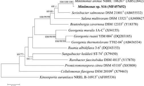
Fig. 1. Neighbour-joining phylogenetic tree based on 16S rRNA gene sequences showing the position of S16 among recognized Miniimonas species and some other related taxa. GenBank accession numbers are indicated in parentheses. Bootstrap values based on 1,000 replicates from neighbor-joining (NJ), maximum-likelihood (ML), and maximum-parsimony (MP) methods are indicated at the branch points. Kineosporia aurantiaca NRRL B-16913T was used as an outgroup. Bar, 0.005 substitutions per nucleotide position.
Luria-Bertani (LB; Difco) agar plates at 30°C under aerobic conditions and preserved as a suspension in an LB broth medium with glycerol suspension (30%, w/v) at -80°C.
For the phylogenetic analysis of strain S16, the genomic DNA (gDNA) was extracted by using a commercial genomic DNA extraction kit (GeneAll Biotechnology Co. Ltd.), as previously described by Koh et al. (2017). The 16S rRNA gene was amplified from gDNA using the universal bacteria primer set 27F (5'-AGAGTTTGATCMTGGCTCAG-3') and 1492R (5'-TACGGYTACCTTGTTACGACTT-3) (Weisburg et al., 1991). The following thermal cycle conditions were used: 94°C for 5 min, followed by 30 cycles of 94°C for 0.5 min, 55°C for 0.5 min, and 72°C for 1 min, and a final extension of 72°C for 5 min. The PCR product was purified using a PCR purification kit (Cosmo Genetech Co. Ltd.) and was sequenced by Macrogen Co. Ltd. The 16S rRNA gene sequence of strain S16, as deter-mined in this study, comprised of 1,515 bases was compiled using SeqMan software (DNASTAR), and the sequence was compared with the related taxa obtained from the GenBank database (www.ncbi.nlm.nih.gov) and EzBioCloud server (https:// www.ezbiocloud.net) (Yoon et al., 2017). The sequences were edited using the BioEdit program and aligned using CLUSTAL_X (Thompson et al., 1997). Evolutionary distances were calculated
using the Kimura two-parameter model (Kimura, 1989). The neighbor-joining method (Saitou and Nei, 1987), maximum- parsimony method (Fitch, 1971), and maximum-likelihood (Felsenstein, 1981) method in MEGA7 software (Kumar et al., 2016) were used to reconstruct phylogenetic trees with bootstrap values based on 1,000 replications. The 16S rRNA gene sequence analysis indicated that strain S16 clustered within the genus Miniimonas (Fig. 1). According to the EzBioCloud server, strain S16 was most closely related to Miniimonas arenae NBRC 106267T (99.79% 16S rRNA gene sequence similarity). As mentioned above, the genus consists of only one species. Therefore, we selected M. arenae NBRC 106267T as a reference strain, which was purchased from the Biological Resource Center, National Institute of Technology and Evaluation (NBRC). Unless stated otherwise, the two strains (S16 and M. arenae NBRC 106267T) were grown on an LB medium under optimal culture conditions.
The morphological, biochemical, and physiological properties of strain S16 were investigated using routine cultivation at 30°C on an LB medium for 3 days. The Gram stain was investigated using a BD Gram staining kit according to the manufacturer’s instructions. Cell morphology and size were observed under a JEOL JEM-1010 Transmission Electron
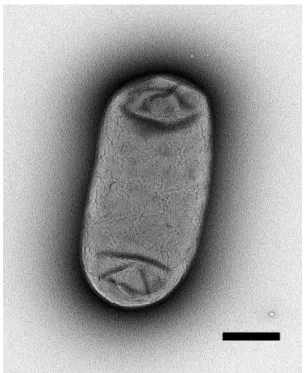
Fig. 2. Morphology of a strain S16 cell as determined by transmission electron microscopy (Bar, 0.5 µm).
Microscope (80 kV) after negative staining with 1% (w/v) phosphotungstic acid (Yashroy, 1990). Motility was tested by hanging-drop (Webley, 1953). Growth under anaerobic condi-tions was determined using the BD GasPakTM EZ Anaerobe pouch system (BD Diagnostics) over a period of 2 weeks. Activity for oxidase and catalase was tested using 1% (w/v) tetramethyl-p-phenylenediamine (Merck), and 3% (v/v) H2O2,
respectively. Growth at different temperatures (0, 5, 10, 15, 20, 25, 30, 40, and 50°C) was measured on an LB plate. The pH range for growth was determined in the LB broth that was adjusted to pH values (pH 4.0~10.0, at intervals of 0.5 pH unit) by adding 1 M of NaOH and HCl. For the pH experiments, four buffers were used (final concentration, 10 mM): Homopiperazine- 1,4-bis (2-ethanesulfonic acid) (Homo-PIPES, pH 4.0~5.0), 2-(N-morpholino) ethanesulfonic acid (MES, pH 5.5~6.5), 1,3-bis [tris (hydroxymethyl)methylamino]propane (Bis-Tris propane, pH 7.0~8.5), and 3-(cyclohexylamino)-1-propanesulfonic acid (CAPS, pH 9.0~10.0), as previously described (Koh et al., 2015). The requirement for NaCl was tested in an LB broth medium supplemented with 0–10% (w/v) NaCl (at intervals of 1% NaCl). API 20NE and API ZYM (bioMérieux), as well as GEN III Microplate systems (Biolog Inc.) tests, were used to determine other physiological and biochemical characteristics according to the manufacturer’s instructions. To perform the peptidoglycan analysis, cells of strain S16 prepared according to the method of Schleifer and Kandler (1972) and were hydrolyzed with HCl at 100°C for 15 h. The hydrolysates were subjected to TLC on cellulose plates using the solvent system as described by Menendez et al. (2017).
To determine genome relatedness, DNA-DNA hybridization (DDH) experiments were carried out with strains S16 and NBRC 106267T using a previously described method (Ezaki et al., 1989). The genomic DNA of two strains was extracted using a genomic DNA extraction kit (GeneAll, Biotechnology Co. Ltd.) and a probe. Salmon sperm DNA was used as negative control. The probe DNA was biotinylated with photobiotin and hybridized with single-stranded, unlabeled genomic DNA fragments from the reference or test microorganisms. The mean of three independent measurements of DNA-DNA hybridization levels was calculated.
Cells of strain S16 were Gram-stain-positive, non-motile, and rod-shaped (1.0~1.2 µm in width and 1.8~3.5 µm length)
(Fig. 2). The activities of oxidase and catalase were positive and negative, respectively. Colonies on the LB agar plate were 1mm in diameter, circular, smooth, and yellow-pigmented after 3 days at 30°C. Cell growth occurred under both aerobic and anaerobic conditions at 15~40°C (optimum, 30°C), a pH of 5.5~9.0 (optimum, pH 7.0~7.5), and 0~9.0% (w/v) NaCl (optimum, 1.0~2.0%). The cell wall peptidoglycan contained alanine, glutamic acid, serine, and ornithine. As shown in Table 1, there are numerous other phenotypic characteristics that could be used to distinguish strains S16 from its closest phylogenetic neighbor: M. arenae NBRC 106267T.
To determine the genomic DNA G + C content of strain S16, the gDNA removed RNAs by using a mixture of RNase A and T1 (20 U/ml each) at 30°C for 1 h, which was also used and analyzed using the method of Gonzalez and Saiz-Jimenez (2002). The genomic DNA G + C content of the strain S16 was 73.7 mol%, which is similar to the value for the recognized species (M. arenae NBRC 106267T, 74.2 mol%) of the genus Miniimonas (Table 1).
For the analysis of fatty acid profiles, strain S16 and M. arenae NBRC 106267T were harvested at the same growth
Table 1. Differential characteristics of strain S16 and closely related type strain of species belonging to the genus Miniimonas
Characteristic 1 2 Motility - -Oxidase + -Catalase - + Cell size (µm): Length 1.8~3.5 0.6~3.7 Width 1.0~1.2 1.0~1.7
Colony color Yellow Vermilion
Growth temperature (°C) 15~40 25~30 pH range 5.5~9.0 5.0~11.0 Enzyme activity: Esterase lipase (C8) + w Valine arylamidase - w Cystine arylamidase + -Trypsin w -α-Chymotrypsin w -β-Glucuronidase - w β-Glucosidase + w α-Mannosidase w -α-Fucosidase w -Assimilation of: α-Ketoglutaric acid - + Acetic acid - + Capric acid - + Formic acid - + Mucic acid + -Phenylacetic acid - + Trisodium citrate + -D-Fructose + -D-Fucose - + D-Galactose + -D-Malic acid - + D-Maltose - + D-Mannose - + D-Saccharic acid + -L-Arabinose - + L-Fucose - +
DNA G + C content (mol%) 73.7 74.2
Strains: 1, S16; 2, Miniimonas arenae NBRC 106267T.
Data were obtained in this study. All strains were Gram-stain-positive and rod-shaped and had MK-8 as the predominant menaquinone. All strains were positive for leucine arylamidase, α-galactosidase, β-galactosidase, α- glucosidase, esculin ferric citrate, D-glucose, citric acid, acetoacetic acid, and propionic acid but negative for alkaline phosphatase, dextrin, esterase (C4), gelatin, glycerol, inosine, lipase (C14), sucrose, urea, D-arabitol, D-cellobiose, D-raffinose, D-serine, D-sorbitol, D-trehalose, L-alanine, L-arginine, L-aspartic acid, L-glutamic acid, L-rhamnose, and L-serine. +, Positive; –, negative; w, weakly positive. Enzyme activity and assimilation data were determined in this study.
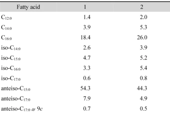
Table 2. Cellular fatty acid content (%) of strain S16 and related type strain of related Miniimonas species
Fatty acid 1 2 C12:0 1.4 2.0 C14:0 3.9 5.3 C16:0 18.4 26.0 iso-C14:0 2.6 3.9 iso-C15:0 4.7 5.2 iso-C16:0 3.3 5.4 iso-C17:0 0.6 0.8 anteiso-C15:0 54.3 44.3 anteiso-C17:0 7.9 4.9 anteiso-C17:0ω9c 0.7 0.5
Strains: 1, S16; 2, M. arenae NBRC 106267T. All data are from this study. Values are percentages of fatty acid. Fatty acids that accounted for < 0.5% of the total are not shown.
cultivation for 5 days at 30°C) from the LB agar plates. The fatty acids were extracted and prepared according to the standard protocol of the Sherlock Microbial Identification System (MIDI). The profiles were also determined according to the MIDI/ Hewlett Packard Microbial Identification System that uses a gas chromatography (6890N and 7683 autosampler, Agilent Technologies) according to the manufacturer’s instructions. The fatty acid profiles of strain S16 and M. arenae NBRC 106267T are shown in Table 2. The major fatty acids of strain S16 were anteiso-C15:0 (54.3%) and C16:0 (18.4%). Also, the
profiles of reference strain M. arenae NBRC 106267T were consistent with that of strain S16. However, we found two differences between strain S16 and M. arenae NBRC 106267T, which are that the ratio of anteiso-C15:0 in strain S16 is higher
than in M. arenae NBRC 106267T, while the ratio of C16:0 is
higher in M. arenae NBRC 106267T than in strain S16(Table 2). Polar lipids were extracted from the freeze-dried cells (100 mg) and analyzed as previously described by Koh et al. (2017). Polar lipids of strain S16included diphosphatidylglycerol (DPG), phosphatidylglycerol (PG), phosphatidylinositol (PI), an unidentified phospholipid (PL), and three unidentified lipids (L1-L3). The profile analyses of the fatty acid and polar lipid supported the conclusion that strain S16 represents a species distinct from the recognized species of the genus Miniimonas. Quinone components were extracted and analyzed as described by Hu et al. (1999). The quinone was eluted by a chloroform/ methanol mixture (2:1, v/v), evaporated under vacuum, and
re-extracted three times with n-hexane/water (1:1, v/v). Then the crude quinones extracted in n-hexane were concentrated and applied to a Sep-Pak Plus silica column (Waters). The isoprenoid quinone of strain S16 was only menaquinone MK-8.
The levels of DNA-DNA relatedness between S16 and NBRC 106267T was more than 70%. This result indicates that strain S16 can be considered as a subspecies of M. arenae NBRC 106267T.
Overall, strain S16 was clearly belonged to the member of the genus Miniimonas as a subspecies of M. arenae NBRC 106267T, despite there was some distinguished results including their isolation habitat. The genus Miniimonas belonging to family Beutenbergiaceae within the phylum Actinobacteria was first described by Ue et al. (2011). At the time of writing, the genus comprises only a single species Miniimonas arenae NBRC 106267T isolated from sea sand. It might infer that the genus Miniimonas should be considered as a rare microorganism in nature. In addition, there is only several physiological characters based on the polyphasic analysis.
As we mentioned above, strain S16 has been isolated from activated sludge not marine sources. The growth of the strain S16 was observed under high salinity culture condition, up to 9% (w/v) of NaCl. The other side, M. arenae NBRC 106267T isolated from sea sand can grow in the absence of NaCl in culture medium. Generally, it has been well known that marine microorganisms as a halophilic organism have been adapted to high concentrations of NaCl, and require sodium ion for their growth (Stolp, 1988). Taken together, there might open an ecological question for their habitat transition between terrestrial and marine environment (Zhang et al., 2019). Therefore, com-parative genome analysis including genome streamlining as a further investigation might be able to address the ecological question and show metabolic potentials or functional roles for strain S16 in WWTP, despite this study have a limitation for only physiological evidences.
Taxonomy
Locality. the activated sludge of a municipal wastewater treat-ment plant in Daejeon metropolitan city, the Republic of Korea.
Characteristics. The cells are Gram-stain-positive, oxidase- positive and catalase-negative, non-motile, 1.0~1.2 µm in
length and 1.8~3.5 µm in width and grow under both aerobic and anaerobic conditions. Colonies are yellow-pigmented, circular, and smooth. Growth occurs at 0~9.0% (w/v) NaCl (optimum, 1.0~2.0%) and at a temperature of 15~40°C (optimum, 30°C) and pH of 5.5~9.0 (optimum, pH 7.0~7.5). Negative for urease activity and indole production. Esculin ferric citrate is hydrolyzed, but gelatin is not hydrolyzed. The following constitutive enzyme activities are detected in API ZYM tests: esterase lipase (C8), leucine arylamidase, cystine arylamidase, α-galactosidase, β-galactosidase, α-glucosidase, β-glucosidase, and weakly positive for trypsin, α-chymotrypsin, acid phosphatase, α-mannosidase, α-fucosidase; but negative for alkaline phosphatase, esterase (C4), lipase (C14), valine arylamidase, naphthol-AS-Bl-phosphohydrolase, β-glucuronidase, and N-acetyl-β-glucosaminidase. The following can be used as sole carbon and energy sources: D-mannose, D-fructose, D- galactose, D-mannitol, mucic acid, D-saccharic acid, citric acid, Tween 40, acetoacetic acid, and propionic acid. The following are not used as sole carbon and energy sources: dextrin, D-maltose, D-trehalose, D-cellobiose, sucrose, D- raffinose, D-fucose, D-sorbitol, D-serine, L-fucose, L-rhamnose, L-alanine, L-arginine, L-serine, gelatin, acetic acid, and formic acid. The menaquinone is only MK-8. Major fatty acids are anteiso-C15:0 and C16:0. The polar lipids are
diphosphatidyl-glycerol, phosphatidyldiphosphatidyl-glycerol, phosphatidylinositol, an uni-dentified phospholipid, and three uniuni-dentified lipids. The cell- wall peptidoglycan contains alanine, glutamic acid, serine, and ornithine.
적 요
폐수 처리 설비에서 생물학적 요인은 유기물 분해 또는 제거 에 필수적인 역할을 수행한다. 본 연구에서는, 폐수처리장의 미생물 기능적 역할을 이해하기 위해, 활성 슬러지 샘플로부터 박테리아 균주를 분리하고 그들의 특성 분석을 시도했다. S16 균주는 대한민국 대전광역시의 폐수처리장의 활성슬 러지로 부터 분리되었다. 세포들은 그람음성, 비운동성, 통성 혐기성 그리고 막대모양이였다. S16 균주는 15~40°C (최적 30°C), 0~9.0% (w/v) NaCl (최적 1.0~2.0%), pH 5.5~9.0 (최적 pH 7.0~7.5)에서 성장하였다. 분자계통학적 분석결과, S16은 Miniimonas 속의 고유종인 Miniimonas areae NBRC 106267T것으로 나타났다. 해양 미생물로 여겨지는 표준균주 NBRC 106267T의 분리 원은 바다 모래이지만 S16 균주는 육상 환경 이다. 이는 서식지 전환에 대한 생태학적 의문을 제기할 수 있 다. 따라서 비교 유전체 분석은 잠재적인 대사 특성 및 유전체 간소화를 밝히기 위한 가치있는 연구가 될 것이다.
Conflict of Interest
The authors declare to have no conflicts of interest.
Acknowledgements
This research was supported by the 2019 scientific promotion program funded by Jeju National University.
References
Bachy C and Worden AZ. 2014. Microbial Ecology: Finding Structure in the Rare Biosphere. Curr. Biol. 24, 315–317.
Ezaki T, Hashimoto Y, and Yabuuchi E. 1989. Fluorometric deoxyri-bonucleic acid-deoxyrideoxyri-bonucleic acid hybridization in microdilution wells as an alternative to membrane filter hybridization in which radioisotope are used to determine genetic relatedness among bacterial strains. Int. J. Syst. Bacteriol. 39, 224–229.
Felsenstein J. 1981. Evolutionary trees from DNA sequences: a maximum likelihood approach. J. Mol. Evol. 17, 368–376.
Fitch WM. 1971. Toward defining the course of evolution: minimum change for a specific tree topology. Syst. Biol. 20, 406–416. Gonzalez JM and Saiz-Jimenez C. 2002. A fluorimetric method for the
estimation of G+C mol% content in microorganisms by thermal denaturation temperature. Environ. Microbiol. 4, 770–773. Hu HY, Fujie K, and Urano K. 1999. Development of a novel solid
phase extraction method for the analysis of bacterial quinones in activated sludge with a higher reliability. J. Biosci. Bioeng. 87, 378–382.
Kimura M. 1989. The neutral theory of molecular evolution and the world view of the neutralists. Genome 31, 24–31.
Koh HW, Hong H, Min UG, Kang MS, Kim SG, Na JG, Rhee SK, and Park SJ. 2015. Rhodanobacter aciditrophus sp. nov., an acidophilic bacterium isolated from mine wastewater. Int. J. Syst. Evol.
Microbiol. 65, 4574–4579.
Koh HW, Rani S, Kim SJ, Moon E, Nam SW, Rhee SK, and Park SJ. 2017. Halomonas aestuarii sp. nov., a moderately halophilic bacterium isolated from a tidal flat. Int. J. Syst. Evol. Microbiol. 67, 4298–4303.
Kumar S, Stecher G, and Tamura K. 2016. MEGA7: Molecular Evolutionary Genetics Analysis Version 7.0 for Bigger Datasets. Mol. Biol. Evol. 33, 1870–1874.
Martin C and Vanrolleghem PA. 2014. Analysing, completing, and generating influent data for WWTP modelling: A critical review. Environ. Model Softw. 60, 188–201.
Menendez E, Flores-Felix JD, Mulas R, Andres FG, Fernandez-Pascual M, Peix A, and Velazquez E. 2017. Paenibacillus tritici sp. nov., isolated from wheat roots. Int. J. Syst. Evol. Microbiol. 67, 2312–2316.
Saitou N and Nei M. 1987. The neighbor-joining method: a new method for reconstructing phylogenetic trees. Mol. Biol. Evol. 4, 406–425.
Schleifer KH and Kandler O. 1972. Peptidoglycan types of bacterial cell walls and their taxonomic implications. Bacteriol. Rev. 36, 407–477.
Stolp H. 1988. Microbial Ecology: Organisms, Habitats, Activities. Cambridge: Cambridge University Press, England.
Thompson JD, Gibson TJ, Plewniak F, Jeanmougin F, and Higgins DG. 1997. The CLUSTAL_X windows interface: flexible strategies for multiple sequence alignment aided by quality analysis tools. Nucleic Acids Res. 25, 4876–4882.
Ue H, Matsuo Y, Kasai H, and Yokota A. 2011. Miniimonas arenae gen. nov., sp. nov., an actinobacterium isolated from sea sand. Int. J. Syst. Evol. Microbiol. 61, 123–127.
Webley DM. 1953. A simple method for producing microcultures in hanging drops with special reference to organisms utilizing oils. J. Gen. Microbiol. 8, 66–71.
Weisburg WG, Barns SM, Pelletier DA, and Lane DJ. 1991. 16S ribosomal DNA amplification for phylogenetic study. J. Bacteriol. 173, 697–703.
Yashroy RC. 1990. Lamellar dispersion and phase separation of chloroplast membrane lipids by negative staining electron microscopy. J. Biosci. 15, 93–98.
Yoon SH, Ha SM, Kwon S, Lim J, Kim Y, Seo H, and Chun J. 2017. Introducing EzBioCloud: a taxonomically united database of 16S rRNA gene sequences and whole-genome assemblies. Int. J. Syst. Evol. Microbiol. 67, 1613–1617.
Zhang H, Yoshizawa S, Sun Y, Huang Y, Chu X, Gonzalez JM, Pinhassi J, and Luo H. 2019. Repeated evolutionary transitions of flavobacteria from marine to non-marine habitats. Environ. Microbiol. 21, 648–666.