This is an Open Access article distributed under the terms of the Creative Commons Attribution Non-Commercial License (http: //creativecommons.org/licenses/by- nc/4.0/) which permits unrestricted non-commercial use, distribution, and reproduction in any medium, provided the original work is properly cited.
© 2018 THE KOREAN SOCIETY OF MYCOLOGY.
https://doi.org/10.4489/KJM.20180035
RESEARCH ARTICLE
A New Record of Nannizziopsis mirabilis from Field Soil in Korea
Mahesh Adhikari
1, Sun Kumar Gurung
1, Sang Woo Kim
1, Hyun Goo Lee
1, Han Jun Ju
1, Byeong Heon Gwon
1, San Kosol
1, Setu Bazie
1, Hyang Burm Lee
2, Youn Su Lee
1*
1
Division of Biological Resources Sciences, Kangwon National University, Chuncheon 24341, Korea
2
Division of Food Technology, Biotechnology and Agrochemistry, College of Agriculture and Life Sciences, Chonnam National University, Gwangju 61186, Korea
*Corresponding author: [email protected]
ABSTRACT
Nannizziopsis mirabilis (KNU17-67) was isolated from a field soil sample collected from Jeju- do in 2017. This fungal isolate was identified and described by its morphological characters and the internal transcribed spacer rDNA gene sequencing. Potato dextrose agar medium was used to study its macro and micromorphology. In addition, a molecular phylogenetic tree was constructed for its precise confirmation as N. mirabilis. This fungal isolate has not officially been reported previously in Korea.
Keywords: Ascomycota, Identification, Nannizziopsis mirabilis , Phylogeny
INTRODUCTION
The genus Nannizziopsis is a member of the family Nannizziopsiaceae, the order Onygenales, and can be obtained from soil. Fungal species belonging to the Nannizziopsiaceae have the peculiar character of causing skin infections in reptiles [1]. In addition, the members of this family produce hyaline, thin, and one-celled conidia [1]. This family lies under the largest phylum of the kingdom Fungi, Ascomycota. There are more than 64,000 species of ascomycetes [2]. The typical feature of the ascomycetes is the presence of the ascus, a microscopic sexual structure containing nonmotile spores called ascospores [2].
The genus Nannizziopsis was first described by Currah [3] in 1985 to represent a single species, Rollandina vriesii Apinis [4]. Apinis’ idea to the genus Rollandina was initially based on the structure of hyphae found on the stalk [4], since this structure appears to be based upon two fungi, the stalk, stipe of a small mushroom, and the ascomata of a Nannizzia and its Microsporum anamorph [5]. According to Uchuyama et al. [6], Currah [3] had proposed the new genus Nannizziopsis and proposed the new combination. This genus is characterized by the presence of white ascomata, peridial asperulate hyphae constricted at the septa, and hyaline and globose ascospores [6].
Accepted: August 28, 2018 Revised: August 28, 2018 Received: June 5, 2018
Kor. J. Mycol. 2018 September, 46(3): 249-254 OPEN ACCESS
pISSN : 0253-651X eISSN : 2383-5249
During a survey of a Korean indigenous fungal species from field soils, Nannizziopsis mirabilis was isolated from Jeju-do field soil in 2017. This fungal species has not been officially reported before, and herein, we are reporting this species as a new record from Korea. The prime objective of this study was to: (1) study the morphology and molecular aspects of N.
mirabilis , and (2) to compare and distinguish this newly recorded species with previously reported N. mirabilis species.
MATERIALS AND METHODS
Soil sample collection and isolation of fungi
Soil samples were brought from agricultural field soils in Jeju-do, Korea. The samples were collected from a depth of 0~15 cm, air dried, and stored in plastic bags at 4°C until use.
Approximately 200~250 g of soil was collected for each sample. Fungal isolates were isolated from a conventional dilution plating technique [7]. One gram of the soil sample was suspended in nine milliliters of sterile distilled water; the mixture was shaken vigorously for 2~3 min. The soil suspensions were diluted serially in sterile distilled water. One milliliter each from the dilutions, 10
-3, 10
-4, 10
-5and 10
-6, was pipetted and streaked onto petri plates containing potato dextrose agar (PDA; Difco, Detroit, MI, USA) supplemented with 100 μg/L chloramphenicol (a bacteriostatic agent) for 5~7 days at 25°C until the growth of fungal colonies was observed.
The pure cultures were maintained on PDA slants at 4°C for future use.
Morphological description
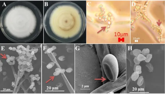
Morphological characteristics of isolates KNU17-67 was observed on PDA (MB Cell, Los Angeles, CA, USA), at 25°C in the dark for 7 days. Medium was prepared as described by Samson et al. [8]. After incubation, the diameter of the colonies was measured, and the degree of sporulation was determined. Colony color (obverse and reverse side) was described using a Methuen handbook of color [9]. Microscopic pictures were taken using an HK 3.1 CMOS digital camera (KOPTIC, Seoul, Korea) attached to an Olympus BX50F-3 microscope (Olympus, Tokyo, Japan) and a scanning electron microscope (LEO Model 1450VP Variable Pressure Scanning Electron Microscope; Carl Zeiss, Oberkochen, Germany). Detailed morphological features of the isolate have been presented in Table 1.
Genomic DNA extraction, sequencing, and data analysis
Total genomic DNA was extracted from isolate KNU17-67 using a DNeasy Plant Mini kit (Qiagen, Germantown, MD, USA), following the manufacturer’s instructions. The internal transcribed spacer (ITS) region of rDNA was amplified by the primers ITS1 (5′-TCCGTAGGTGAACCTGCG-3′) and ITS4 (5′-TCCTCCGCTTATTGATATGC-3′) [10].
The amplified PCR products were sequenced using an ABI Prism 3730 DNA analyzer
(Applied Biosystems, Foster City, CA, USA). The sequences were compared with reference
ITS sequences from GenBank at the National Center for Biotechnology Information (NCBI), using the basic local alignment search tool [11]. The nucleotide sequence of KNU17-67 was deposited at the culture collection facility of the National Institute of Biological Resources (NIBR, NIBRFG0000501850). The nucleotide sequences were also deposited in GenBank and assigned the accession number MH231761. The isolates used in this study to construct the phylogenetic tree were summarized in Table 2 with their strain and GenBank accession numbers. A phylogenetic relationship was analyzed using molecular evolutionary genetic analysis (MEGA 6) software [12]. A neighbor-joining tree was constructed using the Kimura two-parameter substitution model [13]. Bootstrap analysis was performed with 1,000 replications to determine the support for each clade.
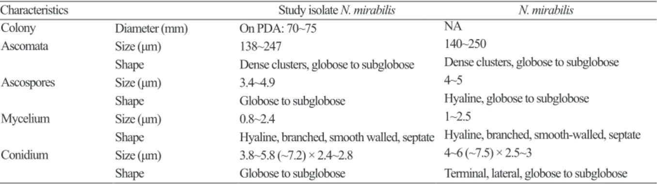
Table 1. Morphological comparison between the studied Nannizziopsis mirabilis isolate and the previously reported N.
mirabilis isolate
Characteristics Study isolate N. mirabilis N. mirabilis
Colony Diameter (mm) On PDA: 70~75 NA
Ascomata Size (μm) 138~247 140~250
Shape Dense clusters, globose to subglobose Dense clusters, globose to subglobose
Ascospores Size (μm) 3.4~4.9 4~5
Shape Globose to subglobose Hyaline, globose to subglobose
Mycelium Size (μm) 0.8~2.4 1~2.5
Shape Hyaline, branched, smooth walled, septate Hyaline, branched, smooth-walled, septate Conidium Size (μm) 3.8~5.8 (~7.2) × 2.4~2.8 4~6 (~7.5) × 2.5~3
Shape Globose to subglobose Terminal, lateral, globose to subglobose PDA, potato dextrose agar; NA, not available.
a