Received: 14 August, 2017 Revised: 16 August, 2017 Accepted: 20 August, 2017
Ⓒ The Korean Society of Mycology
This is an Open Access article distributed under the terms of the Creative Commons Attrib- ution Non-Commercial License (http://creative- commons.org/licenses/by-nc/4.0/) which permits unrestricted non-commercial use, distribution, and reproduction in any medium, provided the original work is properly cited.
Kor. J. Mycol. 2017 September, 45(3): 224-228 https://doi.org/10.4489/KJM.20170026
pISSN : 0253-651X eISSN : 2383-5249
OPEN ACCESS
RESEARCH NOTE
Sporothrix stylites : A New Record from Field Soil in Korea
Sangkyu Park
1, Seung-Yeol Lee
1, Chang-Gi Back
2, In-Kyu Kang
3, Leonid Ten
1, Hyang Burm Lee
4, Hee-Young Jung
1*1
School of Applied Biosciences, College of Agriculture and Life Sciences, Kyungpook National University, Daegu 41566, Korea
2
National Institute of Horticultural and Herbal Science, Wanju 55365, Korea
3
Department of Horticultural Science, College of Agriculture and Life Sciences, Kyungpook National University, Daegu 41566, Korea
4
College of Agriculture and Life Sciences, Chonnam National University, Gwangju 61186, Korea
*Corresponding author: [email protected]
Abstract
The fungal strain, designated KNU16-008, was isolated from field soil in Chungcheongnam-do, Korea. The isolated fungi was characterized by morphological and phylogenetic analyses.
Isolated fungus showed typical morphological characteristics of the genus Sporothrix. Based on its phylogenic analysis using internal transcribed spacer (ITS) of rDNA and β-tubulin gene sequences, the strain KNU16-008 was identified as Sporothrix stylites. This species has not been previously reported in Korea.
Keywords: Phylogenetic analysis, Soil fungi, Sporothrix stylites
The genus Sporothrix was established by Hektoen and Perkins [1] who isolated the fungus from infected boy’s finger, for which the binomial Sporothrix schenckii was introduced. Fungal species that related to the genus Sporothrix have a long history of phylogeny and recently summarized by de Beer et al. [2]. As a result, based on phylogenetic analyses of four gene regions they recognized 51 taxa under the genus Sporothrix, with six species complexes. Members of the Sporothrix originate from a wide variety of environments, including wood, plant debris, peat moss, human clinical materials, but mostly also from soil [2, 3]. Amongst Sporothrix taxa there are clinically relevant species such as S. brasiliensis, S. globosa and S. mexicana [4], insect pathogens such as S.
insectorum [5] as well as saprophytic species such as S. stylites and S. lignivora [3]. A member of S. pallida species complex, S. stylites, was isolated from pine pole at soil level and was characterized as producing micro- to semimacronematous, solitary, straight conidiophores and nonseptate, hyaline, smooth, thin-walled conidia [3]. Dark secondary conidia are absent in S. stylites therefore its cultures do not darken with age.
During our studies of microbial communities in field soil in Buyeo-gun, Chung-
cheongnam-do, Korea, several fungal strains were isolated. With distinctive morphology, one isolate, KNU16-008, was selected for further morphological study and molecular phylogenetic analysis. Based on its morphological characteristics and phylogenetic analysis, this isolate was identified as Sporothrix stylites and named S. stylites KNU16-008.
To the best of our knowledge this fungus has not been previously reported in Korea.
Collected soil sample (1 g) was suspended in 10 mL of sterile distilled water, and prepared suspension was vortexed, serially diluted, and then spread on potato dextrose agar (PDA; Difco, Detroit, MI, USA) plates. The plates were incubated at 25°C for 3 days.
Single colonies on these plates were purified by transferring them onto new plates and subjecting them to an incubation on PDA at 25°C.
During the past decade, species of Sporothrix were distinguished mainly by the molecular phylogenetic anlaysis, primarily using the internal transcribed spacer (ITS) sequences of rDNA [3, 6]. Other gene regions such as β-tubulin (BT) [3, 7], calmodulin (CAL) [4] or ribosomal large subunit (LSU) [8] were also used as markers for phylogenetic analysis in Sporothrix. In the present phylogenetic analysis, ITS regions and BT gene sequences of related Sporothrix taxa were obtained from the GenBank (Table 1). The recovered sequences were aligned with the ITS and BT sequences of isolate KNU16-008 using the program Clustal X. Gaps and 5 ′ and 3′ ends of the alignments were edited
Table 1. Sporothrix and Ophiostoma species used in phylogenetic analysis, with the GenBank accession numbers of their ITS regions of rDNA and β -tubulin gene sequences
Species Strain ITS β-Tubulin
Sporothrix stylites CMW 14544 EF127884 EF139097
Sporothrix stylites CMW 14541 EF127881 EF139094
Sporothrix brasiliensis CBS 130106 KC113212 AM116955
Sporothrix brasiliensis IPEC 17943 FN549902 AM116935
Sporothrix brasiliensis CBS 130107 KC113214 AM116952
Sporothrix inflata CMW 12529 AY495428 AY495439
Sporothrix inflata CMW 12527 AY495426 AY495437
Sporothrix globosa CBS 130104 KC113225 AM116959
Sporothrix globosa CBS 130116 KC113226 AM116962
Sporothrix lignivora CMW 18600 EF127890 EF139104
Sporothrix lignivora CMW 18597 EF127887 EF139101
Sporothrix schenckii CBS 130098 KC113215 AM116917
Sporothrix schenckii CBS 130103 KC113222 AM116915
Sporothrix variecibatus CMW 23060 DQ821569 DQ821572
Sporothrix variecibatus CMW 2543 DQ821567 DQ821573
Sporothrix stylites KNU16-008 MF673228 MF673229
Ophiostoma nigrocarpum CMW 651 AY280490 AY280480
ITS, internal transcribed spacer.
manually using the BioEdit program. Evolutionary distance matrices for the neighbor- joining algorithm were calculated using Kimura’s two-parameter model [9]. Tree topologies were inferred by the neighbour-joining, maximum-likelihood, and maximum- parsimony methods in the program MEGA7 [10], with bootstrap values based on 1,000 replications.
A BLAST search in the NCBI database revealed that ITS regions and BT gene sequences of KNU16-008 matched with those of Sporothrix stylites CMW 14544 (EF127884, EF139097) with 99.6 and 100% similarities, respectively. Phylogenetic tree based on the combined ITS rDNA and BT gene sequences confirmed the affiliation of the isolate to Sporothrix stylites; KNU16-008 was clustered together with S. stylites CMW 14544 and CMW 14541 in a monophyletic clade with the maximum bootstrap value. This phylogenetic relationship was supported by three tree-inferring methods employed in this study (Fig. 1).
Fig. 1. Phylogenic relationship between Sporothrix stylites (KNU16-008) and allied species of Sporothrix and Ophiostoma taxa, constructed using the neighbor-joining method for the combined internal transcribed spacer (ITS) rDNA and β-tubulin gene sequences. Bootstrap values (based on 1,000 replications) greater than 50% are shown at branch points. Filled circles indicate that the corresponding nodes were also recovered in the trees generated with the maximum-likelihood and the maximum-parsimony algorithms. Bar means 0.02 substitutions per nucleotide position.
Morphology of the isolate was examined under an Olympus CX31 light microscope
(Olympus, Tokyo, Japan). The isolate KNU16-008 was cultured at 25°C, and colony
characteristics such as color, shape and size were recorded. After 14 days of incubation on
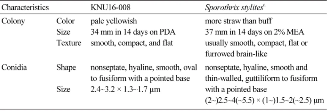
PDA agar, colony was 3.4~3.5 cm in diameter, pale yellowish in color, smooth, compact
and flat (Fig. 2A, 2B). The colony did not darken with age. Conidia were 2.4~3.2 × 1.3~1.7
µm, nonseptate, hyaline, smooth, oval to fusiform with a pointed base (Fig. 2C, 2D). These
morphological characteristics of isolate KNU16-008 are in good agreement with those of
Sporothrix stylites reported by de Meyer et al. [3] (Table 2), which is in line with the phylogenetic results of KNU16-008.
Fig. 2. Cultural and morphological characterization of Sporothrix stylites KNU16-008. A, colony in front; B, colony in reverse; C, D, microscopic pictures of conidia (scale bars: C, D = 10 µm).
Table 2. Morphological characteristics of Sporothrix stylites isolated in this study
Characteristics KNU16-008 Sporothrix stylites
aColony Color
Size Texture
pale yellowish
34 mm in 14 days on PDA smooth, compact, and flat
more straw than buff
37 mm in 14 days on 2% MEA usually smooth, compact, flat or furrowed brain-like
Conidia Shape
Size
nonseptate, hyaline, smooth, oval to fusiform with a pointed base 2.4~3.2 × 1.3~1.7 µm
nonseptate, hyaline, smooth and thin-walled, guttiliform to fusiform with a pointed base
(2~)2.5~4(~5.5) × (1~)1.5~2(~2.5) µm
PDA, potato dextrose agar; MEA, malt extract agar.
a