ISSN 2234-3806 • eISSN 2234-3814
http://dx.doi.org/10.3343/alm.2014.34.3.243 www.annlabmed.org 243
Ann Lab Med 2014;34:243-246
http://dx.doi.org/10.3343/alm.2014.34.3.243
Case Report
Clinical Microbiology
Pulmonary Infection Caused by Mycobacterium neoaurum: The First Case in Korea
Chang-Ki Kim, M.D.1, Soo In Choi, M.D.2, Byung Ryul Jeon, M.D.2, Yong-Wha Lee, M.D.2, You Kyoung Lee, M.D.2, and Hee Bong Shin, M.D.2
Department of Laboratory Medicine1, Korean Institute of Tuberculosis, Osong; Department of Laboratory Medicine and Genetics2, Soonchunhyang University College of Medicine, Cheonan, Korea
Mycobacterium neoaurum is rapidly growing mycobacteria that can cause human infec- tions. It commonly causes bloodstream infections in immunocompromised hosts, and un- like other mycobacteria species, it rarely causes pulmonary infections. We confirmed the first pulmonary infection case in Korea caused by M. neoaurum using full-length 16S rRNA gene sequencing.
Key Words: Nontuberculous mycobacteria, Mycobacterium neoaurum, 16S rRNA, se- quencing
Received: January 6, 2014 Revision received: January 15, 2014 Accepted: February 14, 2014 Corresponding author: Hee Bong Shin Soonchunhyang University Hospital, 170 Jomaro-ro, Wonmi-gu, Bucheon 420-767, Korea Tel: +82-32-621-5942 Fax: +82-32-621-5944 E-mail: [email protected]
© The Korean Society for Laboratory Medicine This is an Open Access article distributed under the terms of the Creative Commons Attribution Non-Commercial License (http://creativecom- mons.org/licenses/by-nc/3.0) which permits unrestricted non-commercial use, distribution, and reproduction in any medium, provided the original work is properly cited.
INTRODUCTION
Rapidly growing mycobacteria are generally defined as nontu- berculous mycobacteria (NTM), which grow within a week on culture media [1]. Currently, there are more than 130 known species of NTM, 70 of which are rapidly growing species, in- cluding Mycobacterium neoaurum [2].
M. neoaurum is commonly associated with catheter-related infections in immunocompromised hosts [3, 4], meningoen- cephalitis [5], and urinary tract infections [6]. Pulmonary infec- tions caused by M. neoaurum are very rare; there has been only one reported case in the literature [7].
M. neoaurum is not easily identified by using phenotypic lab- oratory methods and additional tests may be required to confirm the diagnosis [2]. We report a case of pulmonary infection caused by M. neoaurum, which was identified by using a mo- lecular method. To the best of our knowledge, this is the first re- port of an M. neoaurum infection in Korea.
CASE REPORT
A 25-yr-old woman visited the Department of Pulmonology and Allergy Clinic at a university hospital to evaluate her cough and whitish sputum that began a month ago. She didn’t have any history of chronic illnesses and was not on any medication. To test for common conditions, such as asthma and bronchitis, the physician arranged for allergy and induction tests, and all of the results were negative. Her initial blood cell counts were as fol- lows: hemoglobin, 15.3 g/dL; white blood cells, 8.64 ×10⁹/L (neutrophils, 66.1%; lymphocytes, 20.5%; monocytes, 10.2%;
eosinophils, 2.4%; and basophils, 0.8%); and platelets, 353×
109/L. There was no prominent change in the routine chemistry tests, except for mildly elevated hepatic enzyme levels (AST, 43 IU/L; and ALT, 65 IU/L). An acid-fast bacillus (AFB) smear based on auramine fluorescent stain showed trace result (1-2 AFB/300 fields) according to the Centers for Disease Control and Prevention (CDC) report system. High resolution computed
Kim C-K, et al.
Pulmonary infection caused by M. neoaurum
244 www.annlabmed.org http://dx.doi.org/10.3343/alm.2014.34.3.243 tomography (HRCT) scans were arranged to further investigate
the presence of pulmonary mycobacteria infection. A HRCT scan displayed a well-marginated centrilobular nodule with branching in the left upper lobe, apicoposterior segment, indi- cating the presence of pulmonary tuberculosis in the left upper lobe. The physician performed bronchoscopy, which revealed no specific endobronchial lesions, but cultures of bronchial washing specimens for AFB were positive. An endobronchial washing specimen culture was negative for other bacterial or fungal pathogens. On the basis of these results, the patient was thought to have a mycobacterial lung infection and was given ri- fampicin, ethambutol, and pyrazinamide.
AFB was identified by an auramine fluorescent stain and fur- ther confirmed by Ziehl-Neelsen-stained smears from colonies grown on Mycobacteria Growth Indicator Tube (MGIT; Becton Dickinson, Sparks, MD, USA) liquid medium. On blood agar plates, we observed yellow colonies within 4 days after inocula- tion and incubation at 42°C. The results of the nitrate reduction test were negative. Cultural characteristics indicated that the or- ganism belonged to a group of rapidly growing mycobacteria.
The American Thoracic Society (ATS) has suggested diagnos- tic criteria of nontuberculous mycobacterial lung disease, which require clinical, radiographic, and microbiologic evidence [8]. In our case, the pulmonary symptoms of the patient and HRCT scan findings suffice clinical and radiologic criteria. In terms of the microbiologic criteria, we obtained positive culture results for NTM from at least one bronchial washing specimen without any evidence of an M. tuberculosis complex infection.
To further identify the mycobacteria, reverse hybridization as- says (GenoType Mycobacterium CM/AS, Hain Lifescience, Neh- ren, Germany) were performed. The test results were consistent with Mycobacterium celatum, but cultural characteristics and clinical features were inconsistent with this species. Therefore, full-length 16S rRNA gene sequencing was performed by Mac- rogen (Seoul, Korea) using the following detailed methods:
Using bacterial colonies grown on blood agar plates, bacterial genomic DNA samples were extracted using InstaGene Matrix (BIO-RAD, Hercules, CA, USA). The following primers were used for PCR: forward primer 27F, 5’-AGAGTTTGATCMTGGCT- CAG-3’; and backward primer 1492R, 5’-TACGGYTACCTTGT- TACGACTT-3’. This primer pair amplified a 1,492-bp fragment of the 16S rRNA gene of the Escherichia coli 16S rRNA gene.
The PCR reaction was performed with 20 ng of genomic DNA as a template in a 30 μL reaction mixture using EF-Taq-DNA polymerase (SolGent, Daejeon, Korea). The following PCR pro- gram was used: activation of Taq polymerase at 95°C for 2 min;
35 cycles at 95°C, 55°C, and 72°C for 1 min; and a 10-min step at 72°C.
The amplification products were purified via a multiscreen fil- ter plate (Millipore Corp., Billerica, MA, USA). Sequencing reac- tions were performed using the PRISM BigDye Terminator v3.1 Cycle Sequencing Kit. The DNA samples containing the exten- sion products were added to Hi-Di formamide (Applied Biosys- tems, Foster City, CA, USA). The mixture was incubated at 95°C for 5 min, placed on ice for 5 min, and then analyzed using an ABI Prism 3730XL DNA analyzer (Applied Biosystems).
After sequencing, the raw sequence file (.abi) was processed using Lasergene SeqMan Pro 7.1 (DNASTAR, Madison, WI, USA) to get trimmed nucleotide sequences. We compared the obtained sequences with those deposited in GenBank (http://www.ncbi.
nlm.nih.gov). The sequence of the 16S rRNA gene was consis- tent with that of M. neoaurum ATCC25796 (FJ172307.1) based on having a sequence similarity of 99.8%.
A phylogenetic tree was generated using the Neighbor-Joining method, which is based on nucleotide sequences of the clinical isolate with those of other Mycobacteria reference strain se- quences prepared from the National Center for Biotechnology In- formation (NCBI) database using Mega 5.0 software [9] (Fig. 1).
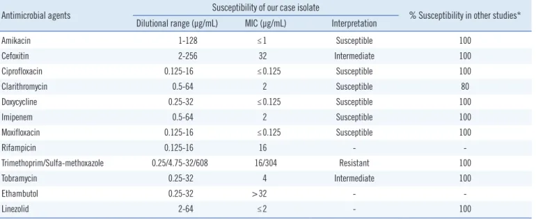
The drug susceptibility test was performed at the Korean Insti- tute of Tuberculosis using first-line and second-line drugs, and the minimal inhibitory concentrations (MIC) were determined us- ing the broth microdilution method in Muller-Hinton media. MICs were interpreted based on the broth microdilution interpretive criteria for rapidly growing mycobacteria [10] (Table 1).
After 1 month of rifampicin, ethambutol, and pyrazinamide treatment, the patient was placed on a clarithromycin regimen for 4 months, and her symptoms improved.
DISCUSSION
NTM are ubiquitous organisms that are found in the environ- ment. Pulmonary infections are the most common manifesta- tion of NTM infections, and they are often caused by M. avium complex (MAC). Currently, the overall incidence of NTM infec- tions in Korea is increasing [11].
M. neoaurum was first isolated from soil and reported in 1972 [12]. Since then, there have been 25 cases of human infec- tions, ranging from sepsis to skin infections. Literature reviews have noted that M. neoaurum-related cases were mostly cathe- ter- or line-related sepsis instead of meningoencephalitis, skin infections [13, 14], or pulmonary infections [7].
Initially, the clinical isolate described in our case was misiden-
Kim C-K, et al.
Pulmonary infection caused by M. neoaurum
http://dx.doi.org/10.3343/alm.2014.34.3.243 www.annlabmed.org 245
tified as M. celatum by the reverse hybridization assay. However, on the basis of sequencing the full-length 16S rRNA gene, the
isolate was re-identified as M. neoaurum.
For accurate Mycobacterium spp. identification, sequencing M.neoaurum ATCC25795 (NR041900.1)
M.neoaurum ATCC25796 (FJ172307.1) M.neoaurum ATCC23072 (FJ172309.1)
M. bacteremicum ATCC25791 (FJ172308.1) M. neoaurum ATCC25801 (FJ172302.1) M. lacticola ATCC9626 (AF480582.1) M. neoauruml lacticola ATCC25799 (FJ172303.1) M. neoauruml lacticola ATCC25800 (FJ172301.1) M. neoauruml lacticola ATCC25803 (FJ172300.1) M. cosmeticum ATCCBAA878 (NR025787.1) M. mucogenicum ATCC49651 (AY457075.1)
M. fortuitum ATCC49404 (AF480581.1)
M. aurum ATCC23366 (FJ172298.1)
M. abscessus ATCC19977 (NR074427.1) M. chelonae ATCC19237 (AY457082.1) M. terrae ATCC15755 (X52925.1)
M. szulgai ATCC35799 (KC951278.1) M. malmoense ATCC29571 (NR044814.1)
M. kansasii ATCC12478 (AF480601.1) M. avium ATCC25291 (EF521895.1) M. tuberculosis ATCC27294 (FJ468345.1)
0.002
Clinical isolate
Fig. 1. Phylogenetic relationships of clinical isolate with related Mycobacterium species on the basis of 16S rRNA gene sequences by neighbor-joining method. Scale bar represents 2 nucleotide substitutions per 1,000 nucleotides.
Table 1. Comparison of the antimicrobial drug susceptibility of this clinical isolate with those from other studies Antimicrobial agents Susceptibility of our case isolate
% Susceptibility in other studies*
Dilutional range (μg/mL) MIC (μg/mL) Interpretation
Amikacin 1-128 ≤1 Susceptible 100
Cefoxitin 2-256 32 Intermediate 100
Ciprofloxacin 0.125-16 ≤0.125 Susceptible 100
Clarithromycin 0.5-64 2 Susceptible 80
Doxycycline 0.25-32 ≤0.125 Susceptible 100
Imipenem 0.5-64 2 Susceptible 100
Moxifloxacin 0.125-16 ≤0.125 Susceptible 100
Rifampicin 0.125-16 16 - -
Trimethoprim/Sulfa-methoxazole 0.25/4.75-32/608 16/304 Resistant 100
Tobramycin 0.25-32 4 Intermediate 100
Ethambutol 0.25-32 >32 - -
Linezolid 2-64 ≤2 - 100
*Adapted from reference [7, 18].
Abbreviation: MIC, minimal inhibitory concentration.
Kim C-K, et al.
Pulmonary infection caused by M. neoaurum
246 www.annlabmed.org http://dx.doi.org/10.3343/alm.2014.34.3.243 multiple housekeeping genes is invaluable. However, we believe
16S rRNA sequencing with phylogenetic analysis would be enough to identify M. neoaurum in this case for several reasons.
First, we performed full-length 16S rRNA gene sequencing (1,492 bp), which was used to confirm its identity in roughly a half of the previously reported cases [4, 7, 15, 16]. Second, M.
neoaurum has a unique 16S rRNA gene sequence that can be used for identification [2]. Finally, and most of all, our phyloge- netic analysis revealed that the clinical isolate forms a tight clus- ter with M. neoaurum ATCC23072, ATCC25795, and ATCC25796 (Fig. 1), which were all confirmed to be M. neoaurum in other studies [17]. The misidentification of M. neoaurum can usually occur due to its similarity to M. lacticola group. However, this clinical isolate was sufficiently discriminated among other rapidly growing mycobacteria [18].
Antimicrobial susceptibility test could be an adjunctive test for identification of rapidly growing mycobacteria. But susceptibility test result for cefoxitin, tobramycin, and trimethoprime/sulfa- methoxazole of this isolate didn’t coincide with other studies, as M. neoaurum is known as pan-susceptible to antimicrobial drugs [2, 7, 18] (Table 1). We are not sure whether this finding has clinical significance, but further study will reveal much more in- formation.
Contrary to other reported cases of M. neoaurum, we de- scribed a pulmonary infection in a fully immunocompetent pa- tient. Any patient with an atypical mycobacteria pulmonary in- fection should be investigated further for a correct identification of NTM.
This is the first reported case of pulmonary infection with M.
neoaurum in Korea, and it emphasizes the importance of proper molecular identification for the diagnosis and treatment of NTM.
Authors’ Disclosures of Potential Conflicts of Interest
No potential conflicts of interest relevant to this article were re- ported.
Acknowledgments
This work was partly supported by the Soonchunhyang Univer- sity Research Fund.
REFERENCES
1. Brown-Elliott BA and Wallace RJ Jr. Clinical and taxonomic status of
pathogenic nonpigmented or late-pigmenting rapidly growing mycobac- teria. Clin Microbiol Rev 2002;15:716-46.
2. Brown-Elliott BA and Wallace RJ Jr. Mycobacterium: Clinical and labo- ratory characteristics of rapidly growing mycobacteria. In: Versalovic J, Carroll KC, Funke G, Jorgensen JH, Landry ML, Warnock DW, eds.
Manual of clinical microbiology. 10th ed. Washington, DC: ASM press, 2010:525-38.
3. Davison MB, McCormack JG, Blacklock ZM, Dawson DJ, Tilse MH, Crimmins FB. Bacteremia caused by Mycobacterium neoaurum. J Clin Microbiol 1988;26:762-4.
4. Lai CC, Tan CK, Chen CC, Hsueh PR. Mycobacterium neoaurum infec- tion in a patient with renal failure. Int J Infect Dis 2009;13:e276-8.
5. Heckman GA, Hawkins C, Morris A, Burrows LL, Bergeron C. Rapidly progressive dementia due to Mycobacterium neoaurum meningoen- cephalitis. Emerg Infect Dis 2004;10:924-7.
6. Zanetti S, Faedda R, Fadda G, Dupré I, Molicotti P, Ortu S, et al. Isola- tion and identification of Mycobacterium neoaurum from a patient with urinary infection. New Microbiol 2001;24:189-92.
7. Morimoto Y, Chan ED, Heifets L, Routes JM. Pulmonary infection with Mycobacterium neoaurum identified by 16S ribosomal DNA sequence.
J Infect 2007;54:e227-31.
8. Griffith DE, Aksamit T, Brown-Elliott BA, Catanzaro A, Daley C, Gordin F, et al. An official ATS/IDSA statement: diagnosis, treatment, and preven- tion of nontuberculous mycobacterial diseases. Am J Respir Crit Care Med 2007;175:367-416.
9. Tamura K, Peterson D, Peterson N, Stecher G, Nei M, Kumar S.
MEGA5: molecular evolutionary genetics analysis using maximum likeli- hood, evolutionary distance, and maximum parsimony methods. Mol Biol Evol 2011;28:2731-9.
10. Clinical and Laboratory Standards Institute. Susceptibility testing of my- cobacteria, nocardiae, and other aerobic actinomycetes; Approved standard, M24-A2E. Wayne, PA: Clinical and Laboratory Standards In- stitute, 2011.
11. Korea Center for Disease Control and Prevention (KCDC). Current status of nontuberculous mycobacterial infection. Public Health Weekly Re- port 2009;2:389-93 (http://www.cdc.go.kr/CDC/info/CdcKrInfo0301.
jsp?menuIds=HOME001-MNU1154-MNU0005-MNU0037&q_type=&
year=2009&cid=12296&pageNum=).
12. Tsukamura M. A new species of rapidly growing, scotochromogenic mycobacteria. Mycobacterium neoaurum. Med Biol 1972;85:229-33.
13. Martin LK, Lawrence R, Kossard S, Murrell DF. Cutaneous Mycobacteri- um neoaurum infection causing scarring alopecia in an immunocom- petent host. Br J Dermatol 2007;157:204-6.
14. Omoruyi OJ, Ip WY, To KK. Hand infection due to Mycobacterium neo- aurum. J Hand Surg Eur Vol 2012;37:574-5.
15. Becker ML, Suchak AA, Wolfe JN, Zarychanski R, Kabani A, Nicolle LE.
Mycobacterium neoaurum bacteremia in a hemodialysis patient. Can J Infect Dis 2003;14:45-8.
16. García-Agudo L, Jesús I, Rodríguez-Iglesias M, García-Martos P. Evalu- ation of INNO-LiPA mycobacteria v2 assay for identification of rapidly growing mycobacteria. Braz J Microbiol 2011;42:1220-6.
17. Simmon KE, Low YY, Brown-Elliott BA, Wallace RJ Jr, Petti CA. Phyloge- netic analysis of Mycobacterium aurum and Mycobacterium neoaurum with redescription of M. aurum culture collection strains. Int J Syst Evol Microbiol 2009;59:1371-5.
18. Brown-Elliott BA, Wallace RJ Jr, Petti CA, Mann LB, McGlasson M, Chi- hara S, et al. Mycobacterium neoaurum and Mycobacterium bactere- micum sp. nov. as causes of mycobacteremia. J Clin Microbiol 2010;
48:4377-85.