Copyright ⓒ 2014, The Microbiological Society of Korea
해면 Callyspongia elegans에 서식하는 세균군집의 계통학적 다양성
박소현1․김지영2․김영주3․허문수1*
1제주대학교 해양의생명과학부, 2제주대학교 기초과학연구소, 3제주대학교 해양과환경연구소
Phylogenetic Diversity of Bacterial Community Inhabited in Callyspongia elegans
So-Hyun Park1, Ji-Young Kim3, Young-Ju Kim2, and Moon-Soo Heo1*
1Department of Aquatic Life Medicine, College of Ocean Science,
2Marine and Environmental Research Institute, Jeju National University, Jeju 690-756, Republic of Korea
3Research Institute for Basic Science, Jeju National University, Jeju 690-756, Republic of Korea
(Received May 12, 2014 / Accepted June 25, 2014)
The aim of this study was to investigate the bacterial community inhabited in Callyspongia elegans. Marine bacteria were isolated from the marine sponge C. elegans using marine agar. The resulting 112 isolated pure cultures were then used for further study. They were characterized by determining morphological characteristics through Gram’s staining and morphological observation. The colony pigments of bacterial isolates were characterized as yellow, brown, ivory, and white. Thirty-seven strains were found to be Gram-positive and 75 strains were Gram-negative.
Seventy-nine strains were coccus-shaped, while 16 strains were rod-shaped. On the basis of the results of the comparative analyses of 16S rDNA gene sequences, the 112 isolated bacteria were divided into 5 major groups:
Alphaproteobacteria (39%), Gammaproteobacteria (22%), Actinobacteria (14%), Fimicutes (9%), and Bacteroidetes (6%). It is strongly suggested that fifteen isolates are candidates for a new genera or species, based on the analyses of 16S rDNA gene sequences.
Keywords: 16S rDNA, Callyspongia elegans, bacterial community, marine sponge, phylogenetic tree
*For correspondence. E-mail: [email protected]; Tel.: +82-64-754- 3473; Fax: +82-64-756-3493
무척추동물로 오랜 진화적 역사를 갖고 있는 해면은 3개의 주 요 강 Calcarea, Demospongiae, Haxactinellida으로 나뉘며, Demospongiae 강에 가장 많은 해면이 속하는 것으로 보고되고 있다(Hentschel et al., 2006; Thomas et al., 2010). 해면은 해양 과 담수에 서식하는 것으로 알려져 있으며, 열대, 온대, 극지 등 다양한 지역에 분포를 나타내고 있다. 또한 해면은 여과 섭식동 물로써 먹이원을 주변의 해수 또는 담수의 여과를 통하여 공급 받는다(Thomas et al., 2010).
이러한 해면 생체량의 40% 이상 세균, 고세균, 시아노박테리아 등 많은 미생물을 세포내 또는 세포외에 포함하고 있으며(Levina et al., 2005), 미생물이 서식하면서 일시적 또는 영구적인 공생 관계를 유지하는 것으로 보고 되어졌다(Park et al., 2009). 그 중 해면에 서식하는 공생미생물 중 세균이 가장 많은 분포를 나타 내며(Thompson et al., 1994), 외부환경의 영향에도 영구적으로 숙주 해면과의 공생관계를 가지는 세균 종이 있다는 보고도 있 다(Friedrich et al., 2001; Muscoll-Silberhorn et al., 2008).
해면에 서식하는 세균은 여과섭식하는 해면의 먹이원이기도 하지만, 소화과정과 면역반응에 저항하며 공생하면서 면역반응에 중요한 역할을 한다(Thoms et al., 2003; Cho and Park, 2009). 현 재 해양에서 유래된 생물의약품 중 무척추동물로부터 대부분이 유래되었으며, 그 중 가장 많은 종류의 생리활성물질이 해면으로 부터 생산된다고 알려져 있다(Thomas et al., 2010). 이러한 생리 활성물질이 해면에 서식하는 공생미생물에 의해 생산된다고 보고 되면서(Lafi et al., 2005; Tamaki et al., 2005; Guangyi, 2006;
Mohamed et al., 2008; Kennedy et al., 2009), 해면의 공생세균의 군집구조와 다양성에 관한 연구가 증가하는 추세이다(Li et al., 2006; Lee et al., 2009; Cho et al., 2010; Radwan et al., 2010).
본 연구에서는 Demospongiae 강에 속하는 해면 Callyspongia elegans에서 배양가능한 세균을 분리·배양하여 탐색하고, 16S rDNA를 비교하여 세균의 군집 구조를 분석하고자 한다.
재료 및 방법 시료채집 및 배양가능한 세균의 분리
본 연구에서는 해면 Callyspogia elegans는 제주도 문섬 앞바
다 수심 10 m에서 스쿠버 다이빙에 의해 채집하였다. 채집한 해 면은 즉시 해수가 담아진 멸균된 용기에 보관하여 운반하였으 며, 4℃에서 보관 후 미생물을 분리하는데 사용하였다.
채집한 해면은 멸균된 인공해수에 세척 후, 해면의 안쪽을 1 g 정도 잘라 멸균된 0.85% NaCl 용액에 넣어 homogenizer로 균 질화 하였다. 균질화된 시료는 연속희석법으로 희석 후, 현탁액 을 marine agar (MA, Difco, USA), R2A (Difco) 배지에 도말 한 후 25℃에 7일간 배양하였다. 각 배지상에 나타난 군락을 육안 으로 관찰했을 때 군락의 모양, 크기, 색깔 등에 따라 형태적으로 분류, 선별하여 순수분리 하였다. 각 분리된 미생물은 20% (v/v) glycerol에 현탁하여 초저온 냉동고(-80℃)에 동결 보관하였다.
배양학적 및 형태학적 특징
순수 배양된 세균들은 MA, R2A 평판배지에 2–3일 배양한 후 집락의 색상을 기록하였다. 또한 방선균은 ISP2 배지에서 7–
10일 배양 후 포자의 색상을 기록하였다.
균주의 형태학적 특징은 그람염색으로 그람양성균과 그람음 성균을 구분하였다. 그람염색은 Color Gram 2 kit (bioMérieux) 를 이용하여 균체를 슬라이드 글라스에 열고정하고, 크리스탈 바이올렛으로 1분 염색 후 세척, 요오드 용액으로 1분간 고정, 알코올로 15초간 탈색 후 사프라닌으로 다시 45초 염색하는 과 정을 통해 수행하였다. 염색 후 광학현미경(AX10SCOPE A1, ZEISS)을 통해서 그람양성균과 그람음성균을 관찰하였다.
Genomic DNA 추출
Genomic DNA는 MINI-PREP 방법을 사용하여 분리하였다.
액체배지에 접종하여 배양 후 원심분리로 상층액을 제거 후 모 아진 pellet을 사용하였다. TE buffer로 용해시킨 후 lysozyme (100 mg/ml)을 첨가하여 반응시켰다. 단백질 제거는 10% SDS와 proteinase K (10 mg/ml)를 첨가하여 37℃에서 1시간 배양하였다.
5 M NaCl를 넣어 잘 섞어주고 CTAB/ NaCl solution을 첨가 후 65℃
에서 10분간 반응시켰다. Phenol/ Chloroform/ Isoamylalcohol (25:24:1)를 첨가 후 원심분리(13,000 rpm, 5분)하여 깨끗한 상 층액을 새 마이크로 튜브에 옮겨주고, 옮겨진 부피의 2배의 chloroform/ isoamylalcohol (24:1)를 첨가하였다. 상층액을 새 튜브에 조심스럽게 옮겨 isopropanol를 첨가하여 DNA를 확인하 였다. 모아진 DNA을 70% ethanol로 세척하여 TE buffer 50‒100 µl와 RNase (10 mg/ml)를 첨가한 후 UV-Vis Spectrophotometer (UV, mini240, Shamdzu)로 DNA 농도를 측정한다.
16S rDNA 유전자 증폭
16S rDNA 분석은 추출된 genomic DNA를 사용하였다. 16S rDNA 유전자 증폭하기 위해 27 Forward (5′-AGAGTTTGATC CTGGCTCAG-3′) primer와 1522 Reverse (5′-AAGGAGGT GATCCAGCCGCA-3′) primer를 사용하였다. PCR 반응의 조 성은 추출된 DNA 1 µl, 10 pmol/primer 1 µl, 10 mM dNTPs, 10X PCR buffer, 5 Unit Taq polymerase (TaKaRa, Japan), DW 를 혼합하여 최종부피 25 µl로 맞추어 수행하였다. PCR 반응은 95℃에서 Initial denaturation 5분, 30 cycle 동안 94℃에서 denaturation
1분, 55℃에서 annealing 1분, 72℃에서 extention 3분을, 72℃
분간 final extention로, PCR (Thermal cycler, Bio-Rad) 조건하 에 실시하였다. 증폭된 PCR 산물은 1% agarose에서 전기영동 (Mupid®-ex, ADVANCE)하여 확인하였다.
유전자 클로닝 및 염기서열 분석
pGEM®-T Easy Vector System (Promega, USA)을 이용하여 ligation 후 클로닝하였다. 클로닝 산물은 ampicillin, IPTG, X-gal 이 포함된 LB agar 배지에서 도말 후 37℃, 16시간 동안 배양하여 흰 집락을 선별하였다. 선별한 집락을 ampicillin이 포함된 LB broth에 접종하여 16시간 배양하여 DNA-spin Plasmid DNA Extraction Kit (iNtRON, Korea)를 사용하여 plasmid DNA를 분리하였다. 분리된 DNA는 1% agarose gel에서 전기영동하여 확인하였다.
분리된 plasmid DNA는 ㈜제노텍(Korea)에 의뢰하여 염기서열 을 분석 하였다. 분석된 염기서열은 EzBioCloud 이용하여 유사한 염기서열을 비교하여 가장 근연속이나 종으로 나타나는 서열을 확 인하였다. 본 연구에 의해서 결정된 염기서열과 EzBioCloud에 서 회수된 표준미생물 염기서열은 Mega 4.0 software (Tamura et al., 2007)에 포함된 Clustal W 프로그램(Thomson et al., 1994) 을 이용하여 multiple alignment로 정렬하였다.
결과 및 고찰
분리균주의 배양상 및 형태학적 특성
평판배양법으로 세균 수를 측정한 결과, MA 배지에서 분리 한 세균은 4.8 × 10⁴ CFU/g, R2A 배지에는 2.0 × 10⁴ CFU/g의 세균수가 관찰되었다. MA보다 R2A 배지에서 세균수가 훨씬 낮 게 계수되었다. C. elegans에서 배양가능한 세균은 총 112균주 가 분리되었다. 그람염색법으로 확인한 결과로 그람양성균은 37 균주, 그람음성균은 75균주로 그람음성균이 더 많게 분포하는 것으로 나타났다. 균주의 집락의 색깔은 아이보리색, 노란색, 흰 색, 갈색 등으로 다소 제한적으로 나타남을 알 수 있었다.
염기서열 분석 및 계통학적 분석
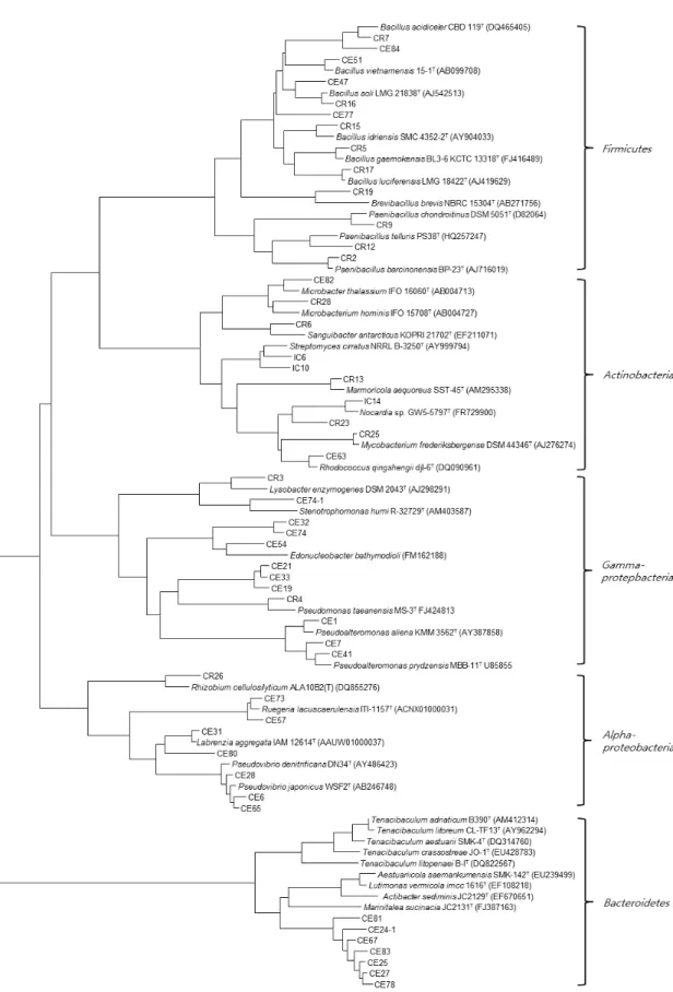
분리된 균주의 16S rDNA 유전자 염기서열은 EzbioCloud를 이용하여 유사한 염기서열을 비교하였다. 계통수(Felsenstein, 1981; Saitou and Nei, 1987)를 작성한 결과는 Fig. 1과 같다.
해면 C. elegans에서 분리된 112균주는 24속 52종으로 동정 되었다. Proteobacteria 그룹은 기존의 염기서열과 93–100%의 상동성을 나타내었다. CR26은 Rhizobium huautlense S02T과 97.1%
의 유사도를 보여주었다. Endozoicomonas 속은 Endozoicomonas elysicola MKT110T와 Endozoicomonas montiporae CL-33T과 93.6‒93.8, 96.2%의 상동성을 보여주었다.
Firmicutes 그룹에 속해 있는 균주는 대부분 기존의 염기서열 과 95.1‒100% 상동성을 보여주었으며, CE84는 Lysinibacillus massiliensis 4400831T과 97.2%, CR9, CR12는 각각 Paenibacillus chondroitinus DSM 5051T, Paenibacillus telluris PS38T와 96.8%와 95.1%의 낮은 유사도를 나타내었다. 그리고 Actinobacteria
Fig. 1. Phylogenetic tree based on comparison of the 16S rDNA gene sequences of bacteria isolated from C. elegans and some other related taxa.
GenBank accession nos. are given in parentheses. Boostrap values (>50%) based on 1,000 replications are shown. Bar 0.05 nucleotide substitutions per nucleotide position.
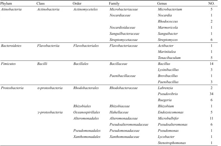
Phylum Class Order Family Genus NO.
Atinobacteria Actinobacteria Actinomyceteles Microbacteriaceae Microbacterium 5
Nocardiaceae Nocardia 1
Rhodococcus 2
Nocardioidaceae Marmoricola 1
Sanguilbacteraceae Sanguibacter 1
Streptomycetaceae Streptomyces 6
Bacteroidetes Flavobacteriia Flavobacteriales Flavobacteriaceae Actibacter 1
Marinitalea 1
Tenacibaculum 5
Fimicutes Bacilli Bacillales Bacillaceae Bacillus 14
Lysinibacillus 3
Paenibacillaceae Brevibacillus 1
Paenibacillus 3
Proteobacteria α-proteobacteria Rhodobacterales Rhodobacteraceae Labrenzia 2
Pseudovibrio 34
Ruegeria 6
Rhizobiales Rhizobiaceae Rhizobium 1
γ-proteobacteria Oceanospirillales Hahellaceae Endozoicomonas 5
Alteromonadales Alteromonadaceae Microbulbifer 11
Pseudoalteromonadaceae Pseudoalteromonas 6
Pseudomonadales Pseudomonadaceae Pseudomonas 1
Xanthomonadales Xanthomonadaceae Lycobacter 1
Stenotrophomonas 1
Table 1. Bacterial diversity associated with C. elegans
그룹에 속하는 균주는 기존의 염기서열과 98.0‒100%의 상동성 을 보여주었다. Bacteroidetes 그룹에 속하는 균주는 다른 그룹 과는 달리 낮은 염기서열 유사도를 보여주었다. CE81는 Actibacter sediminis JC2129T와 91.9%, Tenacibaculum 속(genus) 분리균 주들은 Tenacibaculum crassostreae JO-1T, Tenacibaculum litopenaei B-IT, Tenacibaculum litoreum CL-TF13T 91.8‒94.5%
낮은 상동성이 확인되었다. 이와 같이 16S rDNA 유전자 염기서 열 비교를 통해 분리된 112균주 중 16균주가 표준균주와 유전자 염기서열의 97% 이하의 상동성을 보여 새로운 속 또는 종으로 보고될 가능성이 있다고 판단된다. 향후 표준균주와 함께 실험 이 신종 실험이 수행되어야 할 것이다.
16S rDNA gene 염기서열로부터 계통학적 다양성을 분석해 보 았다. 그 결과, C. elegans의 주요 세균 군집구조로 Proteobacteria (Alphaproteobacteria, Gammaproteobacteria), Actinobacteria, Firmicutes, Bacteroidetes의 4개의 문(Phylum), 5개의 강(Class), 9개의 목(order), 15개의 과(Family), 그리고 23개의 속(Genus) 으로 구성되었다(Table 1). 그 중 Gammaproteobacteria 문 (phylum)에는 4개의 목(order), 5개의 과(family), 5개의 속 (genus)으로 Actionobacteria 문(phylum)에서는 5개의 과, 6개 의 속(genus) 등 다양한 분류군이 나타났다(Table 1).
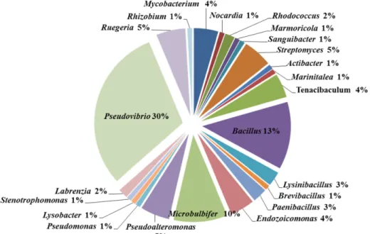
속(genus)은 Pseudovibrio 30%, Bacillus 13%, Microbulbife 10%, Pseudoalteromonas, Streptomyces, Ruegeria 5%,
Mycobacterium와 Tenacibaculum 그리고 Endozoicomonas는 4%, Lysinibacillus, Paenibacillus 3%이며, Rhodococcus 2%, Nocardia, Marmoricola, Sanguibacter, Actibacter, Marinitalea, Brevibacillus, Pseudomonas, Lysobacter, Stenotrophomonas, Labrenzia, Rhizobium는 1%순으로 구성되어 있으며, Pseudovibrio (34균주), Bacillus (14균주), Microbulbifer (11균주)로 나타났 다(Fig. 2).
Proteobacteria 계통군은 다시, Alphaproteobacteria, Gammaproteobacteria 계통에 각각 39%와 22%에 속하였음을 확인하였으며(Fig. 3), Proteobacteria가 다른 분류군에 비해 우 점하는 것을 알 수 있었다. 이는 해양환경에서 일반적으로 서식 하며, 해양 해면에서도 높은 비율로 분포하는 것으로 보고 되었 고(Li et al., 2006), 대다수 해양생태계에서 Proteobacteria와 Bacteroidetes 우점으로 존재한다는 결과와 일치한다(Alfeider et al., 1996; Eilers et al., 2000). 비배양 혹은 배양법에 의해 밝혀 진 해면 공생세균 군집에서 Proteobacteria (Alphaproteobacteria, Gammaproteobacteria)가 우점한다고 보고되고 있으며(Braekman and Daloze, 2004; Lafi et al., 2005), 이 결과와도 일치하였다.
그리고 남태평양에서 서식하는 Callyspongia sp. 주요 공생세균 으로 Proteobacteria, Chloroflexi, Cyanobacteria의 3개의 문에 속하는 세균 그룹이 보고되었으나(Park, 2010), 본 연구에서는 Proteobacteria, Firmicutes, Actinobacteria, Bacteroidetes의 4
Fig. 2. Genus of bacterial community of C. elegans.
Fig. 3. Diversity and a structure of bacterial community of C. elegans.
개의 문에 속하는 세균 그룹이 확인되었다(Fig. 3). 이 결과와 비 교하여 볼 때 공통적으로 나타난 Proteobacteria를 제외하고는 세균군집 구조가 서로 다르게 분류되고 있음을 알 수 있었고, C.
elegans에서는 다양한 세균군집을 나타났음을 확인하였다. 이는 해 면 종에 따라 세균군집 구조가 서로 다르다는 연구(Li et al., 2006;
Thiel et al., 2007; Lee et al., 2009; Cho et al., 2010)와 일치하 는 결과로써 해면 공생세균의 군집구조는 숙주 특이적인 것으로 판단된다. 동일한 종의 해면이라도 온대와 열대의 지리적 분포 에 따라 세균군집(Taylor et al., 2005), 또는 계통 유연관계가 다 르거나 지리적 분포가 다른 해면에서도 유사한 세균 군집구조가 나타난다고 보고되었다(Lafi et al., 2005).
그리고 중요한 대사산물을 생산하는 해면의 세균그룹에서는 Actinobacteria의 비율이 높은 것으로 밝혀진 연구(Li et al., 2006; Graeber et al., 2008)를 볼 때, C. elegans 내 분리균주도 중요한 대사산물을 생산할 가능성이 있다고 보여지며, Pseudovibrio sp.와 Actionobacteria 문에 속한 균주에 대한 연구가 더욱 필요
하고, 특히 우리나라의 해양에 서식하는 해면에 관한 공생 세균 의 군집구조에 관한 보고는 매우 적기 때문에 이에 관하여 많은 연구가 필요한 실정이다.
적 요
이 논문은 Callyspongia elegans에서 서식하는 세균군집에 관 한 내용이다. 해양세균은 marine agar를 사용하여 해면동물 C.
elelgans에서 분리하였다. 그 결과 112균주를 분리하였으며, 본 연구에 사용하였다. 현미경 및 그람 염색을 통해 형태학적 표현 형질을 측정하였다. 분리균주의 집락 색소는 노란색, 갈색, 아이 보리색, 흰색으로 나타났다. 그람염색 결과 37균주는 그람양성 균이였으며, 75균주는 그람 음성균이었다. 균주의 형태는 분리 균주 중 79균주는 구균형태로 관찰되었고, 16균주는 간균이었다.
16S rDNA 유전자 염기서열 분석을 통해 분리균주들의 계통학적 특성을 파악하였다. 그 결과, 분리된 112균주는 5개의 주요 계통군
이 확인되었으며, Alphaproteobacteria는 39%, Gammaproteobacteria 는 22%, Acinobacteria는 14%, Firmicutes는 9%, Bacteroidetes 는 6%에 속하는 것으로 나타났다. 그리고 16S rDNA 유전자 염 기서열을 통해 계통분석 결과 15균주가 새로운 속 또는 종으로 분류될 가능성을 나타났으며, 앞으로 추가적인 실험이 필요한 실정이다.
감사의 말
이 논문은 2014학년도 제주대학교 학술진흥연구비 지원사업 에 의해 연구되었음.
References
Alfreider, A., Pernthhaler, J., Amann, R., Sattler, B., Glöckner, F.O., Wille, A., and Psenner, R. 1996. Community analysis of the bacterial assemblages in the winter cover and pelagic layers of high mountain lake by in situ hybridization. Appl. Environ. Microbiol. 62, 2138–
2144.
Braekman, J. and Daloze, D. 2004, Chemical and biological aspects of sponge secondary metabolites. Phytochem. Rev. 3, 275–282.
Cho, H.H. and Park, J.S. 2009. Comparative analysis of the community of culturable bacteria associated with sponges, Spirastrella abata and Spirastrella panis by 16S rDNA-RFLP. Kor. J. Microbiol. 45, 155–162.
Cho, H.H., Shim, E.J., and Park, J.S. 2010. Phylogenetic diversity of bacteria associated with the marine sponges, Spirastrella abata and Cinachyrella sp. Kor. J. Microbiol. 46, 177–182.
Eilers, H., Pernthaler, J., Glöckner, F.O., and Aman, R. 2000.
Culturability and in situ abundance of pelagic bacteria from the North Sea. Appl. Environ. Microbiol. 66, 3044–3051.
Felsenstein, J. 1981. Evolutionary trees from DNA sequences: a maximum like lihood approach. J. Mol. Evol. 17, 368–376.
Friedrich, A.B., Hacker, J., Fischer, I., Proksch, P., and Hentschel, U.
2001. Temporal variations of the microbial community associated with the Mediterranean sponge Aplysina aerophoba. FEMS Microbiol. Ecol. 38, 105–113.
Graeber, I., Kaesler, I., Borchert, M.S., Dieckmann, R., Page, T., Lurz, R., Nielsen, P., Dohren, H.V., Michaelis, W., and Szewzyk, U. 2008.
Spongiibacter marinus gen. nov., sp. nov., a Halophilic marine bacterium isolated from the boreal sponge Haliclona sp.1. Int. J. Syst.
Evol. Microbiol. 58, 585–590.
Guangyi, W. 2006. Diversity and biotechnological potential of the sponge-associated microbial consortia. J. Ind. Microbiol. Biotechnol.
33, 545–551.
Hentschel, U., Usher, K.M., and Taylor, M.W. 2006. Marine sponges as microbial fermenters. FEMS Microbiol. Ecol. 55, 167–177.
Kennedy, J., Baker, P., Piper, C., Cotter, P.D., Walsh, M., Mooij, M.J., Bourke, M.B., Rea, M.C., O’Connor, P.M., Ross, R.P., and et al.
2009. Isolation and analysis of bacteria with antimicrobial activities from the marine sponge Haliclona simulans collected from Irish Waters. Mar. Biotechnol. 11, 384–396.
Lafi, F.F., Garson, M.J., and Fuerst, J.A. 2005. Culturable bacterial symbionts isolated from two distinct sponge species (Pseudoceratina clavata and Rhabdastrella globostellata) from the great barrier reef display similar phylogenetic diversity. Microb. Ecol. 50, 213–220.
Lee, O.O., Wong, Y.H., and Qian, P.Y. 2009. Inter and intraspecific
variations of bacterial communities associated with marine sponges from San Juan Island, Washington. Appl. Environ. Microbiol. 75, 3513–3521.
Levina, E.V., Kalinovsky, A.I., Andriyashenko, P.V., Dmitrenok, P.S., Aminin, D.L., and Stonik, V.A. 2005. Phrygiasterol, a cytotoxic cyclopropane containing polyhydroxysteroid, and related compounds from the pacific starfish Hippasteria phrygiana. J. Nat. Prod. 68, 1541–
1544.
Li, Z.Y., He, L.M., Wu, J., and Jiang, Q. 2006. Bacterial community diversity associated with four marine sponges from the South China Sea based on 16S rDNA-DGGE fingerprinting. J. Exp. Mar. Biol.
Ecol. 329, 75–85.
Mohamed, N.M., Rao, V., Hamann, M.T., Kelly, M., and Hill, R.T. 2008.
Monitoring bacterial diversity of the marine sponge Ircinia strobilina upon transfer into aquaculture. Appl. Environ. Microbiol.
74, 4133–4143.
Muscholl-Silberhorn, A., Thiel, V., and Imhoff, J.F. 2008. Abundance and bioactivity of cultured sponge-associated bacteria from the Mediterranean Sea. Microbiol. Ecol. 55, 94–106.
Park, J.S. 2010. Bacterial community diversity associated with two marine sponges from the South Pacific Ocean based on 16S rDNA-DGGE analysis. Kor. J. Microbiol. 46, 255–260.
Park, J.S., Sim, J.J., and An, K.D. 2009. Community structure of bacteria associated with two marine sponges from Juju Island based on 16S rDNA-DGGE profile. Kor. J. Microbiol. 45, 170–176.
Radwan, M., Hanora, A., Zan, J., Mohamed, N.M., Abo Elmatty, D.M., Abou-El-Ela, S.H., and Hill, R.T. 2010. Bacterial community analyses of two red sea sponges. Mar. Biotechnol. 12, 350–360.
Saitou, N. and Nei, M. 1987. The neighbor-joining method: a new method for reconstructing phylogenetic trees. Mol. Biol. Evol. 4, 406–425.
Sung, H.R. and Ghim, S.Y. 2010. Bacterial diversity and distribution of cultivable bacteria isolated from Dokdo Island. Kor. J. Microbiol.
Biotechnol. 38, 263–272.
Tamaki, H., Sekiguchi, Y., Hanada, S., Nakamura, K., Nomura, N., Matsumura, M., and Kamagata, Y. 2005. Comparative analysis of bacterial diversity in freshwater sediment of a shallow eutrophic lake by molecular and improved cultivation-based techniques. Appl.
Environ. Microbiol. 71, 2162–2169.
Tamura, K., Dudley, J., Nei, M., and Kumar, S. 2007. MEGA4:
Molecular evolutionary genetics analysis (MEGA) software version 4.0. Mol. Biol. Evol. 24, 1596–1599.
Taylor, M.W., Schupp, P.J., de Nys, R., Kjelleberg, S., and Steinberg, P.D.
2005. Biogeography of bacteria associated with the marine sponge Cymbastela concentrica. Environ. Microbiol. 7, 419–433.
Thiel, V., Leininger, S., Schmaljohann, R., Brummer, F., and Imhoff, J.F.
2007. Sponge- specific bacterial associations of the Mediterranean sponge Chondrilla nucula (Demospongiae, Tetractinomorpha).
Microb. Ecol. 54, 101–111.
Thompson, J.D., Higgins, D.G., and Gibson, T.J. 1994. CLUSTAL W:
improving the sensitivity of progressive multiple sequence alignment through sequence weighting, position-specific gap penalties and weight matrix choice. Nucleic Acids Res. 22, 4673–
4680.
Thomas, T.R.A., Kavlekar, D.P., and LokaBharathi, P.A. 2010. Marine drugs from sponge-microbe association-a review. Mar. Drugs 8, 1417–1468.
Thoms, C., Horn, M., Wagner, W., Hentschel, U., and Proksch, P. 2003.
Monitoring microbial diversity and natural products profiles of the sponge Aplysina cavernicola following trasplantation. Mar. Biol.
142, 685–692.