DGGE 방법과 Pyrosequencing 방법을 이용한 지렁이 장내미생물의 다양성 분석
김은성·홍성욱·정건섭*
연세대학교 생명과학기술학부
Received : August 29, 2011 / Revised : November 22, 2011 / Accepted : November 23, 2011
Comparative Analysis of Bacterial Diversity in the Intestinal Tract of Earthworm (Eisenia fetida) using DGGE and Pyrosequencing. Kim, Eun Sung, Sung Wook Hong, and Kun Sub Chung*. Division of Bio- logical Science and Technology, Yonsei University, Wonju, Korea − The beneficial effects of Eisenia fetida on soil properties have been attributed to their interaction with soil microorganisms. The bacterial diversity of the intestinal tract of E. fetida was investigated by culture-dependent and culture-independent methods including denaturing gradient gel electrophoresis (DGGE) and pyrosequencing analyses. In a pure culture, Lysinibacil- lus fusiformis (51%), Bacillus cereus (30%), Enterobacter aerogenes (21%), and L. sphaericus (15%) were identified as the dominant microorganisms. In the DGGE analyses, B. cereus (15.1%), Enterobacter sp.
(13.6%), an uncultured bacterium (13.1%), and B. stearothermophilus (7.8%) were identified as the dominant microorganisms. In the pyrosequencing analyses, Microbacterium soli (26%), B. cereus (10%), M. esteraro- maticum (6%), and Frigoribacterium sp. (6%) were identified as the dominant microorganisms. The other strains identified were Aeromonas sp., Pseudomonas sp., Borrelia sp., Cellulosimicrobium sp., Klebsiella sp., and Leifsonia sp. The results illustrate that culture independent methods are better able to detect unculturable microorganisms and a wider range of species, as opposed to isolation by culture dependent methods.
Keywords: Bacterial diversity, pure culture, DGGE, pyrosequencing
서 론
지렁이는 전 세계적으로 3,500종에 이르며 사막과 극지를 제외한 대부분의 지역에 분포하고 토양 물리적 특성의 변화, 토양 유기물의 혼합과 분해, 토양 질소의 전이, 유효 인 함 량의 증가, 그리고 중금속의 흡착과 같은 토양의 화학적인 조성에 영향을 준다[25]. 지렁이의 장은 영양물질의 증가, pH, 수분함량 및 온도 등과 같은 미생물 생육에 있어서 최적 의 환경을 제공하므로 미생물의 생육이 촉진된다고 알려져 있다[9]. 지렁이 중에서 Lumbricus terrestiris와 Aporrectodea caliginosa의 장을 통과한 세균은 더 빠르게 증식하며, 세균 뿐만 아니라 곰팡이도 지렁이 장을 통과하면서 개체수가 증 가한다고 보고되어 있다[17, 19].
지렁이는 유기물을 자체적으로 소화 및 흡수하는 소화기 능이 덜 발달되어 있어 미생물의 도움을 받는 것으로 알려 져 있고, 이러한 지렁이 장내 미생물 군집을 조사하기 위해 배양방법을 이용한 연구가 진행되어 왔다[4, 8, 10, 23]. 하지 만 지렁이 장내에는 미확인된 많은 종류의 미생물들이 존재 하며, 지렁이와 장내미생물 간의 상호작용 및 장내미생물이
생산하는 대사산물의 역할, 그리고 지렁이 장내미생물상 (microflora)에 대하여 정확하게 밝혀지지 못하고 있는데 그 이유는 미생물의 분리와 동정이 쉽지 않기 때문이다.
일반적으로 자연계의 미생물 중에서 배양 가능한 미생물 은 전체 미생물의 1% 이하인 것으로 알려져 있어[21], 배양 방법의 한계를 극복하고자 미생물 군집 분석에 분자생물학 적 방법들이 많이 이용되고 있다. 그 중에서 denaturing gra- dient gel electrophoresis(DGGE) 방법은 생물체가 가지고 있는 유전자 중에서 보존적 결합서열(conserved region)인 16S rDNA를 확인 및 비교하여 동정하는 방법으로 하나의 염기 차이까지도 분리가 이루어지기 때문에 군집의 다양성 을 더 정밀하게 표현할 수 있는 방법으로 알려져 있다[6, 15].
또한, pyrosequencing방법은 기존 염기서열 분석방법인 Sanger method가 dideoxynucleotide를 이용하여 DNA 염기 서열을 결정하는데 반해, 염기중합 반응시 방출되는 pyro- phosphate(PPi)와 효소반응을 이용하여 염기서열을 결정하는 방법으로[3], 대량의 염기서열을 얻을 수 있어 최근 하수처 리 생물반응기의 미생물 군집 분석[12] 등의 군집 분석뿐만 아니라 품종 감별[14], 유전자형 검사[24] 등의 다양한 분야 에 널리 응용되고 있다.
본 연구에서는 지렁이 장으로부터 배양방법을 사용하여 장내미생물을 분리 및 동정하고 동시에 비배양방법인 DGGE
*Corresponding author
Tel: +82-33-760-2252, Fax: +82-33-760-2183 E-mail: [email protected]
와 pyrosequencing 방법을 이용한 지렁이 장내미생물 군집 의 분포를 조사하여 배양방법에 의한 미생물 군집조사의 한 계점을 보완하고 지렁이 장내미생물 군집에 대한 기초자료 를 마련하고자 하였다.
재료 및 방법 지렁이와 먹이원
본 실험에서 사용된 지렁이는 지렁이 농장(강원도 홍천) 에서 구입한 Eisenia fetida를 사용하였고, 제지 슬러지, 도 축 폐기물, 키틴, 키토산을 먹이원으로 하여 톱밥과 1:4로 혼 합한 후 5주 동안 교반 발효하여 20~25oC의 온도를 유지하 면서 16주 동안 지렁이를 사육하였다.
지렁이 장내 미생물의 분리 및 동정
지렁이 장을 채취하기 위해 16주 동안 사육하면서 8주와 16주 사육지렁이의 장을 채취하였으며, 지렁이 장내미생물 분리는 지렁이(3개)의 표면을 70% ethanol로 소독하고 화염 멸균한 후, clean bench내에서 지렁이를 메스로 절개하여 장 을 취해 0.85% NaCl에 넣고 현탁하였다[10]. 현탁액 1 mL 를 취하여 멸균 생리식염수로 단계별로 희석한 후 brain heart infusion(BHI; Difco, Sparks, MD, USA) agar plate 에 100 µL씩 도말하고 30oC에서 24시간동안 배양하여 100 개 전후로 얻어진 agar plate의 colony를 모두 분리하였다.
분리한 colony의 16S rDNA 염기서열 분석을 위해 genomic DNA를 추출하여 universal primer인 27F(5'-AGAGTTTGA- TCATGGCTCAG-3')와 1492R(5'-GGATACCTTGTTACG- ACTT-3')를 이용해 PCR증폭하였다. 2% agarose gel에서 전 기영동을 수행 후 관찰된 band에서 DNA를 extraction 하여 염기서열 분석을 하였다. 얻어진 염기서열을 NCBI의 BLAST program(http://blast.ncbi.nlm.nih.gov/Blast.cgi)를 이용하여 GenBank(http://www.ncbi.nlm.nih.gov)의 ribosomal DNA gene sequencing과 비교하여 동정하였다.
지렁이 장내 미생물의 genomic DNA 추출 및 DNA 증폭 지렁이 장을 취하여 0.85% NaCl에 현탁한 상등액을 원심 분리(4,000×g/10 min) 하여 균체를 취하고 멸균된 생리식염 수에 균체를 2회 세척한 후, DNeasy tissue kit(Qiagen, Valencia, CA, USA)를 사용하여 DNA를 추출하였다. 추출 된 DNA의 16S ribosomal DNA gene 증폭을 위하여 GC clamp(5'-CGCCCGGGGCGCGCCCCGGGCGGGGCGGGG- GCACGGGGGG-3')가 부착된 341F(5'-CCTACGGGAGGC- AGCAG-3’)와 518R(5’-ATTACCGCGGCTGCTGG-3')를 이 용하였다[18,25]. PCR 반응은 0.4 mM dNTP, 0.5 units Taq polymerase, 4 mM Mg2+이 함유된 Takara Perfect Premix (Takara, Japan) 10µL에 DNA template(20 µg/mL) 1 µL, 1.0µM forward primer와 1.0 µM reverse primer를 각각 1
µL씩 넣고 나머지는 증류수를 첨가하여 총 부피가 20 µL가 되도록 제조하였다. PCR 조건은 denaturation(95oC, 5분) 시 킨 후, denaturation(94oC, 30초), annealing(54oC, 30초), extension(72oC, 45초) 단계를 40회 반복하고, 마지막으로 extension(72oC, 5분)하는 조건으로 반응시켰으며 얻어진 PCR 산물을 이용하여 DGGE 분석에 사용하였다.
DGGE 분석 및 염기서열 분석
PCR을 이용하여 증폭된 DNA는 Bio-Rad DCodeTM Universal Mutation Detection System(Bio-Rad Laboratories, Hercules, CA, USA)을 이용하여 urea와 formamide의 첨가 량을 달리한 40%(urea, 3.36 g/formamide, 3.2 mL) - 80%
(urea, 6.72 g/formamide, 6.4 mL) 8%(w/v) polyacrylamide gradient gel을 만든 다음, loading하여 60 V, 60oC에서 12시 간 전기영동 하였다. 전기영동이 끝난 후 GreenStarTM Nucleic Acid Staining(1:10,000 dilution)(Bioneer, Seoul, Korea)으로 염색하고 UV-transilluminator(Korea Bio-Tech Co., Korea)를 이용하여 DNA band를 확인하였다. Denatur- ing gradient gel 상에서 DNA 단편들을 회수하기 위하여 각 각의 band를 잘라낸 후, 증류수 50 µL를 첨가하고 4oC에서 하룻밤 동안 방치하였다. 이를 template DNA로 하여 GC clamp가 부착되지 않은 341F와 518R primer를 이용하여 재 증폭을 수행하였으며, PCR 산물은 PCR purification kit (Qiagen, Valencia, CA, USA)로 정제한 후 염기서열 분석을 하였다. 얻어진 염기서열은 NCBI의 BLAST program(http:
//blast.ncbi.nlm.nih.gov/Blast.cgi)를 이용하여 GenBank(http:
//www.ncbi.nlm.nih.gov)의 ribosomal DNA gene sequenc- ing과 비교하여 동정하였다.
Pyrosequencing 분석
지렁이 장으로부터 추출한 DNA를 주형으로 사용하여, 16S rDNA gene의 증폭을 위하여 fusion primer인 B16S- F(5'-CCTATCCCCTGTGTGCCTTGGCAGTCTCAGACG- AGTTTGATCMTGGCTCAG-3')와 B16-7-4(5'-CCATCTCAT- CCCTGCGTGTCTCCGACTCAGAGAGCTGACWTTACC GCGGCTGCTGG-3')을 사용하였다. PCR 반응은 10×buffer + MgCl2 5 µL, 10 mM dNTPs 1 µL, 20 pmole/µL primers (forward/reverse) 2 µL, 5 U/µL Taq Polymerase(Roche, Brandord, USA), 0.25µL, DNA template(100 ng) 1 µL를 넣고 총 부피가 50 µL가 되도록 dH2O를 넣어 PCR 증폭을 하였다. PCR 반응은 Touch-down program을 사용하여 denaturation(94oC, 5분), denaturation(94oC, 30초), anneal- ing(60oC, 45초), extension(72oC, 90초) 단계를 10회 반복하 면서 각 단계마다 annealing 온도를 0.5oC씩 낮추면서 실시 한 후, denaturation(94oC, 30초), annealing(55oC, 45초), extension(72oC, 90초) 단계를 20회 반복하였다. 증폭된 PCR 산물을 template로 한 개의 bead당 한 개의 DNA fragment
가 부착되도록 하여 PCR을 하고 필요한 기질 및 효소와 함 께 PicoTiterPlate의 well에 첨가한 뒤 GS FLX Titanium system(Roche, Brandord, USA) 염기 서열 분석기를 이용하 여 pyrosequencing 반응을 진행시켰다. 얻어진 염기서열은 EzTaxon(http://www.eztaxon.org)의 16S rDNA sequence와 비교하여 동정하였다.
결과 및 고찰
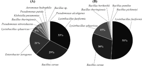
배양방법을 이용한 지렁이 장내미생물의 분리 및 동정 지렁이를 8주와 16주동안 사육한 후 장을 추출하여 BHI agar plate에 배양한 결과, 134개와 97개의 single colony를 각각 분리하였다. 8주동안 사육한 지렁이의 장내미생물을 동 정한 결과, Lysinibacillus fusiformis(33%), Bacillus cereus (29%), Enterobacter aerogenes(21%), L. sphaericus(5%), Pseudomonas nitroreducens(3%), B. thuringiensis(2%), Klebsiella pneumoniae(2%), P. putida(2%), Aeromonas hydrophila(1%), Bacillus sp.(1%), Pseudomonas alcaligenes (1%)로 확인되었고, 16주 사육 지렁이 장내미생물을 동정한 결과, L. fusiformis(51%), B. cereus(30%), L. sphaericus (15%), B. thuringiensis(1%), B. horikoshii(1%), B. pumilus (1%), B. pichinotyi(1%)로 확인되었다(Fig. 1).
지렁이 장에서는 분리한 미생물 중에서 Lysinibacillus와 Bacillus 속으로 동정되어 이들 미생물이 지렁이 장내에 우 점하고 있는 미생물임을 확인하였다. Lysinibacillus 속 균주 는 Ahmed 등[1]이 제안하여 Bacillus 속으로부터 재분류된 미생물로서 대부분의 Bacillus속 세균들은 세포벽 구성 성분 인 peptidoglycan type이 A1γ인 반면에 Lysinibacillus속 세 균들은 A4α type인 lysine(Lys)-aspartic acid(Asp)으로 구성 된 interpeptide bridge를 가지고 있어 Bacillus속으로부터 재
분류 되었으며, 표준 균주로는 L. boronitolerans, L. fusi- formis, L. sphaericus 그리고 L. parviboronicapiens[16, 22] 등이 알려져 있다. Kim 등[10]이 E. fetida의 장내 미생 물을 조사한 결과 B. thuringiensis, B. vallismortis 등 Bacillus 속 미생물이 지렁이 장내 우점미생물이라고 보고하 였으며, 이는 본 연구 결과와 유사하였다. Parthasarathi 등 [19]은 식양토와 가축분을 먹이원으로 사용하여 사육한 E.
fetida의 장내미생물을 분석한 결과, Klebsiella pneumonia와 Morganella morganii미생물이 우점하는 것으로 알려져 있으 나 이는 본 연구와 상이한 결과를 나타내었다. 또한, Lampito mauritii, Eudrius eugeniae, Eisenia fetida, Perionyx exca- vatus 각각 지렁이와 먹이원별 지렁이 장내미생물을 연구한 결과, 지렁이 장을 통과하면서 미생물의 개체수가 증가하는 것과 먹이원에는 존재하지만 지렁이 장과 배설물인 분변토 에서 확인되지 않은 P. aeruginosa, E. aerogenes, B. subtilis, 그리고 B. cereus 등의 미생물은 지렁이 장속에서 완전히 소 화된다고 보고하였다. 반면에, Byzov 등[2]과 Ihssen 등[9]
은 각각 E. fetida와 Aporrectodea caliginosa를 사용한 지렁 이 장내 미생물 분석에서 Aeromonas sp., Pseudomonas sp., 그리고 Bacillus sp. 등의 미생물이 존재한다고 보고하였 다. 이는 동일한 종의 지렁이도 먹이원 속에 포함된 유기물 의 종류와 각 유기물 분해에 관여하는 미생물의 종류가 다양 하기 때문에 지렁이 장내에 우점하는 미생물 군집이 먹이원 에 따라 차이가 있는 것으로 사료된다. 또한, 8주 사육 지렁 이 장내 미생물과 16주 사육 지렁이 장내 미생물의 배양 결 과에서 우점미생물인 L. fusiformis, B. cereus, E. aerogenes 뿐만 아니라 P. nitroreducens, K. pneumoniae가 지렁이의 사육시간이 길어지면서 분리되지 않는 것으로 보아 먹이원의 차이뿐만 아니라 지렁이 사육시간에 의해서도 지렁이 장내에 우점하는 미생물의 종류가 변화되어 지렁이 장내미생물의 군
Fig. 1. Identification and proportion of isolated microorganisms from the intestinal tract of earthworm. Dilutions of the intestinal tract of the earthworm were cultured on brain heart infusion agar plate at 30oC for 24 h after cultivation for 8 weeks (A) and 16 weeks (B).
The 16S rDNA sequences from the all isolated strains were sequenced by use of a PCR-based technique. Sequences were aligned with the GenBank reference sequences for 16S rDNA by using the Basic Local Alignment Search Tool (BLAST).
집이 다르게 나타나는 것으로 사료된다. Lysinibacillus속 세 균들도 Bacillus속 세균들과 마찬가지로 그람 양성, 포자를 형성하는 세균으로 Bacillus 속과 유사한 생화학적 특성을 가진다. 배양방법에서 분리한 세균들의 16S rDNA 분석 결 과 모든 미생물이 Lysinibacillus 혹은 Bacillus 속 세균들로 동정이 되었는데, Parle[17]은 이용 가능한 영양분의 증가, 습도 및 pH 등의 물리적 환경 변화 등으로 Bacillus 속이 지 렁이 장 후반부로 갈수록 포자의 germination이 증가하여 영 양세포의 수가 증가한다고 보고하였다. 따라서 지렁이 사육 기간 동안 포자화된 Bacillus 속 세균들이 지렁이 장을 통과 하면서 포자의 germination이 증가로 인해 분리한 지렁이 장 내 미생물에서 Bacillus와 Lysinibacillus 속 세균들의 비율이 높아진 것으로 사료된다.
DGGE 방법을 이용한 지렁이 장내 미생물의 동정 8주와 16주동안 사육한 지렁이로부터 장내미생물을 배양 하지 않고 직접 DNA를 추출하여 DGGE 전기영동한 결과, 8주 사육 지렁이에서는 10개의 band로 분리되었고 Borrelia burgdorferi, Enterobacter sp., Microbacterium sp., uncul- tured bacterium, Bacillus sp., Gamma proteobacterium, un- cultured soil bacterium, uncultured bacterium으로 확인하였 다. 16주 사육 지렁이에서는 12개의 band로 분리되었고 Photorhabdus luminescens, Enterobacter sp., gamma pro- teobacterium, uncultured Enterobacteriales bacterium, B.
thuringiensis, B. cereus, uncultured gamma proteobacterium, B. stearothermophilus, uncultured bacterium, uncultured Escherichia sp., Sporosarcina pasteurii로 확인하였다(Fig.
2, Table 1).
DGGE band의 intensity가 높다는 것은 개체군의 수가 많 다는 것을 의미하는데, 본 연구결과에서는 DGGE band의 intensity가 높은 Enterobacter sp.와 B. cereus 균주가 지렁 이 장내 우점미생물로 확인되었다. DGGE gel 상에서 band 의 intensity가 변화하는 것으로 보아 지렁이의 사육시간에 따라 우점하는 미생물도 변화하는 것으로 사료된다. 또한 배 양방법과 동일한 genus에 속하는 세균뿐만 아니라 배양방법 에서는 확인되지 않던 미생물인 B. burgdorferi, Microbac- terium sp., S. pasteurii, P. luminescens 등이 확인되었다.
Farnleitner 등[5]은 DGGE 방법은 적은 양의 DNA를 PCR 로 증폭하고 증폭된 DNA를 이용함으로써 종 다양성을 더 정밀하게 확인할 수 있는 방법이라고 보고하였고, Kim 등 [11]은 더 다양한 미생물뿐만 아니라 배양을 통해서는 표현 되지 않는 미생물까지도 확인할 수 있다고 보고하였다.
Knapp 등[13]도 DGGE 방법을 이용하여 먹이원의 종류에 따라 지렁이 장내미생물 군집을 조사하였는데, A. hydrophila, uncultured gamma proteobacterium, Mannheimia sp., Cel- lvibrio fulvus, Spiroplasma sp.가 먹이원의 종류별로 우점하 는 미생물임을 확인하였으며, 먹이원의 종류에 따라 우점하
는 미생물도 변화된다고 보고하였다.
Hong 등[7]은 DGGE 방법을 이용하여 지렁이 장내 미생 물과 효소생산 미생물의 지렁이에 대한 효과를 연구한 결과, 지렁이 장내에 Entomoplasma somnilux와 B. licheniformis 가 우점하는 것으로 확인하였고, 효소생산이 우수한 미생물 을 지렁이의 먹이원으로 섭취하게 하였을 때 지렁이 장내에 지속적으로 존재하는 반면 지렁이의 생육에도 효과가 있다 고 하였다. 또한 DGGE 방법을 이용하여 E. somnilux와 같 은 신규 미생물을 규명하였는데, DGGE 방법은 배양방법의 한계를 극복하여 지렁이 장내의 다양한 미생물 군집을 조사 Fig. 2. Denaturing gradient gel electrophoresis analysis of PCR-amplified 16S rDNA fragments from the intestinal tracts of earthworm. Lane (A), Intestinal tract of earthworm cultured for 8 weeks; Lane (B), Intestinal tract of earthworm cultured for 16 weeks. PCR amplification of the 16S rDNA sequences was per- formed with the primers 341F-GC and 518R for 40 cycles. DGGE anlaysis was performed between 40% and 80% denaturing gradi- ent.
할 수 있을 뿐만 아니라 신규 미생물을 규명하는데도 유용 한 방법이라고 하였다. 배양방법과 동일하게 Parthasarathi 등 [19]이 지렁이 장내에서 완전히 소화된다고 보고하였던 P.
aeruginosa, E. aerogenes, B. subtilis, 그리고 B. cereus가 비배양방법인 DGGE 방법에서도 확인이 되어 지렁이 장내 미생물의 군집과 우점하는 미생물의 종류가 달라진다는 근 거가 되는 것으로 생각된다. 이처럼 DGGE 방법은 미생물 군집을 조사하는데 있어 우점하는 미생물과 그렇지 않은 미생물을 구분할 수 있을 뿐만 아니라 신규 미생물을 확인 할 수 있는 유용한 방법으로 배양방법의 한계점을 보완하 고 미생물 군집의 다양성을 표현하는데 적합한 방법으로 사료된다.
Pyrosequencing방법을 이용한 지렁이 장내미생물의 동정 Pyrosequencing 방법으로 지렁이 장내미생물 군집을 분석 한 결과, 8주 사육 지렁이는 16,803개(average length = 448.6 bp)와 16주 사육 지렁이는 3,204개(average length = 437.1 bp)의 16S rDNA gene sequence를 획득하였다. 그리 고8주와 16주 지렁이 장내미생물 군집의 종 다양성(species richness)을 비교하기 위해 rarefaction curve를 나타내었다 (Fig. 3). Operational taxonomic unit(OTU)은 분류의 대상 이 되는 생물체의 개체 또는 군을 나타내고 일반적으로 종 을 말하는데, 8주와 16주 사육 지렁이 장내미생물 군집의 분
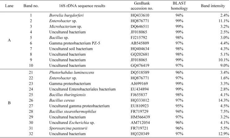
석 결과를 비교하였을 때, 두 그래프의 기울기가 유사한 것 으로 나타났다. Rarefaction curve는 그래프가 수렴하게 되 면 시료 수를 늘려 pyrosequencing을 해도 더 이상 개체군 이 발견되지 않는다는 것을 의미하는데, 두 개의 곡선 모두 그래프가 수렴하지 못해 미생물 군집 분석에 이용된 sequence의 수가 지렁이 장내에 포함된 모든 종을 확인하기 Table 1. Diversity of the microorganisms identified in the intestinal tract of earthworm by denaturing gradient gel electrophoresis analysis.
Lane Band no. 16S rDNA sequence results GenBank
accession no.
BLAST
homology Band intensity
A
1 Borrelia burgdorferi HQ433610 94% 2.4%
2 Enterobacter sp. HQ876771 99% 11.1%
3 Microbacterium sp. DQ646511 99% 3.2%
4 Uncultured bacterium JF018065 99% 2.5%
5 Bacillus sp. FJ215792 98% 3.0%
6 Gamma proteobacterium PZ-5 AB545809 97% 4.4%
7 Uncultured soil bacterium HQ404634 98% 4.3%
8 Uncultured bacterium GQ202681 98% 5.1%
9 Uncultured bacterium JF018065 99% 10.1%
10 Uncultured bacterium GQ476419 97% 9.0%
B
21 Photorhabdus luminescens DQ518589 96% 3.4%
22 Enterobacter sp. HQ876771 97% 1.6%
23 Gamma proteobacterium AJ699169 99% 3.3%
24 Uncultured Enterobacteriales bacterium EU434894 99% 2.8%
25 Bacillus thuringiensis FJ655837 98% 4.1%
26 Bacillus cereus HQ333012 97% 14.3%
27 Uncultured gamma proteobacterium EU810923 95% 4.5%
28 Bacillus stearothermophilus FR719729 94% 7.5%
29 Uncultured bacterium HM566439 97% 3.2%
30 Uncultured Escherichia sp. AM712054 96% 4.1%
31 Sporosarcina pasteurii FR719721 96% 5.5%
32 Uncultured bacterium HQ320349 97% 12.9%
Fig. 3. Rarefaction curves of OTUs defined by sequence varia- tions in the intestinal tract of earthworm. The X-axis shows the number of sequences in each sample, while the Y-axis shows the numbers of operational taxonomic units (OTU) encountered.
에는 부족한 것으로 사료된다.
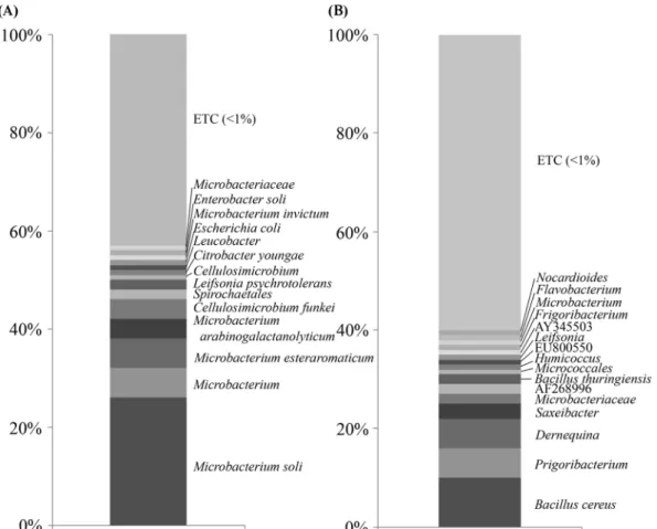
Genus 수준에서 비교한 결과, 8주와 16주 사육 지렁이의 장 내미생물 군집을 비교한 결과, 8주에서 16주까지 사육시간에 따라 Microbacterium이 46%에서 2%로, Cellulosimicrobium 이 5%에서 1%로 감소하였다. 반면에, Bacillus(1%에서 13%
로), Frigoribacterium(1%에서 8%로), 그리고 Demequina (1%에서 6%로)가 사육시간에 따라 개체수가 증가하여 지렁 이 장내 우점미생물은 사육시간에 따라 변화되는 것으로 사 료된다. Species 수준에서 비교한 결과, 8주 사육 지렁이는 Microbacterium soli가 26%로 우점하는 미생물로 확인되었 고, 그 뒤를 이어 Microbacterium_uc 6%, Microbacterium esteraromaticum 6%, Microbacterium arabinogalactanoly- ticum 4%, Cellulosimicrobium funkei 4%, Spirochaetales 2%, Leifsonia psychrotolerans 2%, Cellulosimicrobium 1%, Citrobacter 1%, Leucobacter 1%, Escherichia coli 1%, Microbacterium invictum 1%, Enterobacter soli 1%, Micro- bacteriaceae 1%, 그밖에 1% 미만의 미생물은 전체 중 43%
로 확인되었다. 16주 사육 지렁이는 Bacillus cereus가 10%
로 우점하는 미생물로 확인되었고, 그 뒤를 이어 Frigori-
bacterium 6%, Demequina 6%, Saxeibacter 3%, Micro- bacteriaceae 2%, AF268996 2%, Bacillus thuringiensis 2%, Micrococcales 1%, Humicoccus 1%, EU800550 1%, Leifsonia 1%, AY345503 1%, Frigoribacterium 1%, Micro- bacterium 1%, Flavobacterium 1%, Nocardioides 1%, 그 밖에 1% 미만의 미생물은 전체 중 60%로 확인되었다(Fig.
4).
Pyrosequencing의 분석결과, 지렁이 장내미생물의 전체 군 집 중에서 1% 미만인 균주들이 차지하는 비율이 8주와 16 주 사육 지렁이 장내미생물에서 각각 43%와 60%로 높게 나타났는데, 이는 분석하고자 하는 sequence의 수(8주:
16,803개, 16주: 3,204개)가 많기 때문에 species 수준에서 분석할 경우 1% 미만 균주들의 비율이 높아지는 것으로 생 각되며, 8주에서 16주로 지렁이 사육시간이 경과함에 따라 Bacillus sp. 뿐만 아니라 Frigoribacterium sp., Demequina sp. 등 8주 사육 지렁이에서 낮은 비율의 미생물들이 16주 로 갈수록 점차 증가하는 것을 확인할 수 있었다.
Fig. 4. Composition of bacterial diversity in the intestinal tract of earthworm by pyrosequencing analysis. (A), Intestinal tract of earthworm cultured for 8 weeks; (B), Intestinal tract of earthworm cultured for 16 weeks. Relative abundance of operational taxonomic units classified to each taxon was determined with partial sequences of bacterial 16S rDNA genes from the intestinal tract of earthworm by EzTaxon.
DGGE와 pyrosequencing 방법에 의한 미생물 군집 조사 비교
배양방법에 의한 미생물의 분포조사는 자연계에 존재하는 미생물의 생육환경을 재현할 수 없기 때문에 전체 미생물의 배양이 불가능하고, 시료를 희석하는 과정에서 소수로 존재 하는 미생물은 배양되지 못하기 때문에 전체적인 미생물을 분리할 수 없다. 이러한 단점을 보완하기 위해 시료로부터 DNA를 추출하여 16S rDNA gene sequence를 증폭한 후 이를 분석하는 분자생물학적 방법은 배양 단계를 거치지 않 고 PCR 산물을 이용하여 sequence를 분석하기 때문에 군집 내 미생물의 다양성과 군집 내 우점종 확인이 가능하다[21].
DGGE 방법의 경우 universal primer를 사용하여 16S rDNA gene을 증폭한 후 chemical denaturant인 urea와 formamide를 포함한 denaturing gradient gel에서 분리되는 것을 이용하는 것으로, 배양을 하지 않고 PCR 증폭을 함으 로써 시료 속에 소수로 존재하는 미생물도 검출이 가능하다 는 장점이 있다. 하지만 PCR을 통해 DNA를 증폭을 하여도 상대적으로 개체수가 많은 종들이 존재하고, 전기영동 후 염 색시 적은 양의 DNA fragments는 염색이 되지 않아 검출 되지 않을 수 있으며 염색이 되어도 육안으로 확인이 되지 않는 문제점이 발생할 수 있다. 또한, DGGE의 특성상 PCR 을 통해 증폭된 16S rDNA fragments가 500bp이상이 되면 DNA band의 분리가 잘 이루어지지 않아 DGGE 수행 시 어 려움이 따르고, 이로 인해 증폭된 16S rDNA fragment의 길 이가 제한이 되어 16S rDNA 분석시 정확한 sequencing이 되지 않을 수 있다[9].
Pyrosequencing 방법은 DGGE와 마찬가지로 PCR 증폭한 후 sequence 분석을 하게 되는데, DGGE와는 달리 denatur- ing gradient gel을 이용하지 않고 DNA fragments로부터 직 접적인 sequencing을 함으로써 더 다양한 미생물의 확인이 가능하다. 또한, 16S rDNA gene은 V1-V9로 구분이 되고 각 부분은 50~100 bp 정도의 길이가 되며 이 부위의 sequence를 분석함으로써 미생물의 동정을 할 수 있는데 pyrosequencing은 평균적으로 350 bp 이상의 sequence를 얻 을 수 있어 분석 가능한 sequence의 길이가 길어져 다양한 부위의 분석을 할 수 있기 때문에 종 다양성을 표현하는데 효율적이다[20].
DGGE와 pyrosequencing의 sequencing 결과 genus 단위 혹은 그 보다 더 큰 분류군으로 동정되는 경우도 있는데, 이 는 두 방법 모두 sequencing이 가능한 염기 서열의 길이가 제한적이기 때문에 생겨나는 결과로 보여지며, 앞으로 sequence 길이의 제한점을 보완한다면, species 수준의 감별 력이 더욱 높아져 미생물 군집조사가 더욱 정확해 질 것으 로 생각된다. 또한, DGGE와 pyrosequencing와 같은 비배양 방법은 16S rDNA gene을 이용하여 미생물 군집을 분석하 므로 미생물의 배양이 불가능하다는 단점이 있으며, se- quencing을 위한 database는 이미 알려진 16S rDNA gene
sequence의 database를 이용하여 동정하기 때문에 분자생물 학적 방법도 미생물 군집을 분석하는데 있어서 제한점을 가 지게 된다 [22]. 배양한 미생물을 이용하여 생화학적 특성 조사, 유용물질의 생산 등 직접적인 이용이 가능한 배양방 법과 16S rDNA gene을 이용하여 다양한 미생물의 군집을 조사할 수 있는 비배양 방법은 독립적이지 않고 병행되어야 좀 더 정확한 군집조사가 가능할 것으로 사료된다.
요 약
미생물과의 상호작용을 통하여 토양의 특성을 변화시킬 수 있는 지렁이 Eisenia fetida의 장내미생물 군집을 조사하 기 위하여, 배양방법과 비배양방법인 DGGE와 pyrose- quencing을 이용하여 8주와 16주 사육 지렁이의 장내미생물 군집을 분석하였다. 배양방법에서는 L. fusiformis(51%), B.
cereus(30%), E. aerogenes(21%), 그리고 L. sphaericus (15%) 등이 우점미생물로 확인되었다. DGGE 분석에서는 B.
cereus(15.1%), Enterobacter sp.(13.6%), uncultured bacterium (13.1%), 그리고 B. stearothermophilus(7.8%)가 우점미생물 로 확인되었다. Pyrosequencing 분석에서는 Microbacterium soli(26%), B. cereus(10%), M. esteraromaticum(6%), 그리 고 Frigoribacterium sp.(6%)가 우점미생물로 확인되었다. 그 외에도 Aeromonas sp., Pseudomonas sp., Borrelia sp., Cellulosimicrobium sp., Klebsiella sp., and Leifsonia sp.
등의 미생물도 확인이 되었으며, 비배양 방법을 이용한 장내 미생물 군집 조사는 배양이 불가능한 미생물을 확인할 수 있 을 뿐만 아니라 더 다양한 미생물 군집도 확인할 수 있었다.
Acknowledgement
This research was supported by Basic Science Research Program through the National Research Foundation of Korea (NRF) funded by the Ministry of Education, Science and Technology (2010-0006708).
REFERENCES
1. Ahmed, I., A. Yokota, A. Yamazoe, and T. Fujiwara. 2007.
Proposal of Lysinibacillus boronitolerans gen. nov. sp. nov., and transfer of Bacillus fusiformis to Lysinibacillus fusi- formis comb. nov. and Bacillus sphaericus to Lysinibacillus sphaericus comb. nov. Int. J. Syst. Evol. Microbiol. 57: 1117- 1125.
2. Byzov, B. A., T. Nechitailo, B. K. Bumazhkin, A. V. Kurakov, P. N. Golyshin, and D. G. Zvyagintsev. 2009. Culturable microorganisms from the digestive tract of earthworm.
Mikrobiolgiia. 78: 404-413.
3. Droege, M. and H. Brendon. 2008. The genome sequencer
FLXTMSystem-longer reads, more applications, straight forward bioinformatics and more complete data sets. J. Bio- technol. 136: 3-10.
4. Edwards, C. A. and K. E. Fletcher. 1988. Interactions between earthworms and microorganisms in organic-matter breakdown. Agric. Ecosyst. Environ. 24: 235-247.
5. Farnleitner, A. H., F. Zibuschka, M. M. Burtscher, G. Lindner, G. R. L. Mach. 2004. Eubacterial 16S-rDNA amplicon pro- filing: a rapid technique for comparsion and differentiation of heterotrophic plate count communities from drinking water. Int. J. Food Microbiol. 92: 333-375.
6. Fisher, S. G. and L. S. Lerman. 1983. DNA fragments differ- ing by single base-pair substitutions are separated in dena- turing gradient gels: correspondence with melting theory.
Proc. Natl. Acad. Sci. USA 80: 1579-1583.
7. Hong, S. W., J. S. Lee, and K. S. Chung. 2011. Effect of enzyme producing microorganisms on the biomass of epi- geic earthworms (Eisenia fetida) in vermicompost. Biore- sour. Technol. 102: 6344-6347.
8. Ihssen, J., M. A. Horn, C. Matthies, A. Gössner, A. Schramm, and H. L. Drake. 2003. N2O-Producing microorganisms in the gut of the earthworm Aporrectodea caliginosa are indi- cative of ingested soil bacteria. Appl. Environ. Microbiol. 69:
1655-1661.
9. Kathrin, F., D. Hanhn, W. Honerlage, and J. Zeyer. 1997.
Effect of passage through the gut of the earthworm Lum- bricus terrestris L. on Bacillus megaterium studied by whole cell hybridization. Soil Biol. Biochem. 29: 1149-1152.
10. Kim, H. J., K. H. Shin, C. J. Cha, and H. G. Hur. 2004. An- alysis of aerobic and culturable bacterial community struc- tures in earthworm (Eisenia fetida) intestine. Agric. Chem.
Biotechnol. 47: 137-142.
11. Kim, M. N. and H. J. Bang. 2006. Comparison of culture- dependent and DGGE based methods the analysis of marine bacterial community. Kor. J. Environ. Biol. 24: 307-313.
12. Kim, T. S., H. S. Kim, S. D. Kwon, and H. D. Park. 2010.
Analysis of bacterial community composition in waste water treatment bioreactors using 16 rRNA gene-based pyrose- quencing. Kor. J. Microbiol. 46: 352-358.
13. Knapp, B. A., J. Seeber, S. M. Podmirseg, E. Meyer, and H.
Insam. 2008. Application of denaturing gradient gel electro- phoresis for analysing the gut microflora of Lumbricus rubellus Hoffmeister under different feeding conditions.
Bull. Entomol. Res. 98: 271-279.
14. Kwon, K. R. and J. C. Seo. 2004. Genetical identification of Korean wild ginseng and american wild ginseng by using pyrosequencing method. Kor. J. Herbology 19: 45-50.
15. Miller, K. M, T. J. Ming, A. D. Schulze, and R. E. Withler.
1999. Denaturing gradient gel electrophoresis (DGGE): a rapid and sensitive technique to screen nucleotide sequence variation in populations. Biotechniques 27: 1016-1018.
16. Miwa, H., I. Ahmed, A. Yokota, and T. Fujiwara. 2009.
Lysinibacillus parviboronicapiens sp. nov., a low-boron- containing bacterium isolated from soil. Int. J. Syst. Evol.
Microbiol. 59: 1427-1432.
17. Parle, J. N. 1963. Microorganisms in the intestines of ear- thworms. J. Gen. Microbiol. 31: 1-11.
18. Park, J. S., C. J. Sim, and K. D. An. 2009. Community struc- ture of bacteria associated with two marine sponges from Jeju Island based on 16S rDNA-DGGE profiles. Kor. J.
Microbiol. 45: 170-176.
19. Parthasarathi, K., L. S. Ranganathan, V. Anandi, and J.
Zeyer. 2007. Diversity of microflora in the gut and casts of tropical composting earthworms reared on different sub- strates. J. Environ. Biol. 28: 87-97.
20. Petrosino, J. F., S. Highlander, R. A. Luna, A. G. Richard, and J. Versalovic. 2009. Metagenomic pyrosequencing and microbial identification. Clin. Chem. 55: 856-866.
21. Rondon, M. R., R. M. Goodman, and J. Handelsman. 1999.
The Earth's bounty: assessing and accessing soil microbial diversity. Trends Biotechnol. 17: 403-409.
22. Schleifer, K. H. and O. Kandler. 1972. Peptidoglycan types of bacterial cell walls and their taxonomic implications.
Bacteriol. Rev. 36: 407-477.
23. Shin, K. H., H. Yi, J. S. Chun, C. J. Cha, I. S. Kim, and H.
G. Hur. 2004. Analysis of the anaerobic bacterial community in the Earthworm (Eisenia fetida) intestine. Agric. Chem.
Biotechnol. 47: 147-152.
24. Song, E. Y., J. K. Noh, Y. M. Yoon, Y. S. Choi, S. S. Park, E. K. Ra, and K. S. Han. 2006. ABO genotyping by pyrose- quencing analysis. Korean J. Blood Transfusion. 17: 106- 115.
25. Walter, J., G. W. Tannock, A. Tilsala-Timisjarvi, S. Rodtong, D. M. Loach, K. Munro, and T. Alatossava. 2000. Detection and identification of gastrointestinal Lactobacillus species by using denaturing gradient gel electrophoresis and species specific PCR primers. Appl. Environ. Microbiol. 66: 297- 303.