Copyright ⓒ 2010, The Microbiological Society of Korea
RFLP와 DGGE에 따른 해면 Spirastrella abata 공생세균의 다양성 비교
정은지·임춘수·박진숙*
한남대학교 생명공학과
A Comparison of Bacterial Diversity Associated with the Sponge Spirastrella abata Depending on RFLP and DGGE
Eun-Ji Jeong, Choon-Soo Im, and Jin-sook Park*
Department of Biotechnology, Hannam University, Daejeon 305-811, Republic of Korea (Received November 29, 2010/Accepted December 29, 2010)
Culture-dependent RFLP and culture-independent DGGE were employed to investigate the bacterial community associated with the marine sponge Spirastrella abata. A total of 164 bacterial strains associated with the sponge were cultivated using Zobell and Natural sea salt media. PCR amplicons of the 16S rDNA from the bacterial strains were digested with the restriction enzymes HaeIII and MspI, and then assigned into different groups according to their restriction patterns. The 16S rDNA sequences derived from RFLP patterns showed more than 95% similarities compared with known bacterial species, and the isolates belonged to four phyla, Proteobacteria (Alphaproteobacteria, Gammaproteobacteria), Actinobacteria, Firmicutes, and Bacteriodetes, of which Alphaproteobacteria was dominant. DGGE fingerprinting of 16S rDNAs amplified from the sponge- derived total gDNA showed five major DGGE bands, and their sequences showed more than 96% similarities compared with available sequences. The sequences derived from DGGE bands revealed high similarity with the uncultured bacterial clones. DGGE revealed that bacterial community consisted of four phyla, including Proteobacteria (Alphaproteobacteria, Gammaproteobacteria), Actinobacteria, Spirochetes, and Chloroflexi. Alphaproteobacteria, Gammaproteobacteria, and Actinobacteria were commonly found in bacteria associated with S. abata by both RFLP and DGGE methods; however, overall bacterial community in the sponge differed depending on the analysis methods.
Keywords: DGGE fingerprinting, RFLP, Spirastrella abata, sponge-associated bacteria
해면은 해양에서 우점적으로 존재하는 주요 무척추동물 그 룹 중의 하나로 해양을 비롯한 담수 환경에도 서식할 수 있으며 약 7,000-15,000종이 전세계적으로 분포하고 있는 것으로 알려 져 있다(20, 28). 계통학적으로는 Calcarea, Hexactinellida, Demospongiae 3개의 강(class)으로 나뉘어진다(8, 26). 해면 조직 내 외에 세균, 고세균, 시아노박테리아, 녹조류, 홍조류, 규조류 등 많은 미생물을 포함하며 특히 세균을 다량 포함하는 것으로 알려져 있다(10). 해면은 해양유래의 생리활성 물질의 풍부한 원천으로써, 해양에서 생산되는 천연물 중 40% 이상이 해면에서 생산되는 것으로 보고되고 있다(7). 최근 해면에서 생산되는 여러 가지 생리활성 물질들이 해면에 공생하는 공생
* For correspondence. E-mail: [email protected]; Tel: +82-42-629-8771;
Fax: +82-42-629-8769
세균에 의해 생산되는 것으로 보고되면서 해면 공생세균에 대 한 연구가 급격히 증가하고 있다. Demospongiae 강(class)에 속하는 Spirastrella abata는 우리나라의 제주도를 비롯하여 국 내 해양 환경에 풍부하게 분포하는 해양 해면으로, 특이적이고 다양한 생리활성 물질을 생산한다(1, 2). 이 해면에서 생산되는 생리활성물질로는 Lysophosphatidylcholine과 Sphingosine 4- sulfates 등이 있으며 Lysophosphatidylcholine의 경우 콜레스 테롤 생합성에 대하여 억제 작용을 갖는 것으로 보고되어있다 (2, 23).
일반적으로 자연계의 미생물 중 실제 배양 가능한 종류는 전체 자연계 미생물 군집의 1% 이하인 것으로 알려져 있다.
따라서 전체 해면 공생세균의 다양성 및 군집구조의 확인을 위 해서 배양에 의하지 않고 직접 DNA 추출에 의한 다양한 분자 적 방법들이 적용되고 있다(11, 13, 25). 분자적 분석 방법 중
PCR- DGGE (denaturing gradient gel electrophoresis)의 경우 실험 방법이 비교적 간단하고 주요 세균군집을 알아내는데 효 율적이어서 미생물 다양성을 파악하는데 흔히 이용되고 있다 (15, 20). DGGE는 특정 유전자 부위를 증폭한 PCR 산물을 Polyacrylamide gel상에서 전기 영동하여 확인하는 방법으로 이론적으로는 하나 이상의 염기서열이 다를 경우 서로 다른 band로 구분되어 자연환경의 미생물을 배양하지 않아도 군집 구조를 쉽게 파악할 수 있는 기술이다(9). 반면, 새로운 미생물 종의 자원화와 미생물에 의한 천연물 생산의 산업화 등을 위해 서는 배양이 기초가 되어야 하므로 배양 가능한 해면 공생세균 의 다양성에 관한 연구 또한 중요하다. 배양에 기초한 RFLP (Restriction Fragment Length Polymorphism)는 특정 유전자 부위를 인식하는 제한효소를 처리하여 절단된 유전자의 패턴 을 분석하는 방법으로 신속하며 간편한 방법으로 배양 가능한 해면 공생세균의 다양성을 용이하게 파악할 수 있는 기술이다.
해면의 공생세균 다양성은 종에 따른 차이는 물론, 동일 종 의 해면일지라도 서식하는 장소 혹은 채집시기에 따라 달라지 는 경향이 있다(3, 18). 따라서 서로 다른 시기에 혹은 서로 다 른 장소에서 채집하였을 경우 동일한 종류의 해면 일지라도 서 로 다른 방법을 적용하였을 경우 분석방법 간의 차이를 규명하 기 어렵다. 본 연구에서는 해면의 서식환경의 차이가 아닌 분 석 방법에 따른 해면공생세균의 다양성의 차이를 파악하고자 동일한 장소에서 동시에 채집한 동종 해면을 대상으로 분석 방 법을 달리하였을 경우, 공생세균 군집구조의 차이를 알아보고 자 하였다. 제주도 무릉 연안에서 2010년 5월에 채집한 해양 해면 Spirastrella abata를 대상으로 해면 공생 세균의 주요 군 집구조를 배양법에 기초한 PCR-RFLP와 비배양법에 기초한 16S rDNA-DGGE (denaturing gradient gel electrophoresis)를 이용하여 다양성을 조사하고 결과를 비교 · 분석하였다.
재료 및 방법
해면시료의 채집
해양 해면 Spirastrella abata에 공생하는 세균의 다양성을 조사하기 위하여 제주도 무릉연안에서 2010년 5월 15일에 스 쿠버 다이빙을 이용하여 약 15 m 깊이의 바다에서 해면을 채 집하였다. 채집한 해면은 멸균된 인공해수(ASW)로 3회 세척 후 -20°C에서 동결하여 운반 후 실험에 사용하였다.
해면 공생세균의 배양 및 분리
해면 조각을 멸균된 인공해수(ASW)로 3회 세척 후, 해면의 안쪽을 1 cm3 크기로 잘라 인공해수 3 ml 넣어 균질화시킨 다 음 10분간 초음파 처리하였다. 각각 10-4까지 순차 희석한 후 ZoBell (ZB) 배지(19)와 천일염 배지(Natural sea salt 10 g, Yeast extract 1 g, Agar 15 g, 1 L DW)에 100 μl씩 도말하여 25°C에서 7일간 배양하였다.
ZB 배지에서 169개, 천일염 배지에서 137개의 균주를 분리 하였다. 분리된 균주들의 순수 분리를 위하여 동일한 배지에서 계대 배양하여 단일 콜로니를 얻었다. ZB 배지와 천일염 배지
를 이용하여 분리한 균주 중 164균주를 무작위로 선별하여 16S rDNA의 RFLP분석을 수행하였다.
DNA 추출 및 16S rDNA의 PCR증폭
염색체 DNA는 분리된 세균 균주의 각 colony로부터 gDNA Extraction kit (Solgent, USA)를 사용하여 분리하였으며 분리 된 DNA는 PCR 반응의 주형으로 사용하였다. 16S rDNA의 증폭에는 27f (5′-AGA GTT TGA TCC TGG CTC AG-3′)와 1492r (5′-TAC GGY TAC CTT GTT ACG AC-3′)의 primer 쌍을 사용하였다. PCR 반응 혼합물의 조성은 5 μl 10× reaction buffer, 1 μl의 10 mM dNTPs, 5 unit/μl Taq polymerase (Solgent, Korea), 각각의 primer 10 pmol, 그리고 100 ng의 시료 DNA를 1 μl 첨가하여 최종 부피 50 μl이 되도록 하여 PCR 반응을 수행하였다. GeneAmp PCR system 2700 thermal cycler (Applied Biosystems, Version 2.0, USA)를 이용하여 94°C에서 3분간 초기 변성시킨 후, 94°C에서 40초간 변성, 55°C에서 40초간 냉각, 72°C에서 1분간 신장, 이 과정을 30 cycle 반복 수행한 후 최종적으로 72°C에서 10분간 신장시켰 다. 증폭된 DNA의 확인을 위해서 PCR 반응액 3 μl를 취하여 1% agarose gel (Bio-Rad, USA)을 이용하여 Mupid-ex (ADVANCE, Japan)로 100 V, 25 분간 1× TAE buffer (40 mM Tris-acetate, 1 mM EDTA, pH 8.0)에서 전기영동 하였 다. 전기영동 후, EtBr (ethidium bromide, 50 ng/ml)에 10 분 간 염색하여 Gel Logic 200 (Kodak, USA)을 이용하여 UV하 에서 약 1.5 kb 단편을 확인하였다.
RFLP 분석
PCR 산물의 RFLP 분석을 위해 2종의 제한효소 HaeIII (TaKaRa, Japan)와 MspI (TaKaRa)을 사용하였다. 증폭된 1.5 kb의 DNA 단편에 각각의 제한 효소를 첨가하여 37°C에서 4 시간 반응시켰다. 반응물은 3% agarose gel (Bio-Rad, USA)을 사용하여 1× TAE buffer (40 mM Tris-acetate, 2 mM EDTA, pH 8.0)로 100 V, 30 분간 전기영동한 후 EtBr (50 ng/ml)로 염색하여 Gel Logic 200 (Kodak, USA)을 이용하여 UV 하에서 관찰하여 각 균주의 밴드유형을 확인하였다.
16S rDNA-RFLP fingerprinting type에 따라 각각의 RFLP type 별로 1-2개의 분리 균주들을 선택하여 총 46균주의 부분 염기 서열 (500 bp 이상)을 분석하였다. 증폭된 PCR 산물은 High Pure PCR Product Purification kit (Roche, USA)를 이용 하여 정제하였으며, 27f primer로 ABI PRISM 3100 automated sequencer (PE Applied Biosystems, USA)를 이용하여 염기서 열을 결정하였다.
Total genomic DNA 추출
채집된 해면은 -70°C에서 동결 후 동결건조기에서 -50°C, 0.033 M bar기압으로 14시간 건조하였다. 동결건조된 해면은 멸균된 막자 사발에 넣고 액체질소를 부은 후 분쇄하였으며 G-spin™ Genomic DNA Extraction kit (Intron, Korea)를 이 용하여 추출한 후 DGGE를 위한 PCR 반응의 주형으로 사용
하였다.
16S rDNA의 DGGE-PCR 증폭
GC clamp가 추가된 341f (5′-CGC CCG CCG CGC CCC GCG CCC GGC CCG CCG CCC CCG CCC GCC TAC GGG AGG CAG CAG-3′)와 518r (5′-ATT ACC GCG GCT GCT GG-3′)의 primer쌍을 이용하여 16S rRNA gene의 V3 영 역을 증폭하였다. PCR 반응 혼합물의 조성은 16S rDNA의 PCR 증폭의 경우와 동일하게 하였으며 PCR 조건은 94°C에서 40초 간 변성, 65°C (1 cycle 당 0.5°C touch down)에서 40초간 냉 각, 72°C에서 1분간 신장, 이 과정을 30 cycle 반복 수행한 후 최종적으로 72°C에서 10분간 신장시켰다. 증폭된 DNA의 확 인을 위해 PCR 반응액 2 μl를 취하여 2% agarose gel (Bio-Rad, USA)을 이용하여 Mupid-ex (ADVANCE, Japan)로 100 V에서 25분간 1× TAE buffer (40 mM Tris, 20 mM acetic acid, 50 mM EDTA, pH 8.0)에서 전기 영동하였다. 전 기 영동 후 Ethidium Bromide (50 ng/ml)로 염색하여 Gel Logic 200 (Kodak, USA)을 이용, 자외선 조사로 확인하였다.
증폭된 DNA의 크기를 확인하기 위한 marker로는 100 bp ladder (Intron, Korea)를 사용하였다. GeneAmp PCR system 2700 thermal cycler (Applied Biosystems, version 2.0, USA) 를 이용하였다.
DGGE 전기영동
DGGE는 Bio-Rad Dcode system (Bio-Rad)을 이용하여 수 행하였다(18). 증폭된 PCR 산물은 bis acrylamide (Bio-Rad)를 포함한 10% polyacrylamide를 이용하여 30%에서 70%의 농 도 구배 조성(7 M urea, 40% formamide)으로 전기영동을 수 행하였다. Polyacrylamide gel은 크기 20×13 (W×H cm), 두 께 1 mm로 작성하여 PCR 시료는 2× loading dye와 혼합하 여 40 μl의 시료를 loading하였다. 1× TAE buffer를 사용하여 60°C, 30 V에서 1시간 안정화시킨 후 60 V로 전압을 올려 15 시간 전기 영동을 수행하였다. 전기영동 후 DGGE gel은 Ethidium bromide (50 ng/ml)로 1시간 염색 후 Gel Logic 200 (Kodak, USA)을 이용, 자외선 조사로 확인하였다.
DGGE band의 동정
DGGE gel 상에서 분리된 band 중, 각 band를 gel에서 잘라 내어 Gel Extraction kit (QIAGEN, Germany)을 이용하여 DNA를 회수하였다. 염기서열 분석을 위하여는 band로부터 회 수된 DNA를 주형으로 사용하였다. Primer는 341f와 518r을 이용하여 위와 동일한 조건으로 PCR을 수행하였다. PCR 증폭 산물은 Wizard SV Gel and PCR Clean-Up System (Promega, USA)을 이용하여 정제한 후 염기서열 분석(Macrogen, Korea) 을 의뢰하였다.
계통학적 분석 및 계통수 작성
RFLP type의 패턴 분석을 위해 FPQuestTM (Bio-Rad, Bel- gium) software를 이용하였으며 Similarity coefficient는 Dice
방법에 의해 구하고 Dendrogram은 Neighbor-joining에 의해 작성하였다(3).
RFLP 및 DGGE 결과로 얻어진 염기서열은 NCBI (the National Center for Biotechnology Information)에 등록된 염 기서열을 대상으로 Blast search를 수행하였다. 각 염기서열의 alignment는 CLUSTAL W (27)를 이용하여 정렬하였고 Molecular Evolutionary Genetics Analysis (MEGA) software version 4.0 (24)을 이용하여 neighbor-joining 방법에 의해 각각의 계통수 를 작성하였다. 1,000회 반복 bootstrap 분석에 의해 계통수를 확인하였다.
결과
해면 공생세균의 분리 및 RFLP 분석
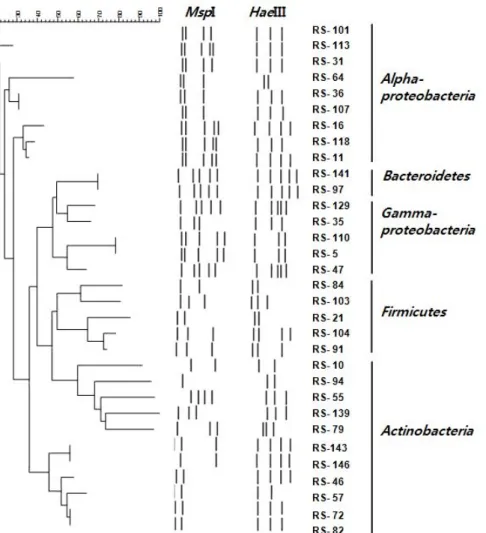
해면 S. abata의 배양 가능한 공생세균 군집구조를 16S rDNA의 PCR-RFLP 방법에 의해 조사하기 위하여 ZoBell 배 지와 천일염 배지를 이용하여 총 306균주를 분리하고 분리된 균주 중 무작위로 선별된 164균주에 대하여 2종류의 제한 효 소를 이용하여 16S rDNA의 RFLP type을 분석하였다. RFLP type은 각각 HaeIII를 이용한 경우 21개, MspI의 경우 27개의 type이 관찰되었다. 두 효소를 이용한 RFLP type을 조합하여 총 30개의 서로 다른 RFLP type이 구분되었다(Fig. 1 and Table 1).
DGGE band 양상
비배양법에 기초한 16S rDNA의 PCR-DGGE를 수행하여 S. abata의 해면 공생세균의 계통학적 다양성을 알아보았다.
해면 시료로부터 추출한 genomic DNA를 주형으로 16S rDNA gene의 V3영역을 증폭하여 194 bp의 예상된 크기의 PCR 산 물을 얻었다. S. abata의 DGGE band pattern 분석 결과, 총 5 개의 band를 확인할 수 있었으며, DGGE는 2회 이상 수행하여 재현성이 있음을 확인하였다(Fig. 2). 이 해면 종의 DGGE band가 나타내는 세균 다양성을 파악하기 위하여 16S rDNA 의 부분 염기서열을 분석하였다.
염기서열 분석 및 계통학적 분석
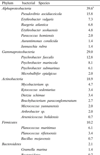
RFLP type의 염기서열 분석: RFLP 분석에 의해 30개의 type으로 나뉜, S. abata에서 분리된 세균들에 대해 각 RFLP type 별로 1-2개의 분리 균주를 선별하여 부분 염기서열을 분 석하였다. 염기서열이 분석된 총 46개의 분리 균주는 모두 기 존에 보고된 세균 종과 95% 이상의 유사도를 나타내었다. 염 기서열이 분석된 30개의 RFLP type은 28개의 세균 종을 나타 내었으며 이 결과를 근거로 처음 RFLP 분석에 쓰여진 164균 주들을 분석하고 이들의 분석 결과에 근거하여 군집의 차이를 분석하였다(Table 2 and Fig. 3). Alphaproteobacteria 39.6%, Gammaproteobacteria 29%, Actinobacteria 18.9%, Firmicutes 10.2%, Bacteroidetes 2.1%를 나타내었다. 가장 많은 분포를 보인 세균 종은 Proteobacteria 문에 속하는 Pseudovibrio 및 Psychrobacter이었다.
Fig. 1. Dendrogram showing the relationship among bacterial isolates based on the 16S rDNA-RFLP profiles.
Fig. 2. DGGE banding patterns of amplified 16S rDNA obtained from Spirastrella abata (DS); DS1 and DS2 indicates the DGGE profiles from independent experiments using the same sponge sample.
DGGE band의 염기서열 분석: 세균군집의 다양성을 분석 하기 위해 DGGE band에서 DNA를 추출하여 341f와 518r을 이용하여 재증폭하여 염기서열을 분석하였다. DGGE band의 염기서열을 분석한 결과, S. abata에서 5개의 band에 대한 염 기서열을 확인할 수 있었으며, 이 염기서열을 이용하여 알려진 염기서열들과 상동성을 비교한 결과는 Table 3과 같다.
DGGE band로부터 결정된 서열들은 모두 알려진 서열들과 96%에서 100%의 상동성을 나타내었으며 모두 배양되지 않은 세균(uncultured bacteria)인 것으로 나타났다.
DGGE band DS-1은 uncultured alpha proteobacterium (EU702822)과 96%의 상동성을 나타내었으며 DS-2는 Chloro- flexus sp. (FJ481347)와 100%의 상동성을 나타내었다. DS-3 은 Uncultured Actinobacterium (EU819009)과 100%의 상동 성을 보였으며 DS-4은 Uncultured gamma proteobacterium (EU816809)과 100%의 상동성을 보였다. DS-5는 Uncultred Spirochetes (FJ529354)와 99%의 상동성을 나타내었다.
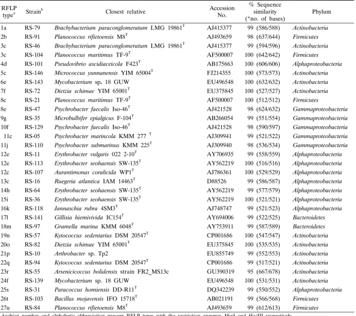
Table 1. RFLP fingerprinting types and sequence similarities of 16S rDNA from bacterial strains isolated from the marine sponge Spirastrella abata (RS)
RFLP
typea Strainb Closest relative Accession
No.
% Sequence similarity
(*no. of bases) Phylum 1a RS-79 Brachybacterium paraconglomeratum LMG 19861T AJ415377 99 (586/588) Actinobacteria 2b RS-91 Planococcus rifietoensis M8T AJ493659 98 (637/644) Firmicutes 3c RS-46 Brachybacterium paraconglomeratum LMG 19861T AJ415377 99 (594/596) Actinobacteria 3c RS-104 Planococcus maritimus TF-9T AF500007 100 (642/642) Firmicutes 4d RS-101 Pseudovibrio ascidiaceicola F423T AB175663 100 (606/606) Alphaproteobacteria 5c RS-146 Micrococcus yunnanensis YIM 65004T FJ214355 100 (573/573) Actinobacteria 6e RS-143 Mycobacterium sp. 18 GUW EU496548 100 (632/632) Actinobacteria 7f RS-72 Dietzia schimae YIM 65001T EU375845 100 (527/527) Actinobacteria 8c RS-21 Planococcus maritimus TF-9T AF500007 100 (512/512) Firmicutes
8e RS-47 Psychrobacter faecalis Iso-46T AJ421528 98 (624/632) Gammaproteobacteria 9g RS-35 Microbulbifer epialgicus F-104T AB266054 99 (551/554) Gammaproteobacteria 10f RS-129 Psychrobacter faecalis Iso-46T AJ421528 98 (590/597) Gammaproteobacteria 11c RS-05 Psychrobacter marincola KMM 277 T AJ309941 99 (521/522) Gammaproteobacteria 11j RS-110 Psychrobacter submarinus KMM 225T AJ309940 98 (536/534) Gammaproteobacteria 12e RS-11 Erythrobacter vulgaris 022 2-10T AY706935 99 (558/559) Alphaproteobacteria 12e RS-113 Erythrobacter seohaensis SW-135T AY562219 100 (516/516) Alphaproteobacteria 12c RS-107 Aurantimonas coralicida WP1T AJ786361 100 (529/529) Alphaproteobacteria 13c RS-16 Ruegeria atlantica IAM 14463T D88526 99 (586/587) Alphaproteobacteria 14h RS-64 Erythrobacter seohaensis SW-135T AY562219 99 (577/579) Alphaproteobacteria 15i RS-36 Erythrobacter seohaensis SW-135T AY562219 100 (521/521) Alphaproteobacteria 16k RS-118 Jannaschia rubra 4SM3T AJ748747 99 (521/523) Alphaproteobacteria 17l RS-141 Gillisia hiemivivida IC154T AY694006 99 (522/525) Bacteroidetes 18m RS-97 Gramella marina KMM 6048T AY753911 99 (587/589) Bacteroidetes 19n RS-57 Kytococcus sedentarius DSM 20547T CP001686 100 (547/547) Actinobacteria 20o RS-82 Dietzia schimae YIM 65001T EU375845 100 (535/535) Actinobacteria 21p RS-10 Arthrobacter sp. Tp2 EU855749 99 (552/553) Actinobacteria 22q RS-94 Kytococcus sedentarius DSM 20547T CP001686 99 (517/521) Actinobacteria 23r RS-55 Arsenicicoccus bolidensis strain FR2_MS13c GU390319 95 (667/678) Actinobacteria 24f RS-139 Mycobacterium sp. 18 GUW EU496548 100 (531/531) Actinobacteria 25s RS-31 Paracoccus homiensis DD-R11T DQ342239 99 (550/552) Alphaproteobacteria 26t RS-103 Bacillus mojavensis IFO 15718T AB021191 99 (566/568) Firmicutes 27u RS-84 Planococcus rifietoensis M8T AJ493659 99 (612/613) Firmicutes
a Arabian number and alphabetic abbreviation present RFLP types with the restriction enzymes MspI and HaeIII respectively.
b Strains used in phylogenetic tree constructed.
* The numbers in parentheses are the part of the total bases used to calculate the levels of sequences similarity.
고찰
제주도 무릉연안에서 채집한 해면 S. abata의 배양 가능한 공생세균은 RFLP type 분석에 의하면 Proteobacteria (Alphapro- teobacteria, Gammaproteobacteria), Firmicutes, Actinobac- teria, Bacteroidetes, 4개 문(phylum), 5개 강(class)에 포함되 는 것으로 나타났다.
Table 2에서 보는 바와 같이 Alphaproteobacteria는 39.6%
로 나타나 S. abata에서 가장 우점하는 세균 군집으로 나타났 다. Alphaproteobacteria의 Pseudovibrio 종은 15.8%로 전체 단일 세균 종 중 가장 우점하는 종이었다. 이는 조 등(3)에 의 한 제주도 모슬포항에 서식하는 해면 S. abata에 대한 배양 가
능한 공생세균 다양성의 분석 결과에서 Alphaproteobacteria가 우점 세균 군집이며 이중 우점하는 세균 종은 Pseudovibrio 속 이라는 결과와 일치하는 것이었다. 이 결과는 군집분석에 사용 한 방법이 동일할 경우 유사한 군집구조를 나타내는 것으로, 서식 환경 등의 또 다른 생태학적 조건이 크게 다르지 않다면 동일한 방법으로 분석한 군집구조의 경우 주요 세균군집에 있 어 일관성을 나타내는 것으로 볼 수 있다.
Alphaproteobacteria가 우점 세균 군집으로 나타난 것은 해 면 공생세균 중 배양 가능한 주요 세균 군집이 Alphaproteo- bacteria라는 많은 연구 결과들과(5, 16, 21) 유사한 결과이다.
Gammaproteobacteria의 경우 29%로 나타나 Alphaproteobac- teria에 이어 우점하는 세균 군집이었다. Alphaproteobacteria
Table 2. Relative abundance of species in major classes deduced from 16S rDNA sequences of bacteria associated with marine sponges, Spirastrella abata
Phylum bacterial Species
Alphaproteobacteria 39.6a
Pseudovibrio ascidiaceicola 15.8 Erythrobacter vulgaris 7.3 Ruegeria atlantica 6.8 Erythrobacter seohaensis 4.8 Paracoccus homiensis 2.0 Aurantimonas coralicida 1.4 Jannaschia rubra 1.4
Gammaproteobacteria 29.0
Psychrobacter faecalis 12.8 Psychrobacter marincola 8.1 Psychrobacter submarinus 6.1 Microbulbifer epialgicus 2.0
Actinobacteria 18.9
Mycobacterium sp. 4.7 Kytococcus sedentarius 3.4 Dietzia schimae 3.4 Brachybacterium paraconglomeratum 2.7
Micrococcus yunnanensis 2.0
Arthrobacter sp. 2.0 Arsenicicoccus bolidensis 0.7
Firmicutes 10.2
Planococcus maritimus 6.1 Planococcus rifietoensis 3.4 Bacillus mojavensis 0.7
Bacteroidetes 2.1
Gramella marina 1.4
Bacteroidetes 0.7
a The number is percent (%) of each species in total bacterial community
와 Gammaproteobacteria를 포함한 Proteobacteria 문은 총 68.6%를 차지하여 S. abata 해면에서 가장 우점하는 세균 문 (phylum)임을 알 수 있었다. Psychrobacter 속 세균은 27% 포 함되어 Gammaproteobacteria 내에서 가장 우점하는 세균 속으 로 나타났다. 전체 세균군집 중 18.9%를 나타내는 Actino- bacteria의 경우 Mycobacterium 속, Kytococcus 속, Dietzia 속, Brachybacterium 속, Micrococcus 속, Arthrobacter 속, Arseni- cicoccus 속 등 다양한 세균 종이 분포하였다. Firmicutes의 경 우 10.2%의 분포를 보였으며 Planococcus 속이 우점하였다.
이는 S. abata에 대한 조 등(3)에 의한 연구 결과에서 해면 공 생 세균 그룹 Firmicutes에서 Planococcus 속이 포함된 결과와 유사한 것이었다. Planococcus 속 세균은 그람양성의 해면 공 생세균 중 비교적 높은 비율로 나타나는 세균 속이다(12).
Bacteroidetes는 2.1%로 S. abata 내에서 가장 적은 분포를 나 타내었다.
본 연구에 사용한 S. abata와 조 등(3)의 연구에서 사용한 S.
abata는 각각 제주도의 무릉 연안과 모슬포항에서 다른 시기에 채집된 동일한 해면 종으로 RFLP 방법에 의한 배양가능한 공
생세균의 군집으로 Alphaproteobacteria, Gammaproteobac- teria, Firmicutes, Actinobacteria가 포함되는 것은 공통적이었 으나 본 연구에 이용한 제주도 무릉연안에서 채집한 해면의 경 우 Bacteroidetes 문이 추가적으로 발견되었다. 이는 본 연구에 서는 서로 다른 2종류의 배양 배지를 이용하여 한 종류의 배양 배지를 이용하였던 이전의 연구 결과(3) 보다 더 다양한 세균 그룹이 검출된 것으로 파악된다.
30개의 RFLP type별로 1-2개씩 중복하여 16S rDNA의 부 분 염기서열(500 bp 이상)을 분석한 결과, 동일한 RFLP type 은 같은 종으로 동정되어(Table 1), 2종의 제한 효소에 의한 RFLP type은 종을 구별하기 위한 간단한 수단임을 확인하였 다. 또한 30개의 RFLP type은 28개의 종으로 나타나 RFLP type은 종 및 종 이하의 분류군에 적합한 분류 지표임을 알 수 있었다.
비배양법에 기초한 DGGE에 의한 fingerprinting 패턴에 의 해 16S rDNA 염기서열로부터 공생세균의 다양성을 분석한 결 과, S. abata의 전체 세균 군집 구조는 Alphaproteobacteria, Gammaproteobacteria, Actinobacteria, Chloroflexi, Spirochetes 로 구성되었다. 일반적으로 배양 가능한 공생세균 중 우점하는 세균군집으로 알려진 Proteobacteria (Alphaproteobacteria, Gammaproteobacteria)를 비롯하여 배양에 기초한 RFLP의 결 과에서는 나타나지 않은 Chloroflexi, Spirochetes를 포함하여 총 4개의 문(phylum)에 속하는 세균 군집이 나타났다(Fig. 4).
Alphaproteobacteria가 우점하는 것은 제주도 운진항에서 2006년 12월(18)과 2009년 4월(4)에 각각 채집되어 DGGE 방 법에 의해 보고된 S. abata의 주요 공생세균 군집의 경우에도 Alphaproteobacteria가 우점하여 일치하는 결과를 나타내었다.
그러나 Alphaproteobacteria를 제외한 세균 군집은 각각 Deltaproteobacteria (4)와 Firmicute (18)였으며, 이러한 차이 는 계절적 차이 혹은 DNA 추출 효율에 있어서의 차이를 나타 낸 것으로 파악된다.
본 연구에서는 해면 공생세균의 다양성 분석에 있어 방법에 따른 차이를 규명하고자 S. abata를 대상으로 배양에 기초한 PCR-RFLP법과 비배양에 기초한 PCR-DGGE법을 적용하였다.
본 연구에서 대상으로 한 S. abata의 경우, 배양가능한 공생세 균 군집과 비배양에 기초한 DGGE법에 의해 밝혀진 공생세균 군집 구조를 비교한 결과, Alphaproteobacteria, Gammaproteo- bacteria 및 Actinobacteria의 그룹이 공통적으로 존재하였다.
한편, PCR-RFLP 방법에 의한 경우, Firmicutes와 Bacteriodetes, 비배양에 기초한 PCR-DGGE에 의한 경우, Chloroflexi와 Spirochetes가 발견되어 방법에 따른 군집 구조의 차이를 나타 내었다. 공통 군집으로 밝혀진 Proteobacteria의 경우 해양 해 면에서 일반적으로 가장 많이 분포하는 것으로 알려있으며 Actinobacteria의 경우, 해양 유래 천연물들의 생산자로 알려져 있다(29). 특히 Spirastrella 속 해면은 다양한 생리활성물질을 생산하는 것으로 알려져 있는 바 본 연구에 이용한 Spirastrella abata에서도 두 가지 방법에 의한 다양성 분석 결과 모두에서 Actinobacteria의 존재를 확인할 수 있었다. 그밖에 Spirastrella 속 해면의 공생세균에 관한 많은 연구들에서 Actinobacteria가
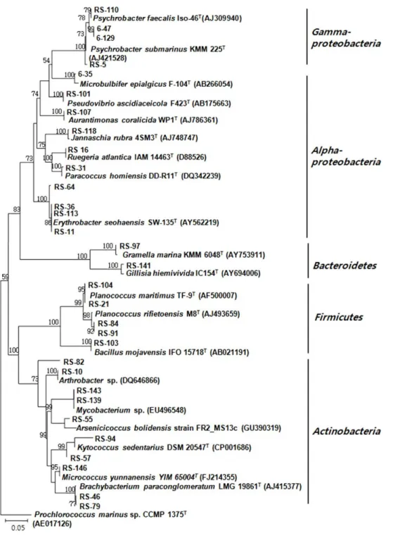
Fig. 3. Phylogenetic relationship determined from the 16S rDNA sequences of bacteria isolated from the marine sponge Spirastrella abata (RS). Bootstrap values calculated from 1,000 resamplings using neighbor-joining are shown at the respective nodes when the calculated values were 50% or greater.
공통적으로 존재하는 것으로 보고되었다(1, 2, 23). 한편 생리 활성물질을 생산하는 것으로 알려진 Hyrtios 속 해면의 경우 (22)에도 Actinobcteria가 발견되어(17) Actinobcteria 세균그룹 은 생리활성 물질의 생산과 연관성이 있는 것으로 추정된다.
남중국해에서 채집된 해양 해면 Craniella austrialiensis에 대하여 서로 다른 분석방법을 적용하여 공생세균 군집을 분석 한 결과, 배양에 기초한 PCR-RFLP법에 의한 경우(14), Alpha-
proteobacteria, Betaproteobacteria, Gammaproteobacteria, Firmicutes, Bacteroidetes, Actinobacteria가 나타났으나 비배 양에 기초한 PCR-DGGE법으로 분석한 경우의 공생세균의 군 집구조에서는 Alphaproteobacteria가 발견되지 않아 군집구조 에서 서로 다른 차이를 나타내었다(12). 또한 DGGE는 배양가 능한 세균을 포함하여 더 다양한 군집구조를 나타낼 것으로 기 대되는 비배양에 기초한 방법임에도 불구하고 PCR-RFLP에
Table 3. Phylogenetic affiliation of re-amplified denaturing gradient gel electrophoresis (DGGE) bands derived from the sponge, Spirastrella abata (DS)
DGGEband Closest relative Accession
no. % Sequence similarity
(*no. of basea) Phylum DS-1 Uncultured alpha proteobacterium clone JBS_E107 EU702822 97 (108/112) Alphaproteobacteria DS-2 Uncultured Chloroflexus sp. clone XC1D07 FJ481347 100 (118/118) Chloroflexi DS-3 Uncultured actinobacterium clone 277Z EU819009 100 (103/103) Actinobacteria DS-4 Uncultured gamma proteobacterium clone 222A EU816809 100 (137/137) Gammaproteobacteria DS-5 Uncultured Spirochete clone E175 FJ529354 99 (123/125) Spirochetes
* The numbers in parentheses are the part of the total bases used to calculate the levels of sequences similarity.
Fig. 4. Phylogenetic tree from analysis of 16S rDNA sequences of DGGE bands from Spirastrella abata (DS). Numbers above branches indicate bootstrap values of neighbor-joining analysis (>50%) from 1,000 replicates. The scale bar represents 0.1 substitution per nucleotide position.
의한 경우 보다 공생세균의 다양성이 강(class) 수준에서 더 낮 은 것으로 보고하고 있다(12).
PCR-RFLP와 PCR-DGGE에 의한 서로 다른 방법에 따른 S. abata의 공생세균 다양성을 분석한 결과, 방법에 따라 공생 세균의 군집구조에 차이를 나타냄을 확인하였다. 또한 해면 공 생세균의 군집구조에 대한 연구방법에서 비배양법의 경우 DNA 의 추출방법에 따라(6, 11), 배양법에서는 배양배지 혹은 배양 조건에 따라(29), 해면 공생세균의 우점 그룹이 달라진다는 연 구결과들이 보고되고 있다. 따라서 방법에 따른 공생세균 군집 구조의 분석에는 각각의 한계가 있으므로 연구 방법에 따른 차 이를 고려하면서 배양법과 비배양법을 동시에 적용하여 수행 하는 것이 효과적일 것으로 사료된다.
적요
해양 해면 Spirastrella abata에 대하여 배양에 근거한 PCR- RFLP와 비배양에 근거한 16S rDNA-DGGE fingerprinting 방
법을 적용하여 공생세균 군집구조를 조사하였다. Zobell 배지 와 천일염 배지를 사용하여 총 164균주를 선별하였다. 이들 균 주로 부터 증폭한 16S rDNA를 제한효소 HaeIII와 MspI을 사 용하여 절단한 후 각각의 다른 RFLP 패턴으로 구분하였다.
RFLP패턴으로부터 유래한 16S rDNA의 염기서열 분석 결과, 알려진 염기서열들과 95% 이상의 유사도를 나타내었으며 Proteo- bacteria (Alphaproteobacteria, Gammaproteobacteria), Actino- bacteria, Firmicutes, Bacteroidetes의 4개의 문이 나타났으며 우점 군집은 Alphaproteobacteria였다. 해면에서 분리한 total gDNA로 부터 증폭한 16S rDNA의 DGGE 분석 결과 5개의 DGGE band가 확인 되었고, 각각의 band의 염기서열 분석 결 과 알려진 염기서열들과 96% 이상의 유사도를 나타내었으며 band로부터 밝혀진 모든 서열들은 배양되지 않은 세균 클론들 과 높은 상동성을 나타내었다. DGGE에 의한 공생세균 군집은 Proteobacteria (Alphaproteobacteria, Gammaproteobacteria), Actinobacteria, Spirochetes, Chloroflexi로 4개의 문(phylum) 으로 나타났다. Spirastrella abata의 공생세균 군집에 대한
RFLP와 DGGE 적용 결과, Alphaproteobacteria, Gamma- proteobacteria, Actinobacteria의 공통 세균 그룹이 발견되었 으나 전체적인 공생세균 군집구조는 분석 방법에 따른 차이를 나타내었다.
감사의 말
이 논문은 2010년 한남대학교 학술연구조성비에 의해 수행 되었으며 이에 감사 드립니다.
참고문헌
1. Alam, N., B.H. Bae, J. Hong, C.O. Lee, B.A. Shin, K.S. Im, and J.H. Jung. 2001. Additional bioactive Lyso-PAF congeners from the sponge Spirastrella abata. J. Nat. Prod. 64, 533-535.
2. Alam, N., W. Wang, J.K. Hong, C.O. Lee, K.S. Im, and J.H.
Jung. 2002. Cytotoxic sphingosine 4-sulfates from the sponge Spirastrella abata. J. Nat. Prod. 65, 944-945.
3. Cho, H.H. and J.S. Park. 2009. Comparative analysis of the community of culturable bacteria associated with sponges, Spira- strella abata and Spirastrella panis by 16S rDNA-RFLP. Kor. J.
Microbiol. 45, 155-162.
4. Cho, H.H., E.J. Sim, and J.S. Park. 2010. Phylogenetic diversity of bacteria associated with the marine sponge, Spirastrella abata and Cinachyrella sp. Kor. J. Microbiol. 46, 177-182.
5. Enticknap, J.J., M. Kelly, O. Peraud, and R.T. Hill. 2006.
Characterization of a culturable alphaproteobacterial symbiont common to many marine sponges and devidence for vertical transmission via sponge Larvae. Appl. Environ. Microbiol. 72, 3724-3732.
6. Hardoim, C.C., R. Costa, F.V. Araujo, E. Hajdu, R. Peixoto, U.
Lins, A.S. Rosado, and J.D. van Elsas. 2009. Diversity of bacteria in the marine sponge Aplysina fulva in Brazilian coastal waters.
Appl. Environ. Microbiol. 75, 3331-3343.
7. Hentschel, U., J. Hopke, M. Horn, A.B. Friedrich, M. Wagner, J.
Hacker, and B.S. Moore. 2002. Molecular evidence for a uniform microbial community in sponge from different oceans. Appl.
Environ. Microbiol. 68, 4431-4440.
8. Hooper, N.J.A. and R.W.M. van Soest. 2002. Systema Porifera:
A guide to the classification of sponges. In N. BouryEsnault and C. Donadey (eds.). Kluwer Academic/Plenum Publisher, New York, USA.
9. Ko, S.R., S.J. Park, C.Y. Ahn, A. Choi, J.S. Lee, H.S. Kim, B.D.
Yoon, and H.M. Oh. 2004. Analysis of microbial communities during cyanobacterial bloom in Daechung Reservoir by DGGE.
Kor. J. Microbiol. 40, 205-210.
10. Levina, E.V., A.I. Kalinovsky, P.V. Andriyashenko, P.S. Dmi- trenok, D.L. Aminin, and V.A. Stonik. 2005. Phrygiasterol, a cytotoxic cyclopropane-containing polyhydroxysteroid, and related compounds from the pacific starfish Hippasteria phrygiana. J.
Nat. Prod. 68, 1541-1544.
11. Li, Z., L. He, and X. Miao. 2007. Cultivable bacterial community from South China Sea sponge as revealed by DGGE finger- printing and 16S rDNA phylogenetic analysis. Curr. Microbiol.
55, 465-472.
12. Li, Z.Y., L.M. He, J. Wu, and Q. Jiang. 2006. Bacterial community diversity associated with four marine sponges from the South China Sea based on 16S rDNA-DGGE fingerprinting.
J. Exp. Mar. Biol. Ecol. 329, 75-85.
13. Li, Z., Y. Hu, Y. liu, Y. Huang, L. He, and X. Miao. 2007. 16S rDNA clone library based bacterial phylogenetic diversity associated with three South China Sea sponges. World J. Microbiol.
Biotechnol. 23, 1265-1272.
14. Li, Z.Y. and Y. Liu. 2006. Marine sponge Craniella austri- aliensis-associated bacterial diversity revelation based on 16S rDNA library and biologically active Actinomycetes screening, phylogenetic analysis. Lett. Appl. Microbiol. 43, 410-416.
15. Mohamed, N.M., V. Rao, M.T. Hamann, M. Kelly, and R.T. Hill.
2008. Monitoring bacterial diversity of the marine sponge Ircinia strobilina upon transfer into aquaculture. Appl. Environ. Microbiol.
74, 4133-4143.
16. Muscholl-Silberhorn, A., V. Thiel, and J.F. Ihoff. 2008. Abun- dance and bioactivity of cultured sponge-associated bacteria from the Mediterranean Sea. Microbial. Ecol. 55, 94-106.
17. Park, J.S. 2010. Bacterial community diversity associated with two marine sponges from the South Pacific Ocean based on 16S rDNA-DGGE analysis. Kor. J. Microbiol. 46, 177-182.
18. Park, J.S., J.J. Sim, and K.D. An. 2009. Community structure of bacteria associated with two marine sponges from Jeju Island based on 16S rDNA-DGGE profile. Kor. J. Microbiol. 45, 170-176.
19. Park, S.H., K.K. Kwon, D.S. Lee, and H.K. Lee. 2002.
Morphological diversity of marine microorganisms on different isolation media. J. Micorobiol. 40, 161-165.
20. Radwan, M., A. Hanora, J. Zan, N.M. Mohamed, D.M. Abo- Elamatty, S.H. Abou-El-Ela, and R.T. Hill. 2009. Bacterial community analyses of two Red Sea sponges. Mar. Biotechnol.
12, 350-360.
21. Ridley, C.P., D.J. Faulkner, and M.G. Haygood. 2005. Investi- gation of Oscillatoria spongeliae-dominated bacterial communities in four dictyoceratid sponges. Appl. Environ. Microbiol. 71, 7366-7375.
22. Salmoun, M., C. Devijver, D. Daloze, J.C. Breakman, R. Gomez, M. de Kluijver, and R.W.M. Van Soest. 2000. New sesquiterpene/
Quinones from two sponges of the genus Hyrtios. J. Nat. Prod.
63, 452-456.
23. Shin, B.A., Y.R. Kim, I.S. Lee, C.K. Sung, J.K. Hong, C.J. Sim, K.S. Im, and J.H. Jung. 1999. Lyso-PAF analogues and lysophosphatidylcholines from the marine sponge Spirastrella abata as inhibitors of cholesterol biosyntehsis. J. Nat. Prod. 62, 1554-1557.
24. Tamura, K., J. Dudley, M. Nei, and S. Kumar. 2007. MEGA 4.
Mol. Biol. Evol. 24, 1596-1599.
25. Thiel, V., S. Leininger, R. Schamljohann, F. Brümer, and J.
Imhoff. 2007. Sponge-specific bacterial associations of the Medi- terranean sponge Chondrilla nucula (Demospongiae, tetractino- morpha). Microbial. Ecol. 54, 101-111.
26. Thiel, V., S.C. Neulinger, T. Saufenberger, R. Schmaljohann, and J.F. Imhoff. 2007. Spatial distribution of sponge-associated bacteria in the Medittanean sponge Tethya aurantium. FEMS Microbiol. Ecol. 59, 47-63.
27. Thomson, C., M. Horn, W. Wagner, U. Hentschel, and P.
Proksch. 2003. Monitoring microbial diversity and natural products profiles of the sponge Aplysina cavericola following transplantation. Mar. Biol. 142, 685-692.
28. Webster, N.S., A.P. Negri, M.M. Munro, and C.N. Battershill.
2004. Diverse microbial communities inhabit Antarctic sponges.
Environ. Microbiol. 6, 288-300.
29. Zhang, H., Y.K. Lee, W. Zhang, and H.K. Lee. 2006. Culturable actinobacteria from the marine sponge Hymeniacidon perleve:
isolation and phylogenetic diversity by 16S rRNA gene-RFLP analysis. Antonie van Leeuwenhoek 90, 159-169.