저작자표시-비영리-변경금지 2.0 대한민국 이용자는 아래의 조건을 따르는 경우에 한하여 자유롭게
l 이 저작물을 복제, 배포, 전송, 전시, 공연 및 방송할 수 있습니다. 다음과 같은 조건을 따라야 합니다:
l 귀하는, 이 저작물의 재이용이나 배포의 경우, 이 저작물에 적용된 이용허락조건 을 명확하게 나타내어야 합니다.
l 저작권자로부터 별도의 허가를 받으면 이러한 조건들은 적용되지 않습니다.
저작권법에 따른 이용자의 권리는 위의 내용에 의하여 영향을 받지 않습니다. 이것은 이용허락규약(Legal Code)을 이해하기 쉽게 요약한 것입니다.
Disclaimer
저작자표시. 귀하는 원저작자를 표시하여야 합니다.
비영리. 귀하는 이 저작물을 영리 목적으로 이용할 수 없습니다.
변경금지. 귀하는 이 저작물을 개작, 변형 또는 가공할 수 없습니다.
공학박사 학위논문
Isolation and characterization of Bacillus sp. 275 exhibiting
lignocellulolytic activities
리그노셀룰로오스 분해 활성 바실러스 275 균주의 분리 및 특성
2017 년 8 월
서울대학교 대학원
공과대학 바이오엔지니어링전공
공 경 택
Abstract
Isolation and characterization of Bacillus sp. 275 exhibiting
lignocellulolytic activities
Gyeongtaek Gong Interdisciplinary Program of Bioengineering The Graduate School Seoul National University
The objective of this research is to isolate lignocellulose-degrading bacteria from environment and characterize their lignocellulolytic activities. In addition, a study on the lignocellulolytic properties and genomic analysis of selected isolates, Bacillussp. 275 were performed. In this study, various environmental soil samples and isolation media were used to acquire isolates. The cell growth, enzyme activities, and the degradation products of cellulose and xylan were analyzed.
First, bacterial isolation from environmental soils and investigation of their cellulolytic, xylanolytic and lignolytic activities and taxonomic distribution were conducted. Finally, 89 bacterial strains were isolated. Lignocellulolytic bacteria
were isolated from not only lignocellulose-rich soils but also lignocellulose-lack soil samples. All the isolates were included in 4 phyla: Firmicutes, Proteobacteria, Actinobacteria and Bacteroidetes. Bacillus and Streptomyces occupied over 70 % of bacteria showing multienzymatic activities and the most abundant genus with only lignolytic activity was Burkholderia. Lignin-containing medium was effective to isolating bacteria with lignolytic activity only, but not favorable to isolating bacteria exhibiting multienzymatic activities.
Second, candidate bacterial isolate exhibiting high lignocellulolytic activities was selected comparing growth and enzyme activity.Bacillussp. 275 was grown in both solid and liquid alkali lignin media, and showed relatively high cellulase, xylanase and peroxidase activities compared to isolates, strain 123 (i.e., Bacillus aryabhattai) and 414 (i.e., Streptomyces durhamensis). As a result, Bacillussp. 275 was selected as final candidate for degradation of lignocellulose among 89 isolates.
Third, whole genome sequencing of Bacillus sp. 275 was conducted and genomic analysis using complete genome sequence was investigated. A lot of genes concerning to the degradation of lignocellulose in the genome of Bacillus sp. 275 were detected. Endoglucanase, β-glucanase, glucohydrolase, glucosidase, 1,4-β- xylosidase, arabinoxylan arabinofuranohydrolase, glucuronoxylanase, dye decolorizing peroxidase and laccase were present in the genome of Bacillussp. 275.
Besides, the genes related to degradation of starch, mannan, galactoside and arabinan were also found.
Finally, the characteristics of lignocellulolytic properties of Bacillus sp. 275 were investigated. The degradation products of cellulose (i.e., cellobiose and cellotriose) and xylan (i.e., xylose, xylobiose, xylotriose, xylotetraose and
xylopentaose) were observed by extracellular proteins secreted by Bacillussp. 275.
In addition, filter paper degradation occurred actively by Bacillus sp. 275 for 11 days. Regarding lignin utilization, the cell growth with alkali lignin added to the culture medium (LB medium) was increased by 78% compared to that without alkali lignin. Moreover, the lag phase was shortened from 12 hours to 6 hours in the case of lignin-added culture medium.
In this research, the isolation condition such as the sampling locations and the isolation medium was significant factors to efficiently isolating bacteria with lignocellulolytic activities. Bacillus sp. 275 showed cellulolytic, xylanolytic and lignolytic activities by extracellularly produced enzymes. In addition, Bacillus sp.
275 is potentially capable of degrading wide ranges of polysaccharides in lignocellulose. Considering only a few reports of bacteria using all major three component of lignocellulose (i.e., cellulose, hemicellulose and lignin) were published, Bacillus sp. 275 is a promising candidate for degrading or utilizing lignocellulosic biomass.
Keywords: bacterial isolation, lignocellulolytic bacteria, lignocellulose degradation, Bacillussp. 275, complete genome sequencing
Student Number: 2008-30288
Contents
Chapter 1. Research background and objective
...1Chapter 2. Literature review
...52.1. Lignocellulosic biomass...6
2.1.1. Cellulose...6
2.1.2. Hemicellulose...8
2.1.3. Lignin...8
2.2. Microbial degradation and utilization of Lignocellulosic biomass...9
2.2.1. Cellulose degradation...9
2.2.2. Hemicellulose and xylan degradation...11
2.2.3. Lignin degradation...12
2.2.4. Degradation for multicomponent of lignocellulose...15
2.3. Screening and isolation of lignocellulolytic bacteria...15
2.4. Lignin degradation pathway...16
Chapter 3. Experimental procedures
...183.1. Acquisition of environmental samples...19
3.2. Isolation of bacteria with potential lignocellulolytic activities...19
3.3. Screening and estimating cellulolytic, xylanolytic and lignolytic activities on agar plates...20
3.4. Identification of isolate with 16S rRNA gene analysis...21
3.5. Selection of high lignocellulolytic bacteria...23
3.5.1. Cellulolytic, xylanolytic and lignolytic activities measurements of cell culture supernatant of the selected isolates...23
3.5.2. Liquid culture in alkali lignin and mixed carbon medium consisting of cellulose, xylan and lignin...24
3.5.3. Analysis of cellulase, xylanase and peroxidase ...24
3.5.4. Oxidation of veratryl alcohol...25
3.6. Genome sequencing...25
3.7. Degradation of lignocellulose by Bacillussp. 275...27
3.7.1. Cell cultures in complex and defined media...27
3.7.2. Gel permeation chromatography (GPC) ...27
3.7.3. Thin layer chromatography (TLC) ...28
3.7.4. High performance liquid chromatography (HPLC) ...28
3.7.5. Cell cultures in xylan-based media...28
3.7.6. Filter paper degradation...29
Chapter 4. Isolation of cellulolytic, xylanolytic and ligninolytic bacteria from various environmental sites
...304.1. Introduction...31
4.2. Overall taxonomic distribution of isolated bacterial strains...32
4.3. Taxonomic distribution of bacterial strains by sampling locations...36 4.4. Taxonomic distribution of bacterial strains by carbon sources in isolation
media...40
4.5. Screening lignocellulolytic activity of isolates and distribution of activities by the isolation media...42
4.6. Taxonomic distribution of isolates revealing abundantly detected lignocellulolytic activities...48
4.7. Conclusion...51
Chapter 5. Selection of high lignocellulolytic potential bacterium Bacillus sp. 275 isolated from mud flat
...525.1. Introduction...53
5.2. Identification of selected isolates exhibiting relatively high cellulolytic, xylanolytic and lignolytic activities in solid media...53
5.3. Bacterial growth in liquid lignin medium and mixed carbon medium consist of cellulose, xylan and lignin...57
5.4. Analysis of lignocellulolytic degrading enzymes of selected isolates...57
5.5. Oxidation of veratryl alcohol in selected isolates and Bacillusspecies...60
5.6. Conclusion...62
Chapter 6. Genomic analysis of Bacillus sp. 275
...656.1. Introduction...66
6.2. Genome summary of Bacillussp. 275...66
6.3. Identification of Bacillussp. 275...69
6.4. Lignocellulose-degrading genes in Bacillussp. 275...69
6.5. Comparative analysis of genes related to lignocellulose degradation in
Bacillusspecies...71
6.6. Conclusion...72
Chapter 7. Lignocellulose degradation by Bacillus sp. 275
...787.1. Introduction...79
7.2. Cellulolytic, xylanolytic and lignolytic activities by decolorization and growth comparison in solid media...79
7.3. Modification and utilization of alkali lignin during growth in complex and defined liquid media...80
7.4. Degradation of polysaccharides by extracellular enzymes...88
7.5. Growth dynamics of Bacillussp. 275in xylan-based media...92
7.6. Filter paper degradation by Bacillussp. 275...96
7.7. Conclusion...96
Chapter 8. Overall discussion and further suggestions
...101Bibliography
...106Appendix
...133Abstract
...136List of Tables
Table 2.1.1Composition of various lignocellulosic biomasses...7
Table 2.1.2Typical linkages of lignin in soft wood and hard wood...10
Table 2.2.1Summary of bacterial enzymes for lignin degradation...14
Table 3.5.1Measurements of enzyme activities in cell culture supernatant...26
Table 4.2.1Information on the sampling locations...34
Table 4.2.2 Taxonomic analysis of 89 bacterial isolates showing a single or multiple lignocellulolytic activities...35
Table 4.6.1 Distribution of bacterial isolates by lignocellulolytic activities at the genus level...50
Table 5.2.1Identification and lignocellulolytic activities of the selected isolates..55
Table 5.2.2 Lignocellulolytic activities of supernatant of the selected isolates grown in liquid medium...56
Table 5.4.1 Lignocellulolytic activities of thee selected isolates grown in different carbon sources...61
Table 6.2.1Genome features of Bacillussp. 275 strain...67
Table 6.5.1 COG functional categories of the complete genome sequences of Bacillus sp. 275, B. siamensis SDLI1, B. amyloliquefaciens DSM 7T and B. velezensisFZB42...73
Table 6.5.2 Comparison of genes encoding lignocellulose-degrading enzymes in Bacillussp. 275 and other Bacillusstrains...75
Table 7.7.1Bacterial strains for multicomponent utilization of lignocellulose....100
List of Figures
Figure 2.4.1 Catabolic pathways for the degradation of lignin-derived aromatic compounds by Sphingobiumsp. SYK-6....17 Figure 3.3.1 Experimental flowchart of a process for isolating, screening and estimating cellulolytic, xylanolytic and lignolytic activities on agar plates....22 Figure 4.3.1Relative abundance of bacterial isolates by the sampling locations at the class level....38 Figure 4.3.2 Taxonomic distribution of isolates between 5 dry mountain soil samples (MS) and 3 wet soil samples (WFS: wetland and mudflat soil samples).
...39 Figure 4.4.1 Relative abundance of bacterial isolates by the combination of lignocellulose components in the isolation media....41 Figure 4.5.1 Distribution of cellulolytic, xylanolytic and lignolytic activities of bacteria isolated from cellulose-only medium, cellulose-xylan medium and cellulose-xylan-lignin medium....44 Figure 4.5.2Venn diagram presenting distribution (%) of bacteria exhibiting single activity and multiple lignocellulolytic activities isolated from cellulose-only medium, cellulose-xylan medium, cellulose-xylan-lignin and all the isolation media.
...47 Figure 5.3.1 Time course of cell growth and pH in CXL (a, b) and alkali lignin medium (c, d)....58 Figure 5.5.1 Veratryl alcohol oxidation by three selected isolates (a) and Bacillus species (b)....63 Figure 6.2.1Circular genome map of Bacillussp. 275....68
Figure 6.3.1 Phylogenetic tree derived from 16S rRNA gene sequence data of Bacillussp. 275 and its relatives....70 Figure 7.2.1Lignocellulolytic activities of Bacillussp. 275 on agar plates....81 Figure 7.3.1 Effect of lignin addition on growth of Bacillus sp. 275 in defined medium....83 Figure 7.3.2 Effect of lignin addition on growth of Bacillus sp. 275 in complex medium ...85 Figure 7.3.3 GPC profiles for supernatant of Bacillus sp. 275 grown in defined medium at initial pH 7 (a) and pH 6 (b), and complex medium at initial pH 7 (c)..
...86 Figure 7.4.1Thin-layer chromatography analysis of hydrolysis products for CMC and xylan using supernatant of Bacillus sp. 275 grown in LB+CMC (A) and LB+xylan (B) medium....89 Figure7.4.2HPLC analysis of the oligosaccharide products of CMC (a) and xylan (b)....91 Figure7.5.1Tine course of growth dynamics on xylan-based carbon sources....93 Figure 7.6.1Filter paper degradation in CX medium supplemented with Whatman filter paper no. 1 (1 x 5cm strip x 4) after 0 day (a), 2 day (b), 4 day (c) and 11 days (d) of incubation....97
Chapter 1.
Research background and objective
Chapter 1. Research background and objective
The use of lignocellulosic biomass is important and promising strategy for a sustainable development inhibiting the problems of fossil resources. In addition, lignocellulosic biomass does not compete with edible biomass such as corn, wheat, sugar cane and sugar beet, and has lower cost than food crops1. In spite of these advantages, efficient utilization of lignocellulosic biomass is limited because of the natural structure (i.e., cross-link in polysaccharides with lignin via ether and ester bonds2) of lignocellulose.
In nature, microbial degradation of lignocellulose is carried out by microorganisms, which have distinct enzymatic systems for degradation of polysaccharides (e.g., cellulose and hemicellulose) and lignin. These cell wall- deconstructing enzymes are unusually classified diversely such as glycoside hydrolases, polysaccharide lyases, carbohydrate esterase and auxiliary activities3. Therefore, the isolation of bacteria which have high lignocellulolytic activities is significant for utilization of lignocellulose. Despite the vast efforts underway to isolate lignocellulose-degrading bacteria, the use of various environmental sources and isolation media to explore taxonomic distributions of lignocellulolytic bacteria and the related lignocellulolytic activities has not been reported yet. The isolation conditions are important factors in the acquisition of efficient lignocellulose degraders.
Currently, studies for degradation of lignocellulose have mainly focused on degradation of cellulose, hemicellulose (or xylan) or lignin, separately. However, it is necessary to use all the main three compositions of lignocellulose.
In this research, isolation of lignocellulolytic bacteria was conducted and their cellulolytic, xylanolytic and lignolytic activities were analyzed on solid culture media. Among 89 isolates from environmental soil samples, Bacillus sp. 275 was selected as a candidate with multienzymatic activities for lignocellulose degradation. Bacillus sp. 275 has genes encoding lignocellulolytic enzymes according to the information of complete genome and also has the cellulolytic, xylanolytic and lignolytic enzyme activities. The characteristics of Bacillus sp. 275 provide insight into bacterial degradation of all main components in lignocellulosic biomass.
In summary, the objectives of this thesis are as follows:
1. Isolation and characterization of cellulolytic, xylanolytic and lignolytic bacteria from environmental sources
2. Selection of candidate bacterial isolate exhibiting high lignocellulolytic activities
3. Analysis of lignocellulose-degrading genes using complete genome sequence of Bacillussp. 275
4. Investigation into characteristics of lignocellulolytic properties of Bacillussp. 275
This thesis provides previously unknown insight into the sampling points and isolation medium for efficient isolation of lignocellulose-degraders. In addition, Bacillus sp. 275 has potentially wide range of use in the degradation of polysaccharide and lignin in lignocellulosic biomasses. Therefore, Bacillussp. 275 can be useful microorganism for conversion of lignocellulose into useful chemicals and fuels.
Chapter 2.
Literature review
Chapter 2. Literature review
2.1. Lignocellulosic biomass
Lignocellulosic biomass is considered the most abundant4-6 and renewable7, 8 resources on earth. Moreover, lignocellulosic biomass does not interfere with food biomass and not threat to food security9. Lignocellulose is the major component of plant cell walls and different lignocellulosic biomasses such as wood, switch grass and agricultural residues can be used for biofuels or chemicals.
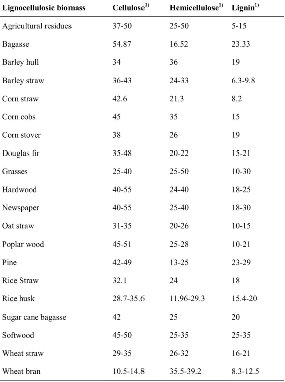
Lignocellulose is composed of three major components which were cellulose, hemicellulose and lignin8. These macromolecules construct a complex structure by aromatic heteropolymer (e.g., lignin) and help resistant from environmental attacks by three-dimensional structures. The ratio of cellulose, hemicellulose and lignin is various depending on the characteristic of the biomass. The composition of various lignocellulosic biomasses was summarized in Table 2.1.110.
2.1.1. Cellulose
Cellulose is the main component of lignocellulose and a homo-polymer of glucose linked by beta(1-4) linkages4. In general, the proportion of cellulose is 15 ~ 50%11 of the dry weight of biomass. Cellulose is difficult for degradation to glucose because of its crystalline structure12, 13. In contrast, amorphous cellulose is more easily attacked by enzymes14. Cellulose fibers are used in the paper industry.
Table 2.1.1Composition of various lignocellulosic biomasses10
Lignocellulosic biomass Cellulose1) Hemicellulose1) Lignin1)
Agricultural residues 37-50 25-50 5-15
Bagasse 54.87 16.52 23.33
Barley hull 34 36 19
Barley straw 36-43 24-33 6.3-9.8
Corn straw 42.6 21.3 8.2
Corn cobs 45 35 15
Corn stover 38 26 19
Douglas fir 35-48 20-22 15-21
Grasses 25-40 25-50 10-30
Hardwood 40-55 24-40 18-25
Newspaper 40-55 25-40 18-30
Oat straw 31-35 20-26 10-15
Poplar wood 45-51 25-28 10-21
Pine 42-49 13-25 23-29
Rice Straw 32.1 24 18
Rice husk 28.7-35.6 11.96-29.3 15.4-20
Sugar cane bagasse 42 25 20
Softwood 45-50 25-35 25-35
Wheat straw 29-35 26-32 16-21
Wheat bran 10.5-14.8 35.5-39.2 8.3-12.5
1) Dry weight percentage
2.1.2. Hemicellulose
Hemicellulose is hetero-polysaccharides composed of pentoses such as xylose and arabinose, hexoses such as glucose, mannose and galactose, and acetylated sugars. The composition and structure are different according to the lignocellulosic materials4. For example, O-Acetyl-4-O-methylglucuronoxylans in hardwoods constitute 10–35% of the hemicelluloses, and arabino-4-O-methylglucuronoxylans in softwoods constitute 10–15 % of the hemicelluloses.
However, xylan (the polymer of xylose) is the major component of hemicellulose up to 50% and the second most abundant polysaccharide after cellulose14, 15. A various linkages such as alpha-1,2, alpha-1,3 are used for connect to the other sugars. These complex structures provide a barrier from microbial and mechanical forces. To link hemicellulose and cellulose, hydrogen and covalent bonding is used. Deconstruction of hemicellulose is easier than cellulose because of a low molecular weight of hemicellulose.
2.1.3. Lignin
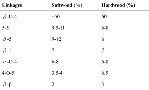
Lignin is an amorphous highly branched aromatic polymer made by the random coupling with three types of monolignols (i.e., coumaryl, coniferyl and sinapyl alcohols)4. These monolignols are linked by β-aryl ether, di-aryl propane, biphenyl, diaryl ether, phenylcoumarane, spirodienone and pinoresinol bonds14. The proportion of lignin is 15 ~ 40% of the dry weight of biomass16. However, the composition of lignin is different. In general, hardwoods are composed of 56%
coniferyl alcohols and 40 % sinapyl alcohols while softwood is mainly composed of coniferyl alcohols (80 %)17. Table 2.1.2 shows the typical linkages of lignin in softwood and hardwood18. Lignin is difficult to breakdown because of its complex chemical nature (protecting cellulose and hemicellulose polymers from the external attack by other microbes)10, 19. Because of the difficult use of lignin, it is mainly burned as a source of energy in the pulp and paper industry8. Lignin also used as adhesives, food additives and production of phenolic compounds14. In addition, lignin and lignin derivatives can be used to plastics and resins as biopolymers20, 21. However, recalcitrant to degradation, lignin does not increase in nature.
2.2. Microbial degradation and utilization of Lignocellulosic biomass
Although lignocellulose is an abundant and available renewable resource, utilization of lignocellulose as substrates by microbial cells has been known as one of the most difficult processes22. Because of difficulty from complex substrates and diversity of various enzymes, microbial community or consortia were also used for lignocellulose degradation23-28. Moreover, a fungal-bacterial microbial consortium was used for enhancement of lignocellulolytic activity29.
However, the major research for bacterial degradation of lignocellulose is focused on single bacterial strain.
2.2.1. Cellulose degradation
Table 2.1.2Typical linkages of lignin in soft wood and hard wood18
Linkages Softwood (%) Hardwood (%)
β-O-4 ~50 60
5-5 9.5-11 6-8
β-5 9-12 6
β-1 7 7
α-O-4 6-8 6-8
4-O-5 3.5-4 6.5
β-β 2 3
Cellulose was typically degraded by three types of enzymes: endoglucanase, cellobiohydrolase and beta-glucosidase30. Endoglucanase break the internal bonds of the amorphous regions of cellulose polymer. Cellobiohydrolases hydrolyze β (1–4)-d- glucosidic linkages in cellulose, generating cellobiose from either the reducing or the non-reducing ends of the polymer. Next, β-glucosidases degrade the disaccharide cellobiose to glucose. Recently, lytic polysaccharide monooxygenases (LPMOs) can be oxidized cellulose12, 31-35. These enzymes are disrupted the cellulose fiber structure, thereby motivating a release of elementary nonofibrils. Therefore, cellulase can be degraded more efficiently by oxidative cleavages of LPMOs.
2.2.2. Hemicellulose and xylan degradation
As the same as cellulose degradation, the enzymatic breakdown of hemicellulose needs an consortium of various enzymes such as endo-xylanase, beta-xylosidase, 1,2-alpha-glucuronosidase, beta-galactosidase and etc36. The degradation of xylan, the major component of hemicellulose, the existence of enzymes which break backbone and various side-chains is essential15. Xylanase is one of the major enzyme related to hemicellulose degradation, which cleaves the beta-1,4-linked D-xylopyranose units from the backbone structure of xylan.
Xylanase is widely distributed in bacteria, fungi, plants and insects. According to the carbohydrate-active enzymes (CAZy) database (http:/www.cazy.org), more than six glycoside hydrolases (GH) families (e.g., 5, 7, 8, 10, 11, 26, 30 and 43) are related to xylanase. Among various glycoside hydrolases families, xylanases from
GH10 and GH11 families are widely studied. Xylanase from GH11 family are only acted to xylan, compared to xylanase from GH10 are functioned to not only xylan but also substituted linkages of the xylan backbone15. The dominant studies about optimum condition for xylanases are mesophilic temperature and neutral or some acidic pH. However, xylanases which are active at 5~105°C, pH from 2 to 11 and NaCl concentrations as high as 30% have been reported37.
2.2.3. Lignin degradation
Enzymatic degradation of lignin is involved in enzymes of oxidative reaction.
The famous enzymes of lignin degradation were laccase and peroxidases such as lignin peroxidase, manganese peroxidase, versatile peroxidase and dye- decolorizing peroxidase. Lignin peroxidase degrades through a mechanism based on the formation of radicals in the presence of hydrogen peroxide. Lignin peroxidase is capable of cleaving alkyl side chains in lignin and lignin model compounds. Manganese peroxidase uses Mn2+ as an electron donor which is oxidized to Mn3+ through peroxide dependent oxidation. Manganese peroxidase is considered a familiar mechanism for the degradation of non-phenolic structures.
Versatile peroxidase can also oxidize Mn2+ and non-phenolic lignin model compounds like manganese peroxidase. However, the versatility of versatile peroxidase could be possible for oxidation of both low and high redox potential aromatic substrates. Dye-decolorizing peroxidase is a new superfamily of heme- containing peroxidase which was found in both bacteria and fungi38. The catalytic cycle is not well-known yet. Laccase is a multicopper-containing enzyme with
oxygen as an electron acceptor. In general, laccase can oxidize the wide spectrum of phenolic compounds18. Recently, superoxide dismutase is considered as a candidate for degradation of lignin39, 40.
Among various kinds of bacteria, lignin degradation using Rhodococcus41-47, Pseudomonas48-54and Streptomyces55-63 genus were widely studied.R.jostiiRHA1 is a famous strain degrading polychlorinated biphenyl and lignin. 96 mg/L of vanillin46 and 80-125 mg/L of pyridine 2, 4-dicarboxylic acid (2, 4-PDCA) or pyridine 2,5-dicarboxylic acid (2,5-PDCA) were produced by these strain. In addition, DyP-type peroxidase in R.jostiiRHA1 is related to lignin degradation by beta-aryl ether cleavage64. R. opacus DSM 1069 and PD630 were also studied about production of lipid from lignin65, 66. Pseudomonas sp. PKE11753, 54, P.
fluorescens Pf-550, P. putidaKT244048and MET9451were reported degradation of lignin and pretreated lignocellulosic biomass in Pseudomonasgenus. Especially,P.
putida KT2440 is the most studied strain in this genus. This strain can be produced bioplastics and polyhydroxalkanate (PHA). More than 55% of cell dry weight of mutant of P. putida was PHA67. Among Streptomyces genus, Streptomyces viridosporusT7A is one of the most well studied. S.viridosporusT7A can degrade wide range of lignin (e.g., softwood, hardwood and grass lignin) and secret the ligninolytic enzymes such as lignin peroxidase and laccase62, 68-70. Some Bacillus species are reported as lignin or lignin analogue degraders39, 71-75. Besides these bacteria, Pandoraea sp. B-676, Novosphingobium sp. B-777 and Pandoraea sp.
ISTKB78 can be degraded kraft lignin as a sole carbon source. Enterobacter lignolyticus SCF179, 80 and Burkholderia sp. LIG3081 are also known as lignin- degrading bacteria.
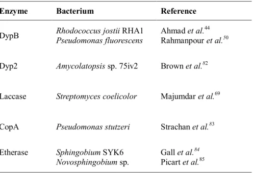
Table 2.2.1Summary of bacterial enzymes for lignin degradation
Enzyme Bacterium Reference
DypB Rhodococcus jostiiRHA1 Pseudomonas fluorescens
Ahmad et al.44 Rahmanpour et al.50 Dyp2 Amycolatopsissp. 75iv2 Brown et al.82
Laccase Streptomyces coelicolor Majumdar et al.69
CopA Pseudomonas stutzeri Strachan et al.83 Etherase SphingobiumSYK6
Novosphingobiumsp.
Gall et al.84 Picart et al.85
2.2.4. Degradation for multicomponent of lignocellulose
Some bacteria can degrade multicomponent of lignocellulosic biomass such as cellulose and hemicellulose (or xylan), cellulose and lignin, hemicellulose and lignin, or all components of lignocellulose. The isolates named as Bacillussp. CS-1 was decomposed cellulose, hemicellulose and lignin in rice straw72.
Similarly, the combination of cellulase, hemicellulase and ligninase is studied for increase of degradation yield. Synergistic interaction of cellulase and xylanase was detected on unpretereated lignocellulosic substrates. The hydrolysis yields of lignocellulose treated by cellulase and xylanase mixture was improved by 133-545%
than only treated with cellulase86. Similarly, the synergistic effect of cellulase and xylanase enzymes was shown by improvement of cellulose accessibility87. Moreover, cellulase, xylanase and laccase in the designed system (e.g., designer cellulosome) were enhanced decomposition of lignocellulose88.
2.3. Screening and isolation of lignocellulolytic bacteria
There are two screening methods for isolation of ligninolytic prokaryotes:
culture-dependent and culture-independent techniques89. Culture-dependent screening method detects bacterial growth or utilization of natural lignin, lignin model compounds90 or aromatic dyes. In contrast to culture-dependent method, culture-independent screening use genetic information such as metagenomics, metatranscriptomics and bioinformatics83, 91-96.
Vanillin is regarded one of the major valuable products from lignin97. The
vanillin-sensing cell was developed in E. coli98that did not show cross-activity of lignin, lignin degradation products and vanillin analogues. High-throughput screening of lignin-degrading enzymes (i.e., vanillin producing enzymes) is possible by this microorganism. In addition, screening of bacterial isolate capable of producing vanillin and vanillic acid was developed by the colorimetric method using ferulic acid as a sole carbon source99.
2.4. Lignin degradation pathway
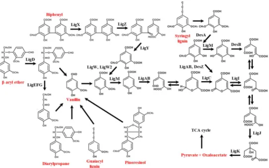
From various lignin degradation pathways, beta-aryl ether and biphenyl catabolic pathways are important because of highly dominant linkages in lignin (e.g., β-O-4, 5-5). Through these pathways, one of the valuable products likes vanillin or 4-hyddroxybenzoic acid was produced.
Degradation pathway of beta-aryl ether has been reported mainly in Sphingobium sp. SYK-6100-102. Dehydrogenase (LigD), beta-etherase (LigE and LigF) and other enzymes such as LigG, LigL, LigN and LigO were participated in the degradation of beta-aryl ether linkage. Regarding biphenyl degradation pathway, extensive studies were conducted because of polychlorinated biphenyl which is a major pollutant in environment. Biphenyl 2,3-dioxygenase, LigX, LigZ, LigY, LigW and LigW2 were associated with this pathway.
Figure 2.4.1 Catabolic pathways for the degradation of lignin-derived aromatic compounds by Sphingobiumsp. SYK-6103.
Chapter 3.
Experimental procedures
3.1. Acquisition of environmental samples
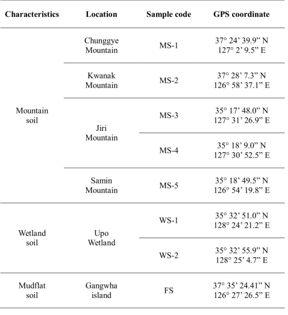
Environmental soil samples were collected from mountains, a wetland and mudflat. Eight soil samples were collected including 5 soil samples from 4 mountains, 2 samples from a wetland and 1 sample from a mudflat. The mountain soil samples were taken from under rotten wood at a depth of 20-30 cm. The wetland soil was taken from the boundary between fresh water and land at a depth of 10-20 cm. The mudflat soil was taken at a depth of 10-20 cm during the ebb tide.
The soil samples were used for bacterial isolation within 3-5 hours of collection.
3.2. Isolation of bacteria with potential lignocellulolytic activities
One gram of each environmental sample was suspended in 10 mL of 0.9%
sodium chloride solution and the suspension was used as an inoculum for agar plates to isolate lignocellulolytic microorganisms. The isolation medium contained (in 1 L of distilled water) 0.3 g of NH4Cl, 0.3 g of MgCl2, 0.3 g of CaCl2, 0.3 g of KH2PO4, 0.5 g of yeast extract, 10 mL minimal trace element solution (DSM medium 318) and 5 g of carboxymethyl cellulose (CMC, soluble in water) or 5 g of microcrystalline cellulose (CRY, insoluble in water) as a carbon source.
Additionally, instead of cellulose, 2 (cellulose and xylan) or 3 (cellulose, xylan and lignin) components of lignocellulose at a total concentration of 5 g/L were used as carbon sources: 2.5 g/L of CMC + 2.5 g/L of xylan (CX), 1.7 g/L of CMC + 1.7 g/L
of xylan + 1.6 g/L of alkali lignin (CXL). To prepare agar plates, agar powder was added at a final concentration of 15 g/L.
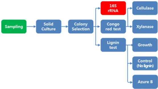
Isolating bacteria with potential lignocellulolytic activities was performed by directly spreading each suspended environmental sample (0.2 mL) on the isolation medium agar plate (CMC, CRY, CX and CXL) after serial dilution of 10-4 ~ 10-7 with 0.9% sodium chloride solution. Morphologically different colonies on the agar plates were transferred to fresh agar medium 3-4 times until acquiring a single bacterial colony. All the agar plates were incubated at 30°C until a colony appeared and then stored at 4°C prior to use.
3.3. Screening and estimating cellulolytic, xylanolytic and lignolytic activities on agar plates
Cellulase and xylanase activities were determined with the agar medium containing 5 g/L of CMC or xylan as a carbon source using the Congo red staining method to detect the clear zone around the colony104. To estimate cellulolytic and xylanolytic activities, a single colony of each isolate from agar plate was steaked onto CMC or xylan agar plate. When the colony appeared after 5 days of incubation on CMC or xylan agar plates, 0.1% of Congo red solution was flooded on the agar plate for 15 minutes. Then, the agar plate was washed with 1M NaCl solution for 15 minutes. The enzyme activity was calculated by the “Ratio of relative enzyme activity, ICMC or Ixylan=diameter of halo zone/diameter of colony”
105. The intensity of cellulolytic and xylanolytic activities was classified into four
groups: strong (+++: Indices of relative enzyme activity was more than 10), medium (++: Indices of relative enzyme activity was between 10 and 3), weak (+:
Indices of relative enzyme activity was below 3) and none (no halo zone detected).
To screen for a lignin degradation ability, a single colony of each isolate was picked and streaked onto an agar plate containing a lignin analogue, Azure B (0.01 w/v %) with 1 g/L of glucose and xylose as carbon sources. After 7 days of incubation at 30°C, the lignolytic activity was detected by observing a decolorizing zone around a colony. Additionally, the capability of lignin utilization was screened by growth on alkali lignin agar plate without any supplemented carbon sources and yeast extract. The growth of the isolate was also compared with the growth on a plate deficient in alkali lignin. After incubating 7 days at 30°C, colony formation and size were compared between the lignin agar plate and the agar plate with no carbon source (lignin-deficient agar plate). The intensity of the lignolytic activity was classified into four groups: strong (+++: Azure B decolorization and growth on the lignin agar plate compared with the lignin-deficient agar plate), medium (++:
no decolorization of Azure B and growth on the lignin agar plate compared with the lignin-deficient agar plate) , weak (+:decolorization of Azure B and no growth on the lignin agar plate compared with the lignin-deficient agar plate) and none (no decolorization of Azure B and no growth on the lignin agar plate compared with the lignin-deficient agar plate).
3.4. Identification of isolate with 16S rRNA gene analysis
The genomic DNA of the isolate was extracted with a G-spin genomic DNA
Figure 3.3.1 Experimental flowchart of a process for isolating, screening and estimating cellulolytic, xylanolytic and lignolytic activities on agar plates.
extraction kit (Intron, Republic of Korea) following the manufacturer’s instructions.
The 16S rRNA gene was amplified using AccuPower PCR PreMix kit (Bioneer, Republic of Korea) and the primers 27F (AGAGTTTGATCMTGGCTCAG) and 1492R (TACGGYTACCTTGTTACGACTT). The sequence of 16S rRNA was analyzed by Macrogen Inc. (Republic of Korea). The isolates were identified using the EzTaxon server (http://www.ezbiocloud.net106) based on the 16S rRNA sequence data. Phylogenetic tree was generated by the Neighbor-Joining method.
Evolutionary analyses were conducted in MEGA7107 and the distances were computed using the Maximum Composite Likelihood method.
3.5. Selection of high lignocellulolytic bacteria
3.5.1. Cellulolytic, xylanolytic and lignolytic activities measurements of cell culture supernatant of the selected isolates
The isolates were pre-cultured by inoculated single colony in 2 mL of LB medium containing 10 g/L of tryptone, 5 g/L of yeast extract and 5 g/L of NaCl at 30°C on a shaking incubator at 200 rpm. After one day incubation, 1% of cell broth was inoculated in 20 mL of LB medium in 150 mL straight neck flask for other one day. Culture supernatant was acquired by centrifuged at 14,000×g for 5 min of cell broth. Protein concentration was measured by Bradford protein assay reagent (Bio- Rad) and Cellulase (endo-cellulase) activity was quantification by Cellulase assay kit (CELLG5 method) (Megazyme). Xylanase activity was measured with xylan as
substrate and detected by 3,5-dinitrosalicylic acid (DNS) colorimetric method at 50°C. Peroxidase and laccase activities was assayed by detecting oxidation of ABTS (ε420nm = 3.6 mM–1 cm–1) in 100 mM acetate buffer at pH 3 and room temperature with or without H2O2, respectively. Peroxidase activity was calculated by differences between two measuring conditions. One unit of enzyme activity was defined as the amount of enzyme required to release one micromole of product per minute.
3.5.2. Liquid culture in alkali lignin and mixed carbon medium consisting of cellulose, xylan and lignin
Ten selected isolates were pre-cultured in 2 mL of isolation medium contained (in 1 L of distilled water) 0.3 g of NH4Cl, 0.3 g of MgCl2, 0.3 g of CaCl2, 0.3 g of KH2PO4, 0.5 g of yeast extract, 10 mL minimal trace element solution (DSM medium 318). After one day incubation, the supernatant was removed by centrifugation. The cell pellet was resuspened with isolation medium except yeast extract. Cell broth was inoculated in isolation medium except yeast extract with CXL (1.7 g/L of CMC + 1.7 g/L of xylan + 1.6 g/L of alkali lignin) and 5 g/L of alkali lignin (LGN) as a sole carbon sources, respectively.
3.5.3. Analysis of cellulase, xylanase and peroxidase
The composition of CXL and LGN media was the same as described in
section 3.5.2. The composition of CMC and XYL media were the same as CXL and LGN media except the carbon sources changed to CMC and xylan.
The peroxidase activity was measured by oxidation of 2,4- dichlorophenol (2,4-DCP). The supernatants of cell broth was reacted with a mixture of 50 mM of potassium phosphate buffer (pH 7.0), 3 mM 2,4-DCP, 0.164 mM
aminoantipyrine and 4.0 mM hydrogen peroxide. After 1 hour of incubation at 37°C, the different of absorbance at 510 nm in the reaction mixture was detected. The cellulase and xylanase activities were detected using DNS methods as mentioned in section 3.5.1.3.5.4. Oxidation of veratryl alcohol
2.0 mM of veratryl alcohol and 0.5 mL of the supernatant were mixed, and then added of 0.4 mM of hydrogen peroxide in total volume of 1 mL (pH 3.0 and 30°C). The product of oxidation of veratryl alcohol (e.g., veratryl aldehyde) can detected at 310 nm using UV spectrophotometer.
3.6. Genome sequencing
The genome of Bacillus sp. 275 was sequenced using a combination of the PacBio RSII system (Pacific Biosciences, CA, USA), Illumina MiSeq (100-bp paired-end) and the Roche 454 sequencing TITAN technology. All the reads were assembled using the Roche gsAssembler 2.6, CLC Genomics Workbench 6.5.1 and
Table 3.5.1Measurements of enzyme activities in cell culture supernatant
Enzyme Method
Cellulase Cellulase assay kit (CELLG5)
Xylanase 3,5-dinitrosalicylic acid (DNS) colorimetric method
Laccase ABTS oxidation
Peroxidase ABTS oxidation
2,4-dichlorophenol (2,4-DCP) oxidation
veratryl alcohol oxidationPacBio SMRT Analysis 2.1 with a genome coverage of 276 folds.
3.7. Degradation of lignocellulose by Bacillus sp. 275
3.7.1. Cell cultures in complex and defined media
The cell growth in complex medium was studied in
20 mL of LB medium in 150 mL straight neck flask. Alkali lignin was added from 2% stock solution to final concentration of alkali lignin at 0.05% and 0.1%.The defined medium based on M9 was composed of 12.8 g/L of Na2HPO4∙7H2O, 3.0 g/L of KH2PO4, 10.0 g/L of NaCl, 1.0 g/L of NH4Cl, 0.492 g/L of MgSO4∙7H2O, 0.111 g/L of CaCl2 and trace element (0.1 mg/L of MnCl2∙4H2O, 0.07 mg/L of ZnCl2, 0.06 mg/L of H3BO3, 0.04 mg/L of NaMoO4∙2H2O, 0.2 mg/L of CoCl2∙6H2O, 0.02 mg/L of NiCl2∙6H2O, 0.02 mg/L of CuCl2∙2H2O, 2.78 mg/L of FeSO4∙7H2O and 0.1 ul/L of HCl). The carbon source was glucose (3 g/L) and the final concentration of added lignin was 0.05%. Glucose concentration was detected by reflectometer Merck RQflex plus 10 meter using glucose test strips.
3.7.2. Gel permeation chromatography (GPC)
The distribution of molecular weight of lignin was analyzed by GPC on HPLC (Agilent) with a Photodiode Array (PDA) detector and TSK-GEL W3000G column (300 x 7.5 mm). 50 mM NaOH containing 20% acetonitrile was used as eluent. The
eluent flow rate was set to 0.6 mL/min.
3.7.3. Thin layer chromatography (TLC)
The hydrolysis products of cellulose and xylan were analyzed by thin layer chromatography (TLC) on Silica Gel 60 plates (Merck). One microliter of samples was spotted on plate, and dried at room temperature around 5 min. The mixture of 63 ml of 2-propanol, 16 mL of 1-butanol and 21 mL of water was used as a developing solution (100 mL total). After developing, a visualization solution (10% sulfuric acid in ethanol) was sprayed using TLC sprayer. The products were visualized by heating with heat gun.
3.7.4. High performance liquid chromatography (HPLC)
The sugars released by cellulose and xylan were detected using high performance liquid chromatography (Agilent HPLC, USA) equipped with an Aminex HPX-87P column [300×7.8mm] (Bio-rad, USA). The column temperature was 80°C and flow rate of the mobile phase (water for HPLC) was 0.15 mL/min.
3.7.5. Cell cultures in xylan-based media
The composition of a xylan-based medium was based on M9 medium
as mentioned in section 3.7.1. The difference was the concentration and combination of carbon sources. Three kinds of medium composition were 5.0 g/L of xylan (X), 5.0 g/L of xylan + 5.0 g/L of CMC (CX) and 5.0 g/L of xylan + 5.0 g/L of CMC + 0.5 g/L of alkali lignin (CXL).
Cellulase and xylanase activities were measured CELLG5 method and DNS method, respectively, as described in section 3.5.1.
3.7.6. Filter paper degradation
Filter paper degradation was conducted by supplemented with Whatman no. 1 filter paper (1 × 4 cm strip × 4ea) in
5.0 g/L of xylan + 5.0 g/L of CMC (CX)
medium as mentioned in section 3.7.5.Chapter 4.
Isolation of cellulolytic, xylanolytic and ligninolytic bacteria
from various environmental sites
4.1. Introduction
Recently, diverse lignocellulolytic bacterial strains have been isolated from environmental sources such as tropical forest soil108 and wood chips109 in which natural degradation of lignocellulose was occurring. In addition, taxonomic characterization of lignocellulolytic bacteria and enzymatic activities has been investigated. Industrial lignocellulose streams including black liquor, paper mill effluent and bamboo slips110, 111 have been also tried as environmental sources to enrich lignocellulolytic bacteria. Likewise, most previous reports for isolation of lignocellulolytic bacteria have been conducted using lignocellulose-abundant or lignocellulose-contained environmental sources112-114 and the solid agar media containing a single lignocellulose component (cellulose, xylan or lignin) to assay lignocellulolytic activity. However, despite the vast efforts underway to isolate lignocellulose-degrading bacteria, use of various environmental sources and isolation media to explore taxonomic distributions of lignocellulolytic bacteria and the related lignocellulolytic activities has not been reported yet. Because the degradation of lignocellulose is the first key step in lignocellulose utilization, special attention is necessary to screen lignocellulose-degrading bacteria and enzymes with effective methodology.
In this work, the isolation and taxonomic identification of cellulolytic, xylanolytic and lignolytic bacteria were performed using: i) various environmental sources including lignocellulose-rich environmental samples (mountain forest soils near rotten wood) and lignocellulose-rare samples (mudflat soil with a high salt concentration and wetland soil with freshwater) and ii) the isolation medium with a
combination of cellulose, xylan and lignin. In addition, the bacterial taxonomic distribution of the isolates was analyzed with respect to environmental sources and isolation media. This study shows the importance of isolation conditions such as environmental sampling locations and isolation media to effectively obtain lignocellulolytic bacteria.
4.2. Overall taxonomic distribution of isolated bacterial strains
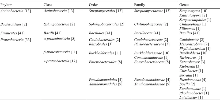
For the isolation of lignocellulose-degrading bacteria, 8 sampling points were chosen from various environmental conditions: 5 mountain soils under rotten wood, 2 wetland samples and 1 mudflat sample (Table 4.2.1). Colonies on the agar plates with CMC, CRY, CX and CXL media were selected based on different morphological characteristics (e.g., size, shape and color). Among 124 isolates, 18 isolates could not be identified by 16S rRNA gene analysis because of either indecipherable overlapping chromatograms or PCR failure. Additionally, 17 isolates from CMC (2 isolates), CRY (4 isolates), CX (3 isolates) and CXL (8 isolates) media were excluded for further study because neither cellulolytic, xylanolytic nor lignolytic activity was shown on the agar plates. Therefore, 89 isolates were used for the 16S rRNA sequence-based taxonomic analysis and assay of the bacterial lignocellulolytic properties.
Table 4.2.2 shows the taxonomic distribution of the 89 isolates. All the isolates were included in 4 phyla, 6 classes, 9 orders, 10 families and 20 genera. Most of
the isolates (98%) belonged to 3 phyla: Firmicutes (46%), Proteobacteria (37%) and Actinobacteria (15%). Bacteroidetes accounted for only 2% of the total isolates. Interestingly, only a single class, order and family were observed with the phyla Actinobacteria, Bacteroidetes and Firmicutes. Moreover, only a single genus, Bacillus, was identified in Firmicutes. Notably, Bacillaceaeand Bacillus were the most dominant family and genus, respectively, comprising up to 46% of the total isolates affiliated to B. aryabhattai (10 isolates), B. anthracis (7 isolates), B.
thuringiensis (7 isolates), B. acidiceler (6 isolates) and other Bacillus species (11 isolates). Unlike the isolates belonging to Firmicutes, Actinobacteria and Bacteroidetes, 33 isolates in Proteobacteria were distributed over 3 classes, 6 orders, 7 families and 14 genera. Overall, in the order of abundance, the most dominant family was Bacillaceae (41 isolates, 46%) followed by Streptomycetaceae (13 isolates, 15%), Burkholderiaceae (10 isolates, 11%) and Enterobacteriaceae (8 isolates, 9%).
Four phyla (Actinobacteria, Bacteroidetes, Firmicutes and Proteobacteria) and 6 classes (Actinobacteria, Sphingobacteria, Bacilli, α-proteobacteria, β- proteobacteria and γ-proteobacteria) have been reported as the taxonomic ranks of cellulolytic bacteria109, lignin-degrading bacteria115 and lignocellulolytic bacteria108, 116. Woo et al.108 investigated the diversity of lignocellulolytic Puerto Rican tropical forest soil bacteria which were screened based on the growth on either cellulose or lignin. However, the genus distributions of isolated bacteria in Woo et al.108 were significantly different from our results. In this work, the major genus of Actinobacteriawas Streptomyces, while the genus Gordoniaconsisted of over 90% of the total Actinobacteria(15 out of 16 isolates) in a Puerto Rican rain
Table 4.2.1Information on the sampling locations
Characteristics Location Sample code GPS coordinate
Mountain soil
Chunggye
Mountain MS-1 37° 24’ 39.9” N
127° 2’ 9.5” E Kwanak
Mountain MS-2 37° 28’ 7.3” N
126° 58’ 37.1” E
Jiri Mountain
MS-3 35° 17’ 48.0” N
127° 31’ 26.9” E
MS-4 35° 18’ 9.0” N
127° 30’ 52.5” E Samin
Mountain MS-5 35° 18’ 49.5” N
126° 54’ 19.8” E
Wetland soil
Upo Wetland
WS-1 35° 32’ 51.0” N
128° 24’ 21.2” E
WS-2 35° 32’ 55.9” N
128° 25’ 4.7” E Mudflat
soil
Gangwha
island FS 37° 35’ 24.41” N
126° 27’ 26.5” E
Table 4.2.2Taxonomic analysis of 89 bacterial isolates showing a single or multiple lignocellulolytic activities. The number in parenthesis is the number of isolates affiliated to the corresponding taxonomic rank
Phylum Class Order Family Genus
Actinobacteria[13] Actinobacteria[13] Streptomycetales[13] Streptomycetaceae[13] Streptomyces[10]
Kitasatospora[2]
Streptacidiphilus[1]
Bacteroidetes[2] Sphingobacteria[2] Sphingobacteriales[2] Chitinophagaceae[2] Chitinophaga[1]
Filimonas[1]
Firmicutes[41] Bacilli[41] Bacillales[41] Bacillaceae[41] Bacillus[41]
Proteobacteria[33] α-proteobacteria[5] Caulobacterales[2]
Rhizobiales[3]
Caulobacteraceae[2]
Phyllobacteriaceae[3]
Caulobacter[2]
Mesorhizobium[2]
Phyllobacterium[1]
β-proteobacteria[11] Burkholderiales[11] Burkholderiaceae[10]
Comamonadaceae[1]
Burkholderia[10]
Variovorax[1]
γ-proteobacteria[17] Enterobacteriales[8]
Pseudomonadales[4]
Xanthomonadales[5]
Enterobacteriaceae[8]
Pseudomonadaceae[4]
Xanthomonadaceae[5]
Enterobacter[3]
Klebsiella[3]
Citrobacter[1]
Serratia[1]
Pseudomonas[4]
Dyella[2]
Xanthomonas[1]
Rhodanobacter[1]
Luteibacter[1]
forest soil sample108. In addition, Aquitalea in Proteobacteria was reported to account for 15% of total isolates (75 isolates)108, but in this work, there was no Aquitalea isolate. Those distinct genus level distributions might be attributed to experimental condition variances such as microbial inoculum sources (e.g., sample collecting locations) and the combination of lignocellulose component materials in isolating media (either of cellulose or lignin in Woo et al.’s work108 vs. including multiple components in this work).
4.3. Taxonomic distribution of bacterial strains by sampling locations
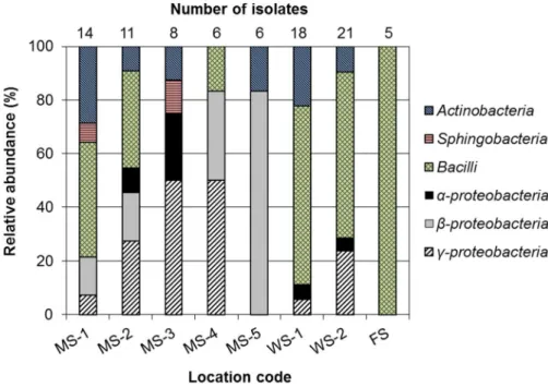
To investigate the influences of ecological conditions in sampling sites on taxonomic distribution of lignocellulolytic bacteria, 4 mountains and 2 wet areas were chosen as lignocellulose-rich sites and lignocellulose-rare sites, respectively.
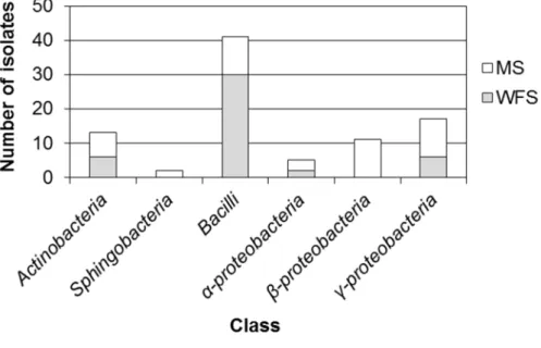
The class-level taxonomic distribution and the number of isolates with respect to the sampling points are shown in Figure 4.3.1. The isolates were distributed widely in 4 classes (MS-1, MS-3, WS-1 and WS-2) or 5 classes (MS-2) except for the isolates from MS-4, MS-5 and FS. Notably, 5 isolates from FS were all Bacilli.
Although the taxonomic distributions of 5 mountain soil samples were different from those of WS and FS, the class Bacilli exhibited the highest abundance in mountain soils (MS-1, MS-2) as well as WS-1, WS-2 and FS. The distributions obtained with the 5 mountain soil samples appeared to be site-specific with different profiles. In particular, even the isolates from MS-3 and MS-4 (1.1 km
apart) sampled from Jiri Mountain showed significantly different taxonomic distributions except for the relative abundance of γ-proteobacter(50%). In contrast, the isolates from WS-1 and WS-2 (1.1 km apart) had a similar taxonomic diversity comprising 4 identical classes with Bacillias the most dominant class (over 60% of the isolates from the wetland). When the overall bacterial distributions of lignocellulose-rich dry soil samples (mountain soil samples) were compared with those of lignocellulose-rare wet soil samples (WS-1, WS-2 and FS) (Figure 4.3.2), the isolates belonging to Actinobacteria, Bacilli, α-proteobacteria and γ- proteobacteria were found in both the mountain soils and wet soil samples. In contrast, Sphingobacteria and β-proteobacteria were isolated only from the mountain soil samples. Bacilli had a 3-fold higher isolation possibility from the collected wet soils than from the mountain soils, while there was a 2-fold higher chance to isolate γ-proteobacteriafrom mountain soils than from wet soils (Figure 4.3.2).
In the previous reports, isolation and taxonomic analysis of lignocellulose- degrading bacteria have been conducted mainly with environmental microbial samples adapted under lignocellulose-rich conditions108, 109. In this work, lignocellulolytic bacteria were isolated from not only lignocellulose-rich mountain soils but also lignocellulose-rare environmental samples such as wetland soils and mudflat soils. This result implies that microbial inoculum sources for the isolation of lignocellulolytic bacteria are not necessary to be from lignocellulose-abundant (and likely lignocellulose-decaying) sites. Furthermore, considering the different taxonomic abundance and distribution with respect to soil characteristics, use of lignocellulose-lack environmental samples might enable to obtain more diverse
Figure 4.3.1Relative abundance of bacterial isolates by the sampling locations at the class level.
Figure 4.3.2 Taxonomic distribution of isolates between 5 dry mountain soil samples (MS) and 3 wet soil samples (WFS: wetland and mudflat soil samples).
lignocellulolytic bacteria than those previously identified from lignocellulose-rich sites.
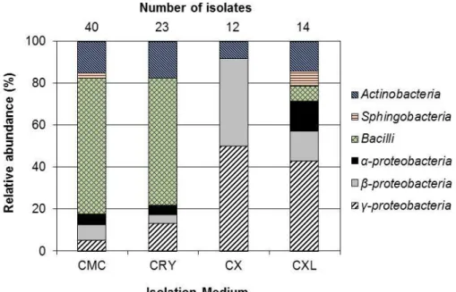
4.4. Taxonomic distribution of bacterial strains by carbon sources in isolation media
Next, the microbial distribution of the 89 isolates was analyzed in terms of the carbon sources used in the agar plates: CMC, CRY, CX and CXL. The number of isolates from the CMC, CRY, CX and CXL media was 40, 23, 12 and 14, respectively. As shown in Figure 4.4.1, taxonomic distributions of the isolates from the CMC and CRY media were not much different. The most dominant bacterial class in the cellulose-only media (CMC and CRY media: hereafter CEL) was Bacilli (40 isolates), which accounted for over 97% of the total Bacilliisolates (41 isolates). However, the class-level distribution of the isolates derived from the CX and CXL media was much different from that of the CEL media indicating a significant effect of the carbon source combinations on the taxonomic distribution.
In the case of the CX medium, Bacilli isolate was not detected, and γ- proteobacteria was the most dominant class (6 isolates; 50% of the CX isolates) followed by β-proteobacteria (5 isolates, 42%). Regarding the isolates from the CXL medium, all 6 classes were detected, and γ-proteobacteria(6 isolates; 43% of the CXL isolates) was the main class. Because 97% of Bacillus (40 out of 41 isolates) and 77% of Actinobacteria (10 out of 13 isolates) were isolated from CMC and CRY media (Figure 4.4.1), the CEL media were likely advantageous for isolating BacilliandActinobacteriaover the CX or CXL medium. In contrast, with
Figure 4.4.1 Relative abundance of bacterial isolates by the combination of lignocellulose components (total concentration of 5 g/L) in the isolation media.
CMC, carboxymethyl cellulose; CRY, microcrystalline cellulose; CX, CMC+xylan;
CXL, CMC+xylan+alkali lignin
the multiple lignocellulose components (e.g., CX and CXL media), the possibility of isolating β-proteobacteria (7 out of 11 isolates) and γ-proteobacteria(12 out of 17 isolates) might be higher than with the CEL media.
Isolation of lignocellulolytic bacteria has been previously attempted using agar plates containing a single component of lignocellulose (cellulose, xylan and lignin)108, 109, 117. In this study, the mixed lignocellulose components (CX and CXL) as well as cellulose-only media were used as carbon sources in the agar plates for the isolation of lignocellulolytic bacteria. Notably, as shown in Figure 4.4.1, the components of carbon sources in agar plates appeared to greatly affect the taxonomic distributions of lignocellulolytic bacteria, suggesting that the use of mixed lignocellulose components could effective to get lignocellulolytic bacteria rarely discovered with cellulose-only media.
4.5. Screening lignocellulolytic activity of isolates and distribution of activities by the isolation media
Because 89 isolates were able to grow on CMC, CRY, CX and CXL agar media, the cellulolytic (Cs), xylanolytic (Xs) and lignolytic (Ls) activities of the isolates on solid agar plates were assayed. The Cs and Xs were evaluated using Congo red method by detecting halo zones around the colonies on CMC and xylan agar plates. The Ls was estimated by Azure B decolorization and lignin utilization by growth on alkali lignin agar plate as the sole carbon source compared with the growth on agar plate with no added carbon source.
The distributions of the isolates showing Cs, Xs and Ls with respect to the CEL, CX and CXL media are shown in Figure 4.5.1. The sum of the percentage in each medium was over 100% because some isolates exhibited multiple lignocellulolytic activities. As expected, the CEL agar plates were favorable for isolating Cs-exhibiting bacteria, which accounted for over 87% of the CEL isolates.
Interestingly, over 80% and 58% of the isolates from the CEL media had Xs and Ls even though there was no xylan and lignin. All Bacilliisolates from the CEL media revealed Cs, and up to 50% ofBacilliisolates showed Ls. Similarly, Xu and Yang57 have also found that S. griseorubens C-5 isolated using cellulose-based isolation medium exhibited not only cellulase but also xylanase and lignolytic activities such as laccase and peroxidase activities. Unexpectedly, β-proteobacteria grown on CEL media exhibited Ls, but did not show Cs probably because of unspecific growth using other nutrients in the agar plates rather than using cellulose. In the case of the CX medium, 75% and 67% of the CX isolates were found to have Cs and Xs, respectively. The portion of Ls-exhibiting isolates was also high (67%), but the taxonomic distribution of isolates with Ls was different from that with Xs: the relative abundance of β-proteobacteria significantly increased by 400% while γ- proteobacteria population decreased by 50%. Considering that all β-proteobacteria bacteria were isolated from the mountain soil samples (Figure 4.3.2) and the relative abundance of β-proteobacteria with Ls was higher than those with Cs, the mountain samples were likely more effective for isolating Ls-possessing β- proteobacteria.
Regarding the isolates from the CXL medium, a large portion of the isolates (71%) had Ls, whereas only 36% exhibited Cs and Xs in spite of the cellulose and
(a)
(b)
0 20 40 60 80 100
Cs Xs Ls CEL Relative abundamce of isolates from CEL (%)
Isolation Medium
Actinobacteria Sphingobacteria Bacilli
α-proteobacteria β-proteobacteria γ-proteobacteria
0 20 40 60 80 100
Cs Xs Ls CX Relative abundamce of isolates from CX (%)
Isolation Medium
Actinobacteria Sphingobacteria Bacilli
α-proteobacteria β-proteobacteria γ-proteobacteria
(c)
Figure 4.5.1 Distribution of cellulolytic, xylanolytic and lignolytic activities of bacteria isolated from (a) cellulose-only medium (CEL), (b) cellulose-xylan medium (CX) and (c) cellulose-xylan-lignin medium (CXL). Cs, cellulolytic activity; Xs, xylanolytic activity; Ls, lignolytic activity
0 20 40 60 80 100
Cs Xs Ls CXL Relative abundamce of isolates from CXL (%)
Isolation Medium
Actinobacteria Sphingobacteria Bacilli
α-proteobacteria β-proteobacteria γ-proteobacteria
xylan in the CXL medium. A possible reason for this result could be a toxic effect of lignin on the bacteria. Because lignin is known to be toxic to some bacteria118, 119, the prerequisite for bacteria to survive on CXL medium is likely a tolerance to lignin rather than having Cs and Xs. Therefore, lignin-absent media might be effective for isolating a wide range of cellulose-degrading bacteria.
The percent compositions of a single activity as well as the multiple lignocellulolytic activities of the isolates from different media are presented as Venn diagrams (Figure 4.5.2 a, b and c). A Venn diagram with all the isolates is shown in Figure 4.5.2d. Regardless of the isolation media, most of the isolates exhibiting Cs were found to have other lignocellulolytic activity such as Xs and Ls.
In particular, among Cs-revealing isolates from the CEL media (55 isolates; 87% of CEL media isolates) (Figure 4.5.2a), 54 isolates exhibited multiple activities: 51 isolates (81% of CEL media isolates) with both Cs and Xs (hereafter CsXs); 29 isolates (46%) with both Cs and Ls (hereafter CsLs); and 26 isolates (41%) with Cs, Xs and Ls (hereafter C