저작자표시-비영리-변경금지 2.0 대한민국 이용자는 아래의 조건을 따르는 경우에 한하여 자유롭게
l 이 저작물을 복제, 배포, 전송, 전시, 공연 및 방송할 수 있습니다. 다음과 같은 조건을 따라야 합니다:
l 귀하는, 이 저작물의 재이용이나 배포의 경우, 이 저작물에 적용된 이용허락조건 을 명확하게 나타내어야 합니다.
l 저작권자로부터 별도의 허가를 받으면 이러한 조건들은 적용되지 않습니다.
저작권법에 따른 이용자의 권리는 위의 내용에 의하여 영향을 받지 않습니다. 이것은 이용허락규약(Legal Code)을 이해하기 쉽게 요약한 것입니다.
Disclaimer
저작자표시. 귀하는 원저작자를 표시하여야 합니다.
비영리. 귀하는 이 저작물을 영리 목적으로 이용할 수 없습니다.
변경금지. 귀하는 이 저작물을 개작, 변형 또는 가공할 수 없습니다.
A Dissertation for the Degree of Master
Antimicrobial effects and mechanisms of plasma-activated acetic acid against
Salmonella Typhimurium and its application for chicken meat
Salmonella Typhimurium에 대한 플라즈마 활성 초산의 항균 효능 및 메커니즘과 계육의 적용
August 2021
Taemin Kang
Department of Agricultural Biotechnology Graduate School
Seoul National University
Antimicrobial effects and mechanisms of plasma-activated acetic acid against Salmonella
Typhimurium and its application for chicken meat
Advisor: Prof. Cheorun Jo, Ph.D.
Submitting a Master’s Dissertation of Agriculture June 2021
Department of Agricultural Biotechnology Graduate School
Seoul National University Taemin Kang
Confirming the Master’s Dissertation written by Taemin Kang
June 2021
)%;5, 2/2 98;
:7;
&1!.*;10;';!&0"0;*;
-0#141;1;; &01; $+( ; 6-!3.3!;';10;--1*&;*.;';
"1;
# !!# "
# #
!# !# !#
#
# !# "#
백 명 기
조 철 훈
김 영 훈
i
Abstract
Antimicrobial effects and mechanisms of plasma-activated acetic acid against Salmonella
Typhimurium and its application for chicken meat
Taemin Kang Program in Animal Science and Biotechnology Department of Agricultural Biotechnology Graduate School of Seoul National University
We conducted research on plasma-activated acetic acid (PAAA) to improve the sterilization efficiency of each individual treatment of plasma-activated water (PAW) and acetic acid (AA) and apply it to poultry meats. PAAA was prepared by addition of different concentrations of AA into deionized water before plasma treatment. The aim of this study was to investigate growth-inhibitory activities of PAAA against Salmonella Typhimurium cells and biofilm and the underlying mechanisms, as well as identify the effect of PAAA on reduction of S. Typhimurium and quality traits of chicken meats.
ii Experiment Ⅰ.
Growth-inhibitory activities of plasma-activated acetic acid against Salmonella Typhimurium cells and biofilm and the underlying mechanisms
This study aimed to examine the growth-inhibitory activities of plasma- activated acetic acid (PAAA) against Salmonella Typhimurium cells and biofilm and elucidate the underlying mechanism. PAAA was prepared by discharging plasma to 20 mL of 0.2% (v/v) acetic acid (AA) for 20 min (2.2 kHz and 8.4 kVpp) after the optimization test. The cells and biofilms on stainless steel were incubated with AA, and PAAA. The count of S. Typhimurium cells decreased by 5.71 log CFU/mL after 10 min of incubation with 0.2% PAAA compared with control (without any treatment). The S. Typhimurium count in the biofilms decreased by 4 log CFU/cm2 upon treatment with 0.4% PAAA for 10 min when compared with the initial S.
Typhimurium count in the biofilms (control) (p < 0.05). Confocal laser scanning microscopy (CLSM) revealed that 0.4% PAAA synergistically decreased the viability of S. Typhimurium in the biofilms. In in 0.2% PAAA, the concentrations of H2O2 and NO3− were directly proportional to the plasma discharge time, while NO2−
was not detected. However, the pH values of both 0.2% PAAA and plasma-activated water (PAW) were inversely proportional to the plasma discharge time. Additionally, treatment with catalase, L-histidine, D-mannitol, and sodium azide inhibited the antibacterial activity of PAAA, which indicated that H2O2, 1O2, ∙OH, and NO2− are involved in the generation and decomposition of peroxynitrous acid (ONOOH).
Therefore, ONOOH, which functions as an intermediate under acidic conditions, plays a key role in the bactericidal effects of PAAA. This study demonstrated that
iii
PAAA has potential applications as a decontaminant in the food industry.
Experiment Ⅱ.
Effect of plasma-activated acetic acid on reduction of Salmonella Typhimurium and quality traits of chicken meats
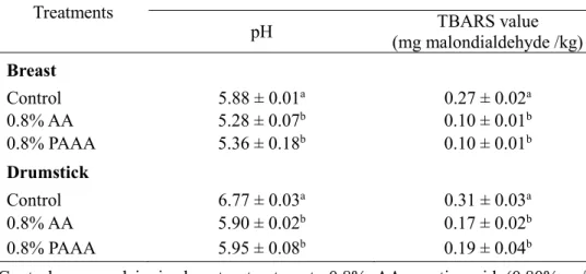
This study investigated the bactericidal effects of plasma-activated acetic acid (PAAA) on reduction of Salmonella Typhimurium and its impact on the physicochemical traits of chicken meats. Twenty mL of 0.8% (v/v) acetic acid (AA) was treated with plasma (2.2 kHz and 8.4 kVpp) for 30 min. The chicken skins, breasts, and drumsticks, inoculated with S. Typhimurium, were immersed in AA or PAAA and incubated for 10 min. The S. Typhimurium on the breasts and drumsticks were significantly susceptible to treatment with AA and PAAA, compared to the control (deionized water treatment), and the population of bacterial cells in PAAA- treated chicken breasts and drumsticks decreased by 0.98 and 1.19 log CFU/g, respectively, compared with AA. The values for pH and 2-thiobarbituric acid reactive substances (TBARS) of PAAA-treated samples decreased significantly compared to the control. The lightness (L*) values of the chicken breasts after AA and PAAA treatments increased compared to the control, whereas the value for yellowness (b*) decreased. The scanning electron microscopic images and the results for volatile compounds in chicken meat revealed similar patterns, with no significant differences between AA and PAAA treatments. Thus, the bactericidal effects and the potential industrial applications of PAAA were confirmed.
iv
Keywords: Plasma-activated acetic acid; Salmonella Typhimurium; Antibacterial effect; Antibiofilm effect; Peroxynitrous acid; Chicken meats
Student Number: 2019-26240
v
Contents
Abstract ··· i
Contents ··· v
List of Tables ··· viii
List of Figures ··· ix
List of Abbreviations ··· xi
Chapter I. General Introduction ··· 1
1.1. Food safety ··· 1
1.2. Salmonella Typhimurium ··· 1
1.3. Antimicrobial agents ··· 2
1.3. Plasma technology ··· 3
1.3. Plasma-activated acetic acid (PAAA) ··· 3
Chapter II. Growth-inhibitory activities of plasma-activated acetic acid against Salmonella Typhimurium cells and biofilm and the underlying mechanisms 2.1. Introduction ··· 5
2.2. Materials and methods ··· 8
2.2.1. Bacterial strains and culture preparation ··· 8
2.2.2. Preparation of biofilm cells ··· 8
2.2.3. Preparation and treatment of PAAA ··· 9
vi
2.2.4. Growth-inhibitory activity of PAAA against S. Typhimurium ····11
2.2.5. Chemical characterization of PAAA ···11
2.2.6. Treatment with reactive oxygen and nitrogen species (RONS) scavengers ··· 12
2.2.7. Measurement of cell membrane integrity ··· 12
2.2.8. Statistical analysis ··· 13
2.3. Results and discussion ··· 14
2.3.1. Growth-inhibitory activity of PAAA against S. Typhimurium··· 14
2.3.2. Antibiofilm activity of PAAA ··· 17
2.3.3. Confocal laser scanning microscopy (CLSM) ··· 19
2.3.4. Chemical characterization of PAAA ··· 21
2.3.5. Effect of RONS scavengers on the antibacterial effect of PAAA ·· 26
2.3.6. Detection of peroxynitrous acid (ONOOH) in PAAA ··· 29
2.4. Conclusion ··· 32
Chapter Ⅲ. Effect of plasma-activated acetic acid on reduction of Salmonella Typhimurium and quality traits of chicken meats 3.1. Introduction ··· 33
3.2. Materials and methods ··· 36
3.2.1. Antibacterial activity of acetic acid (AA) and PAAA treatments on chicken meats ··· 36
3.2.1.1. Bacterial strains and culture preparation ··· 36
3.2.1.2. Sample preparation, sterilization and inoculation ··· 36
vii
3.2.1.3. Preparation of PAAA ··· 37
3.2.1.4. Microbial analysis ··· 37
3.2.2. pH measurement ··· 38
3.2.3. Lipid oxidation measurement··· 38
3.2.4. Color measurement ··· 39
3.2.5. Scanning electron microscopy (SEM) analysis ··· 39
3.2.6. Electronic nose analysis ··· 40
3.2.7. Statistical analysis ··· 41
3.3. Results and discussion ··· 42
3.3.1. Antibacterial effect of PAAA on chicken meats ··· 42
3.3.2. pH ··· 47
3.3.3. TBARS ··· 47
3.3.4. Color ··· 50
3.3.5. SEM ··· 53
3.3.6. Electronic nose ··· 55
3.4. Conclusion ··· 57
References ··· 58
Summary in Korean ··· 71
viii
List of Tables
Chapter II.
Table 1. Chemical composition of 0.2% plasma-activated acetic acid (PAAA) according to plasma discharge time and plasma-activated water (PAW) ··· 24
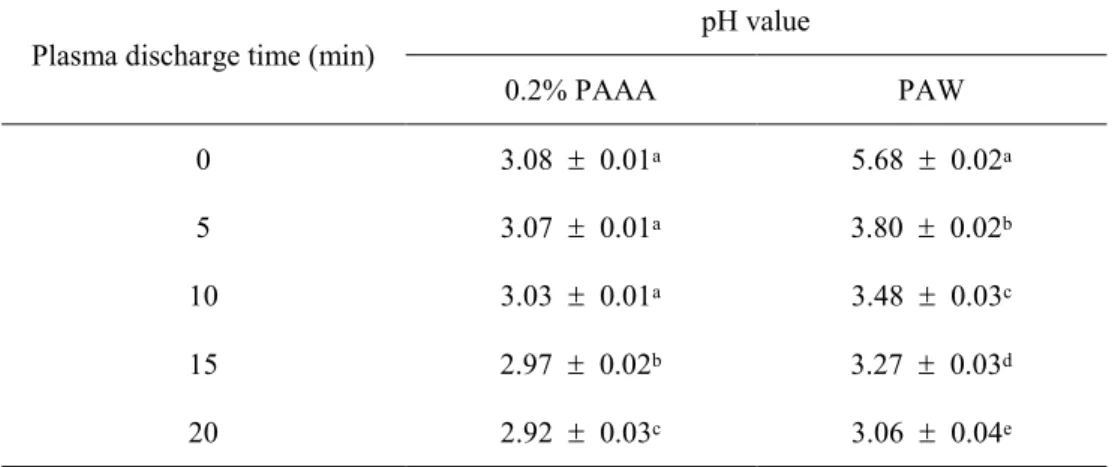
Table 2. The pH value of 0.2% plasma-activated acetic acid (PAAA) and plasma- activated water (PAW) according to plasma discharge time ··· 25
Chapter Ⅲ.
Table 3. pH and lipid oxidation of breasts and drumsticks treated with acetic acid (AA) and plasma-activated acetic acid (PAAA) ··· 49
Table 4. Surface color values of breasts and drumsticks treated with acetic acid (AA) and plasma-activated acetic acid (PAAA) ··· 52
ix
List of Figures
Chapter II.
Figure 1. Schematic diagram of the experimental setup for the preparation of dielectric barrier discharge plasma ··· 10
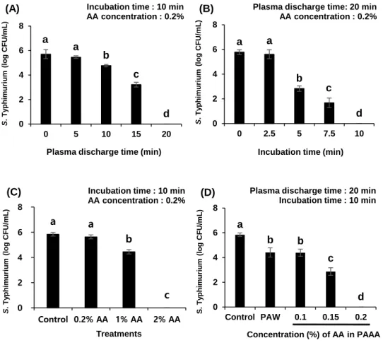
Figure 2. Effect of acetic acid (AA) and plasma-activated acetic acid (PAAA) on the viability of bacterial cells according to (A) plasma discharge time, (B) incubation time, (C) concentrations of AA and (D) concentrations of AA in PAAA ··· 16
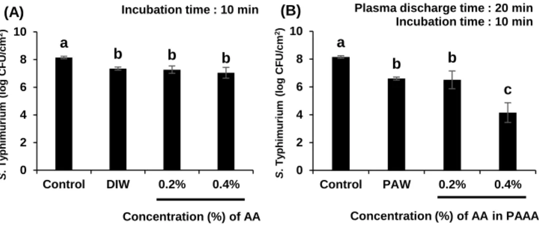
Figure 3. Effect of acetic acid (AA) and plasma-activated acetic acid (PAAA) on the viability of Salmonella Typhimurium cells in the biofilm according to (A) different concentrations of AA and (B) different concentrations of AA in PAAA ··· 18
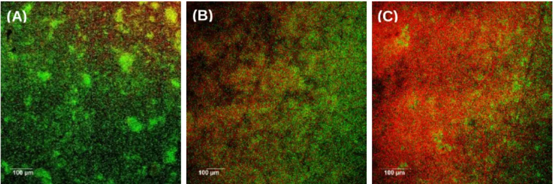
Figure 4. Evaluation of membrane integrity of cells in the Salmonella Typhimurium biofilm using confocal laser scanning microscopy. (A) Control group cells; (B) 0.4%
acetic acid (AA; for 10 min)-treated cells; (C) Cells treated with 0.4% plasma- activated acetic acid (PAAA; obtained after plasma discharge for 20 min) for 10 min ··· 20
Figure 5. Effect of reactive oxygen and nitrogen species (RONS) scavengers added before (A) or after (B) plasma discharge for 20 min on the viability of bacterial cells ··· 28
x
Figure 6. (A) Detection of peroxynitrous acid (ONOOH) in 0.2% plasma-activated acetic acid (PAAA) during plasma discharge using the fluorescence dye 2,7- dichlorodihydrofluorescein diacetate (H2DCFDA); (B) Detection of ONOOH in 0.2%
PAAA post-plasma discharge; (C) The viable bacterial cell count post-plasma discharge ··· 31
Chapter Ⅲ.
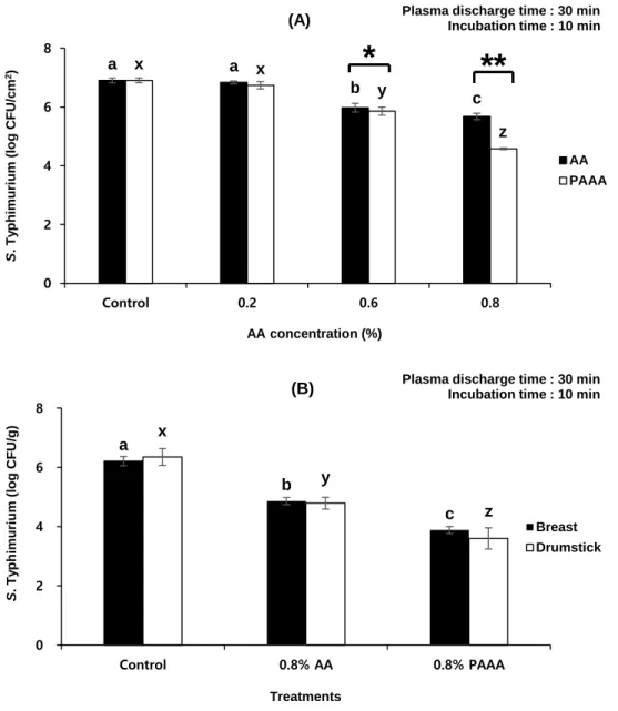
Figure 7. Effect of acetic acid (AA) and plasma-activated acetic acid (PAAA) on population of Salmonella Typhimurium according to (A) the concentration of AA on chicken skins and (B) the antibacterial effects of 0.8% AA and PAAA on chicken breasts and drumsticks ··· 45
Figure 8. Evaluation of morphological images of chicken meats using scanning electron microscope (SEM). (A) Control group (breasts); (B) 0.8% AA treatment (breasts); (C) 0.8% PAAA treatment (breasts); (D) Control group (drumsticks); (E) 0.8% AA treatment (drumsticks); (F) 0.8% PAAA treatment (drumsticks) ·· 54
Figure 9. Principal component analysis (PCA) score plots for all samples in different type of treatments. (A) Chicken breasts; (B) Drumsticks ··· 56
xi
List of Abbreviations
∙OH : Hydroxyl radical
1O2 : Singlet oxygen
a* : Redness
AA : Acetic acid
b* : Yellowness
CFU : Colony-forming unit
CLSM : Confocal laser scanning microscopy
FDA : Food and Drug Administration
h : Hour
H2DCFDA : 2,7 –dichlorodihydrofluorescein diacetate
H2O2 : Hydrogen peroxide
L* : Lightness
min : Minutes
NO : Nitric oxide
NO2 : Nitrogen dioxide
NO2− : Nitrite
NO3− : Nitrate
O2∙− : Superoxide anion radical
O3 : Ozone
ONOOH : Peroxynitrous acid
xii
PAAA : Plasma-activated acetic acid
PALA : Plasma-activated lactic acid
PAW : Plasma-activated water
PI : Propidium iodide
PTFE : polytetrafluoroethylene
RONS : Reactive oxygen and nitrogen species
ROS : Reactive oxygen species
s : Second
Saline : 0.85% NaCl solutions
SEM : Scanning electron microscopy
TBARS : 2-thiobarbituric acid reactive substances
XLD : Xylose lysine deoxycholate agar
1
Chapter I.
General introduction
1.1. Food safety
As the food industry is growing and food consumption is increasing, consumers are paying more attention to fresher, safer, higher quality foods (Ji et al., 2018). Thus, consumer’s concerns of food-contamination also have increased. With regard to food safety, the Food and Drug Administration (FDA) reported that the incidence of food- borne pathogens have continued to increase over the years, causing the increases in outbreaks of food-borne illness throughout the world (FDA, 2008). Salmonella spp., Escherichia coli O157:H7, and Listeria monocytogenes are general food-borne pathogens that cause severe illness in food industry (Reij et al., 2004). Above all, outbreaks of Salmonella infection are generally linked to chicken meat and eggs.
(Wang et al., 2011). Common presenting symptoms of Salmonella in human include diarrhea, fever, chills, nausea and vomiting (Costerton et al., 1999). Hence, microbiological safety of the chicken meat has been a challenge for the poultry industry due to the contamination of Salmonella.
1.2. Salmonella Typhimurium
Salmonella Typhimurium, as the one of the most important food-borne
2
pathogens, is the most commonly isolated serotype of Salmonella spp. For this reason, it often causes the problems in animal-based foods, such as beef, pork and poultry meats (Ferrari et al., 2019). S. Typhimurium is able to produce biofilms which is defined as the assemblage of microbial cells (Merino et al., 2019). Thus, it is essential to develop a promising technology for decontamination of S.
Typhimurium on chicken meats.
1.3. Antimicrobial agents
Antimicrobial agents have been developed and applied for many years in order to control spoilage and pathogenic microorganisms (Xu et al., 2016). Over the past few years, there were numerous efforts to inactivate microbial contaminants by utilizing various substances (Baek et al., 2020). The efficacy of chlorine-containing sanitizer was conducted to inactivate bacteria and the efficiency of slightly acidic electrolyzed water at different temperatures was also investigated for microbial inactivation on the surface of shell eggs (Cao et al., 2009; Chen et al., 2017). Actually, chlorine solution has widely been utilized to reduce the bacteria in chicken meat (Royintarat et al., 2020). In addition, various technologies including UV-C light, ultrasound with a chemical immersion and electrolyzed water, as a potential decontaminant of food-borne pathogenic bacteria, have already been studied for chicken products (Yang et al., 2017; Royintarat et al., 2020; Rahman et al., 2012).
However, some technologies including the chlorine-based solutions still have some limitations with low efficiency, chemical residues and high cost of application
3 (Rahman et al., 2012).
1.4. Plasma technology
Recently, plasma has gained increasing attention as a novel technology for microbial inactivation among the non-thermal technology. In particular, plasma- activated water (PAW) could be produced by the treatment of non-thermal plasma for microbial decontamination against food-borne pathogens (Baek et al., 2020;
Thirumdas et al., 2018). The antibacterial activity of the PAW is contributed by the presence of reactive chemical species, such as H2O2, ·OH, O3, NO2- and NO3-, under acidic condition (Lu et al., 2017). The pH of PAW decreases with an increasing plasma discharge time due to the dissolution of nitrogen oxides, resulting that the H2O2, NO2-, NO3- is subsequently formed via various reactions (Zhou et al., 2018).
In order to improve the antibacterial effect of PAW, various solvents were added into the water before plasma treatment. There were some studies for stronger bactericidal ability by adding 0.9% saline solution (Su et al., 2018), H2O2 solution (Wu et al., 2017) and phosphate buffer solution (Laurita et al., 2015). However, some solvents still have some problems because the efficiency and antibacterial activity of these methods are still low and limited, respectively (Oehmigen et al., 2011; Tian et al., 2015).
1.5. Plasma-activated acetic acid (PAAA)
4
Organic acids, as common preservative for decontamination of the chicken meats, were also used in combination treatments with other methods (Yuk et al., 2006;
Lang et al., 2000; Park et al., 2013). Among the organic acids, several countries have studied and mainly used the lactic acid, citric acid and acetic acid for hygienic meat production (Kang et al., 2002; Seol et al., 2012). Based on this, some studies that the synergistic bactericidal effects of lactic acid with plasma treatment have already conducted by adding lactic acid into the water before plasma treatment (Qian et al., 2020; Qian et al., 2021). So far, no study on the synergistic bactericidal effects using plasma-activated acetic acid (PAAA) against chicken is reported. Here, we prepared PAAA by addition of different concentrations of acetic acid (AA) into deionized water before plasma treatment to enhance the sterilization efficiency of PAW and apply to chicken. The objective of this study was to investigate growth-inhibitory activities of PAAA against S. Typhimurium cells and biofilm and the underlying mechanisms, as well as investigated the effect of PAAA on reduction of S.
Typhimurium and quality traits of chicken meats.
5
Chapter II.
Growth-inhibitory activities of plasma- activated acetic acid against Salmonella Typhimurium cells and biofilm and the underlying mechanisms
This manuscript consists of part of a paper submitted to Food Control as partial fulfillment of the Master's program of Taemin Kang.
2.1. Introduction
In the last two decades, consumer awareness of food safety has increased (Nerin et al., 2016). Food-borne diseases are typically caused by pathogenic bacteria, such as Escherichia coli O157:H7, Salmonella spp., and Listeria monocytogenes, which must be controlled to avoid the outbreak of food-borne illnesses (Reij et al., 2004).
In particular, the consumption of food contaminated with S. Typhimurium, a major food-borne pathogen that adversely affects human health, causes diarrhea, fever, chills, nausea, and vomiting. Additionally, S. Typhimurium can form biofilms in a hydrated extracellular polymeric matrix (Costerton et al., 1999). In the biofilm, the
6
microbial cells are attached to a substratum (Giaouris et al., 2014). The formation of biofilms on food surfaces and processing equipment results in cross-contamination and the spread of food-borne pathogens (Kim et al., 2012). Thus, the presence of Salmonella spp. biofilms can lead to food contamination.
In the food industry, disinfectants are used to control food-borne pathogens (Greene et al., 1993). Previous studies have reported the bactericidal and biofilm- inhibitory properties of agents, such as chlorine-containing chemicals, electrolyzed water, organic acids, and ozone generators (Kreske et al., 2006; Cao et al., 2009; Lee et al., 2010; Fan et al., 2020). PAW, an effective antibacterial agent, is prepared by treating water with non-thermal plasma discharge above or beneath the water surface (Kim et al., 2018; Ma et al., 2015; Porto et al., 2018). The advantages of PAW include easy preparation, safety, cost-effectiveness, and enhanced bactericidal properties (Qian et al., 2020). The antimicrobial properties of PAW can be attributed to the generation of reactive oxygen and nitrogen species (RONS) whose production is upregulated under acidic conditions (Jung et al., 2017; Kovačević et al., 2016; Xu et al., 2016). Additionally, the generation of peroxynitrite in PAW contributes to the antimicrobial properties of PAW (An et al., 2019; Lukes et al., 2014).
Several solvents are added to the water to enhance the antibacterial effect of PAW (Xu et al., 2016). Recently, Baek et al. (2021) enhanced the growth-inhibitory effect of PAW against Staphylococcus aureus through pretreatment with blue light.
However, some solvents are associated with several disadvantages, such as low efficiency and limited antibacterial activity (Qian et al., 2020; Xu et al., 2016). In this study, PAAA was prepared by supplementing different concentrations of AA to deionized water to enhance the sterilization efficiency of PAW. Previous studies have
7
reported the antibacterial effect of plasma-activated lactic acid (PALA) (Qian et al., 2019; Qian et al., 2020). However, the growth-inhibitory effects of PAAA against S.
Typhimurium viable cells and biofilm and the underlying mechanisms have not been reported previously. Therefore, this study aimed to examine the antibacterial and antibiofilm efficacies of PAAA and elucidate the underlying mechanisms to enable its industrial application.
8
2.2. Materials and methods
2.2.1. Bacterial strains and culture preparation
S. Typhimurium (ATCC 13311), which was obtained from the Korean Culture Center of Microorganisms (Seoul, Korea), was cultured in nutrient broth (Difco, Becton Dickinson Co., Sparks, MD, USA) at 37°C for 48 h. The cultures were transferred to a 50-mL tube and centrifuged at 2,266 ×g and 2°C for 14 min (UNION 32R, Hanil Science Industrial, Co. Ltd, Gimpo, Korea). The pellets were washed twice with sterile 0.85% NaCl solution (saline) and the cells were resuspended in saline solution to a final concentration of approximately 108–109 colony-forming units (CFU)/mL.
2.2.2. Preparation of biofilm cells
Stainless steel coupons (20 × 20 × 10 mm3) were used for the preparation of biofilms (An et al., 2019). The coupons were immersed in 70% ethanol for 10 min, rinsed with sterile deionized, and autoclaved at 121°C for 15 min as described previously (Ban et al., 2016). The sterile coupons were transferred to a Petri dish (60
× 15 mm) using sterile forceps. The cell suspension (4 mL; 108–109 CFU/mL) was transferred to the Petri dish containing the coupons. The samples were incubated at 4°C for 24 h to allow the cells to attach to the coupons. The coupons were aseptically removed from the Petri dish and gently agitated in 500 mL of sterile deionized water for 5 s (Kim et al., 2006). Next, the coupons were transferred to Petri dishes containing 4 mL of nutrient broth and incubated at room temperature (25°C) for 5
9 days without the replacement of broth.
2.2.3. Preparation and treatment of PAAA
An encapsulated atmospheric pressure dielectric barrier discharge plasma was used to prepare PAAA. The device was constructed using a rectangular plastic container (137 × 104 × 53 mm) containing copper electrodes with a polytetrafluoroethylene (PTFE) sheet attached to the inner walls (Figure 1). To prepare PAAA, 300 mL of deionized water was supplemented with 0.6, 1.2, 3.0, or 6.0 mL of 100% AA to obtain a final AA concentration of 0.2, 0.4, 1.0, or 2.0%, respectively. A glass dish containing AA was placed at the center of the container and exposed to plasma generated inside the container. Finally, 20 mL of AA was exposed to plasma discharge for 5, 10, 15, and 20 min. Various concentrations of PAAA (0.2%, 0.4%, 1.0%, and 2.0%) represent the AA concentrations in PAAA.
Atmospheric air was used as a carrier gas and plasma discharge conditions were 2.2 kHz and 8.4 kVpp, which were the previously reported modified conditions (Yoo et al., 2021). The microbial and biofilm cells without any treatment were prepared as a control.
10
Figure 1. Schematic diagram of the experimental setup for the preparation of dielectric barrier discharge plasma.
11
2.2.4. Growth-inhibitory activity of PAAA against S. Typhimurium
AA or PAAA (9.9 mL) was added to a sterile 50-mL conical tube containing 0.1 mL bacterial cells (approximately 8–9 log CFU/mL). The suspension was mixed thoroughly by vortexing for 5 s and incubated at room temperature (25°C) for different time (Baek et al., 2020; Xiang et al., 2018). The cell suspension (100 μL) was collected at the indicated time points, serially diluted 10-fold, plated onto nutrient agar plates, and incubated at 37°C for 24 h. The bacterial colonies were counted, and the results were presented as log CFU/mL.
2.2.5. Chemical characterization of PAAA
The concentrations of hydrogen peroxide (H2O2), nitrate anions (NO3−
), and nitrite anions (NO2−
) in 0.2% PAAA prepared after exposure to plasma discharge for various time and PAW prepared after exposure to plasma discharge for 20 min were measured. The H2O2 concentrations were measured using the titanium oxysulfate assay with a TiOSO4 reagent. This assay is based on the reaction of H2O2 with titanium (IV) ions under acidic conditions. Additionally, sodium azide was added to prevent the decomposition of H2O2 under acidic conditions. The concentrations of nitrites and nitrates were measured using an ion chromatography system (ICS-3000, Dionex Co. Ltd., Sunnyvale, CA, USA). The pH values were measured using a pH meter (Seven 2Go, Mettler-Toledo International Inc., Schwerzenbach, Switzerland).
The ONOOH in 0.2% PAAA was measured using fluorescence spectroscopy with the fluorescent dye 2,7-dichlorodihydrofluorescein diacetate (H2DCFDA) (Kooy et
12
al., 1997). The fluorescence intensity of the dye was measured using a fluorescence spectrometer (SpectraMax M2e; Molecular Devices, San Jose, CA, USA) with peak excitation and emission wavelengths of 495 and 521 nm, respectively (Tarabová et al., 2019).
2.2.6. Treatment with reactive oxygen and nitrogen species (RONS) scavengers
The RONS scavengers tiron (O2∙− scavenger), catalase (H2O2 scavenger), L- histidine (1O2 scavenger), D-mannitol (∙OH scavenger), and sodium azide (NO2−
scavenger) were used in this study. Tiron (20 mM), catalase (1000 U/mL), L- histidine (150 mM), D-mannitol (150 mM), and sodium azide (10 mM) were added before or after plasma discharge based on the final concentration of the solution containing 0.2% AA or 0.2% PAAA obtained after exposure to plasma discharge for 20 min. Next, the solution was incubated with bacterial cells for 10 min after vortexing for 5 s as described above (Aboubakr et al., 2016; Guo et al., 2018).
2.2.7. Measurement of cell membrane integrity
The viability of biofilm cells was determined using the BacLightTM Live/Dead bacterial viability kit (L-7012; Molecular Probes, Eugene, OR, USA). The dye mixture (300 μL) containing propidium iodide (PI; red fluorescence), SYTO9 (green fluorescence), and sterile deionized was dripped on the surface of the coupons with biofilm. The red fluorescence of PI indicates bacterial membrane damage, whereas
13
the green fluorescence of SYTO9 indicates an intact bacterial membrane (Baek et al., 2020). The coupons were incubated for 20 min at room temperature (25°C) in the dark. Each coupon was dropped onto a glass slide (Paul Marienfeld GmbH & Co.
KG, Laud-konigshofen, Germany) and covered with a cover glass (Marienfled Superior). The stained biofilm was examined under a CLSM (Leica TCS SP8X, Wetzlar, Germany) using appropriate filters with excitation/emission wavelengths of 483/490–540 nm for SYTO9 and 535/590–680 nm for PI.
2.2.8. Statistical analysis
All experiments were independently performed in triplicates. The data were analyzed using one-way analysis of variance, followed by Tukey’s multiple range test and Student’s t-test. The differences were considered significant at p < 0.05. All statistical analyses were performed using the SAS software program (version 9.4, SAS Institute Inc., Cary, NC, USA).
14
2.3. Results and discussion
2.3.1. Growth-inhibitory activity of PAAA against S. Typhimurium
The growth-inhibitory activities of PAAA against S. Typhimurium under different treatment conditions are shown in Figure 2. As shown in Figure 2A, the plasma discharge time significantly affected the S. Typhimurium inactivation efficiency of PAAA (p < 0.05). The initial number of bacterial cells was 5.71 log CFU/mL. The viability of bacterial cells was inversely proportional to the plasma discharge time. Additionally, the incubation time significantly affected the S.
Typhimurium inactivation efficiency of PAAA (p < 0.05).
The viability of bacterial cells was inversely proportional to the incubation time (Figure 2B). The bacterial cells were not detected after incubation with 0.2% PAAA (obtained after a plasma discharge time of 20 min) for 10 min. These results were consistent with those of Xiang et al. (2018) who demonstrated that the incubation time and plasma discharge time affected the antibacterial effect of PAW.
Figure 2C shows the inactivation patterns of various concentrations of AA (0.2%–2%) after incubated for 10 min. The viability was not significantly different between control and 0.2% AA treatment groups. However, the bacterial count decreased by 1.40 and 5.84 log CFU/mL in the 1% and 2% AA treatment groups, respectively, when compared with the initial bacterial count (p < 0.05).
Figure 2D shows the antibacterial activity of PAW and 0.1%–0.2% PAAA (obtained after plasma discharge for 20 min) after 10 min of incubation. The viable population of bacterial cells decreased by 1.43, 1.45, 2.97, and 5.84 log CFU/mL in
15
the PAW, 0.1% PAAA, 0.15% PAAA, and 0.2% PAAA treatment groups, respectively (p < 0.05). AA at a concentration of 0.2% did not exhibit antibacterial activity (Figure 2C). However, the percentage viability of bacterial cells in the 0.2%
PAAA treatment group was 0%. These findings are consistent with those of Qian et al. (2020) who reported the viable count of Salmonella Enteritidis decreased with the increase in both treatment time and lactic acid concentration. Similarly, Qian et al.
(2019) reported that PALA (0.2%) exhibited the highest antibacterial activity. The antibacterial activity of PAAA with lower concentrations of AA significantly decreased. In this study, 0.2% PAAA exhibited the most potent growth-inhibitory activity against S. Typhimurium.
16
Figure 2. Effect of acetic acid (AA) and plasma-activated acetic acid (PAAA) on the viability of bacterial cells according to (A) plasma discharge time, (B) incubation time, (C) concentrations of AA and (D) concentrations of AA in PAAA. Control group, without ant treatment; AA, AA treatment; PAW, plasma-activated water treatment; PAAA, PAAA treatment. Error bars represent standard deviation. a-dDifferent letters indicate a significant difference (p < 0.05) among the treatments.
a a
b c
d 0
2 4 6 8
0 5 10 15 20
S. Typhimurium (log CFU/mL)
Plasma discharge time (min) Incubation time : 10 min AA concentration : 0.2%
(A)
a a
b c
d 0
2 4 6 8
0 2.5 5 7.5 10
S. Typhimurium (log CFU/mL)
Incubation time (min) Plasma discharge time: 20 min
AA concentration : 0.2%
(B)
a a
b
c 0
2 4 6 8
Control 0.2% AA 1% AA 2% AA
S. Typhimurium (log CFU/mL)
Treatments
Incubation time : 10 min AA concentration : 0.2%
(C)
a
b b
c
d 0
2 4 6 8
Control PAW 0.1 0.15 0.2
S. Typhimurium (log CFU/mL)
Concentration (%) of AA in PAAA Plasma discharge time : 20 min
Incubation time : 10 min (D)
17
2.3.2. Antibiofilm activity of PAAA
To determine the potential applications of PAAA as a disinfectant in the industry, the growth-inhibitory activity of PAAA against S. Typhimurium biofilms formed on stainless steel surfaces was examined. Figure 3A shows the viability of biofilm cells after treatment with various concentrations of AA (0.2%–0.4%) or deionized water for 10 min. The initial number of cells in the biofilm (control) was 8.15 log CFU/cm2. The number of cells in the biofilm of the deionized water, 0.2% AA, and 0.4% AA treatment groups was slightly lower than that in the biofilm of the control (p < 0.05).
The antibiofilm effects of PAW, 0.2% PAAA, and 0.4% PAAA are shown in Figure 3B. The number of cells in the biofilm of the 0.2% PAAA treatment group decreased by 1.64 log CFU/cm2 when compared with that in the biofilm of the control. The 0.2%
PAAA and PAW treatment groups exhibited a similar number of cells in the biofilm (Figure 3B). However, the number of cells in the biofilm significantly decreased by 4 log CFU/cm2 upon treatment with 0.4% PAAA (p < 0.05). This suggests that PAAA induced cell detachment (Khan et al., 2016). In this study, treatment with 0.4%
PAAA optimally decreased the cell count in the S. Typhimurium biofilm.
18
Figure 3. Effect of acetic acid (AA) and plasma-activated acetic acid (PAAA) on the viability of Salmonella Typhimurium cells in the biofilm according to (A) different concentrations of AA and (B) different concentrations of AA in PAAA. Control group, without ant treatment; DIW, deionized water treatment; PAW, plasma-activated water treatment; AA, acetic acid treatment; PAAA, plasma-activated acetic acid treatment.
Error bars represent standard deviation. a-eDifferent letters indicate a significant difference (p < 0.05) among the treatments.
a b b b
0 2 4 6 8 10
Control DIW 0.2% 0.4%
S. Typhimurium (log CFU/cm2)
Concentration (%) of AA Incubation time : 10 min (A)
a
b b
c
0 2 4 6 8 10
Control PAW 0.2% 0.4%
S. Typhimurium (log CFU/cm2)
Concentration (%) of AA in PAAA Plasma discharge time : 20 min
Incubation time : 10 min (B)
19
2.3.3. Confocal laser scanning microscopy (CLSM)
The membrane integrity of cells in the S. Typhimurium biofilm was examined using CLSM after staining with SYTO9 and PI. The result of control and the effects of 0.4% AA treatment for 10 min on the membrane integrity are shown in Figures.
4A and 4B, respectively. Green fluorescence was detected in both groups, which indicated the presence of live cells. However, only some red fluorescence signals were detected (Figure 4B). Figure 4C presents the effect of 0.4% PAAA treatment for 10 min on the cells in the biofilm. The red fluorescence intensity in the 0.4%
PAAA treatment group was higher than that in the control and 0.4% AA treatment groups. The 0.4% AA treatment group mostly exhibited green fluorescence signals with some red fluorescence signals (Figure 4B). However, 0.4% PAAA damaged most of the cells. An et al. (2019) examined the growth-inhibitory activity of sodium hypochlorite solution and plasma combination against the microbial biofilm based on membrane integrity using CLSM. The uptake of PI after plasma treatment indicates the formation of pores in the bacterial membrane, which increases the bacterial membrane permeability to PI (Dolezalova et al., 2015). Thus, the S.
Typhimurium biofilm inactivation efficiency of 0.4% PAAA was higher than that of 0.4% AA.
20
Figure 4. Evaluation of membrane integrity of cells in the S. Typhimurium biofilm using confocal laser scanning microscopy. (A) Control group cells; (B) 0.4% acetic acid (for 10 min)-treated cells; (C) cells treated with 0.4% plasma-activated acetic acid (PAAA;
obtained after plasma discharge for 20 min) for 10 min. Control group, without ant treatment; AA, 0.4% acetic acid treatment; PAAA, 0.4% PAAA treatment. The fluorescence of red, damage of bacterial membrane; The fluorescence of green, intactness of bacterial membrane
(A) )
(B) (C)
)
21
2.3.4. Chemical characterization of PAAA
To further investigate the antimicrobial mechanisms of PAAA, the concentrations of H2O2, NO2−, and NO3− were measured. Reactive oxygen species (ROS) with a long half-life, such as H2O2, NO2−, and NO3− may be the major antimicrobial agents and mediate a series of complex chemical reactions in PAW (Bruggeman et al., 2016; Samukawa et al., 2012). This study considered H2O2 as an indicator of ROS formation as it is stable with a long half-life in aqueous conditions (Baek et al., 2020). Table 1 presents the concentrations of H2O2, NO2−, and NO3− in 0.2% PAAA produced after exposure to plasma discharge for various time and PAW prepared after plasma discharge for 20 min. The concentrations of H2O2 and NO3−
increased significantly in 0.2% PAAA with the increase in plasma discharge time (p
< 0.05) but NO2− was not detected. During plasma discharge, H2O2 is produced from
∙OH radicals through reaction (1) (Lukes et al., 2014).
∙OH + ∙OH → H2O2 (1)
Under acidic conditions, NO2− is converted to a relatively stable NO3− through reaction (2) and undergoes decomposition into nitric oxide (NO∙) and nitrogen dioxide (NO2∙), which are acidified nitrites generated through reactions (3), (4), and (5) (Babaeva et al., 2012; Oehmigen et al., 2011; Park et al., 2015). As shown in Table 2, the pH value of 0.2% PAAA was less than pKa = 3.29 (HNO2 ↔ NO2−
+ H+) even before plasma discharge (Ryu et al., 2009). Additionally, NO2−
contributes
22
to the formation of ONOOH, which is highly cytotoxic under acidic conditions (Lukes et al., 2014). This explains the reason for the non-detection of NO2−
and the increased concentrations of NO3−
and H2O2 with the increase in plasma discharge time.
3NO2− + 3H+ + H2O → 2NO+ NO3− + H3O (2)
NO2− + H+ → HNO2 (3)
HNO2 + H+ → NO+ + H2O (4)
2HNO2 → NO∙ + NO2∙ + H2O (5)
The acidity of 0.2% PAAA obtained after plasma discharge for 20 min was more than that of PAW (Table 2). Thus, the concentrations of H2O2 and NO3−
in 0.2%
PAAA were lower than those in PAW exposed for the same plasma discharge time (20 min) although 0.2% PAAA exhibited potent antibacterial activity. Oehmigen et al. (2010) suggested that the activity of reactive species is potentiated under acidic conditions, which leads to enhanced antibacterial activity. During plasma discharge, H2O2 is utilized for the NO2−-dependent generation of ONOOH under acidic conditions through reaction (6) (Laurita et al., 2015; Lukes et al., 2014). The concentration of H2O2 in 0.2% PAAA obtained after plasma discharge for 20 min was lower than that of PAW. Thus, the results presented in Table 2 support the results of antibacterial activity presented in Figure 2D. The enhanced concentration of NO3
− may partially contribute to low pH values of PAW (Jung et al., 2017), which is consistent with the results of this study. However, Qian et al. (2019) reported that
23 NO3−
did not play a key role in the bactericidal effect of PALA. The pH values of 0.2% PAAA and PAW were inversely proportional to the plasma discharge time (p <
0.05), which is due to the dissolution of nitrogen oxides through reactions (7) and (8) (Table 2) (Zhou et al., 2018). Several studies have demonstrated that the pH value alone may not play a key role in the antibacterial activity of PAW. However, the pH value affects the activity of reactive species in plasma-activated liquid (Oehmigen et al., 2010; Qian et al., 2019).
NO2− + H2O2 + H+ → O=NOOH + H2O (6) NO2 + NO2 + H2O → NO2− + NO3− + 2H+ (7) NO + NO2 + H2O → 2NO2− + 2H+ (8)
24
Table 1. Chemical composition of 0.2% plasma-activated acetic acid (PAAA) according to plasma discharge time and plasma-activated water (PAW)
Plasma discharge time (min) H2O2 (μM) NO3− (mg/L) NO2− (mg/L)
0 12.44 ± 2.78d ND ND
5 33.56 ± 5.48d 2.35 ± 0.54d ND
10 83.00 ± 7.62c 7.99 ± 1.79cd ND
15 98.22 ± 14.61c 18.60 ± 6.84bc ND
20 126.78 ± 13.18b 27.70 ± 4.19ab ND
PAW* 195.00 ± 5.24a 35.02 ± 4.19a ND
PAW*, PAW obtained after plasma discharge for 20 min.
ND, not detected (the detection limit for analysis of nitrites and nitrates was 1 mg/L).
The results are expressed as the mean ± standard deviation (n=3).
a-dDifferent letters within the same column differ significantly (P < 0.05).
25
Table 2. The pH value of 0.2% plasma-activated acetic acid (PAAA) and plasma- activated water (PAW) according to plasma discharge time
Plasma discharge time (min) pH value
0.2% PAAA PAW
0 3.08 ± 0.01a 5.68 ± 0.02a
5 3.07 ± 0.01a 3.80 ± 0.02b
10 3.03 ± 0.01a 3.48 ± 0.03c
15 2.97 ± 0.02b 3.27 ± 0.03d
20 2.92 ± 0.03c 3.06 ± 0.04e
The results are expressed as mean ± standard deviation (n=3).
a-eDifferent letters within the same column differ significantly (P < 0.05).
26
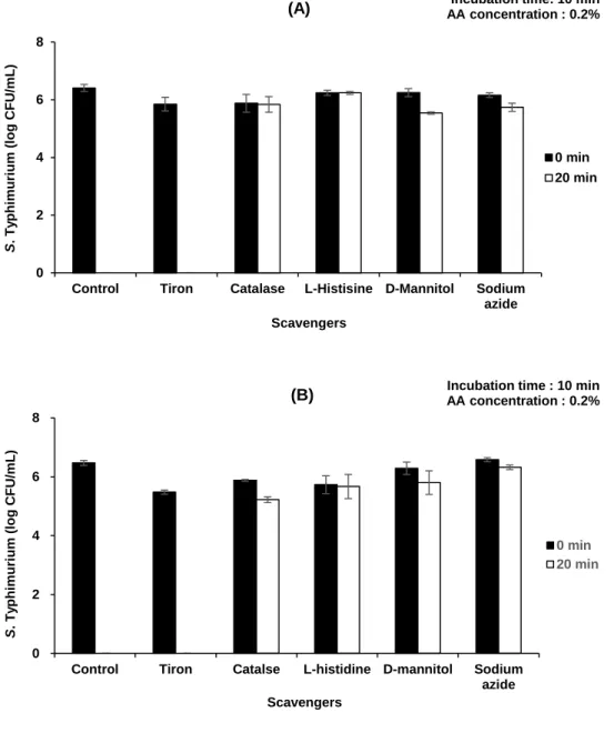
2.3.5. Effect of RONS scavengers on the antibacterial effect of PAAA
RONS scavengers were used to identify the major reactive species involved in the antibacterial activity of 0.2% PAAA. As shown in Figure 5A, the antibacterial activity of 0.2% PAAA was inhibited upon treatment with RONS scavengers, except tiron (O2∙−scavenger). This indicated that H2O2, 1O2, ∙OH, and NO2− mediated the antibacterial activity of 0.2% PAAA during plasma discharge for 20 min. Catalase and sodium azide scavenge H2O2 and NO2−needed for the formation of ONOOH through reaction (6), which impairs ONOOH formation and consequently inhibits the activity of 0.2% PAAA during plasma discharge. Thus, the role of ONOOH in the antibacterial activity of 0.2% PAAA is also supported by the data presented in Figure 5A. Tarabová et. al. (2019) demonstrated the formation and activity of ONOOH using the fluorescent dye H2DCFDA and RONS scavengers. ONOOH is a powerful oxidant that can diffuse through cell membranes, damage cells, and promote cell death through apoptosis and necrosis (Denicola et al., 1998; Huie et al., 1993). However, ONOOH damages the bacteria through its decomposition products, such as ∙OH and ∙NO2 free radicals under acidic conditions due to their short half- life (Oehmigen et al., 2011). The free radicals mentioned above are generated by the decomposition of ONOOH through reactions (9) and (10) during plasma discharge (Lukes et al., 2014; Radi et al., 2000).
ONOOH ↔ ∙OH + ∙NO2 (9)
ONOOH → HNO3 → NO3− + H+ (10)
27
Lukes et al. (2014) suggested that ONOOH can react directly or indirectly through the activity of ∙OH and ∙NO2 radicals, which are formed by the H+-catalyzed decomposition of ONOOH through reaction (9) at pH < 6.8. Additionally, ONOOH interacts with H2O2 to generate O2NOOH, which can be dissociated into 1O2, which plays a key role in the bactericidal properties of PAW through reactions (11)–(13) (Ikawa et al., 2016; Ma et al., 2020).
ONOOH + H2O2 → O2NOOH + H2O (11)
O2NOOH → O2NOO− (12)
O2NOO− → 1O2 + NO2− (13)
Thus, the dissociated species may be continuously generated and accumulated in 0.2% PAAA and consequently mediate the growth-inhibitory effect against S.
Typhimurium. This was consistent with the results of the scavenger analysis presented in Figure 5B, which exhibited a similar pattern to that shown in Figure 5A.
As shown in Figure 5B, the activity of ONOOH continued for a short period even after the cessation of plasma discharge. Piskarev et al. (2014) reported that the half- life of ONOOH is approximately 1.3 s under acidic conditions because ONOOH is continuously produced in PAW due to the presence of H2O2 and NO2−. As shown in Figure 5A, a series of activities that occurred during the plasma discharge also occurred for a short time immediately after the end of plasma discharge. Thus, the generation and decomposition of ONOOH are also considered to mediate the bactericidal effect of 0.2% PAAA immediately after plasma discharge.
28
Figure 5. Effect of reactive oxygen and nitrogen species scavengers added before (A) or after (B) plasma discharge for 20 min on the viability of bacterial cells. Control group, without ant treatment. Error bars represent standard deviation.
0 2 4 6 8
Control Tiron Catalase L-Histisine D-Mannitol Sodium azide
S. Typhimurium (log CFU/mL)
Scavengers
0 min 20 min
(A) Incubation time: 10 min
AA concentration : 0.2%
0 2 4 6 8
Control Tiron Catalse L-histidine D-mannitol Sodium azide
S. Typhimurium (log CFU/mL)
Scavengers
0 min 20 min
(B) Incubation time : 10 min
AA concentration : 0.2%
29
2.3.6. Detection of peroxynitrous acid (ONOOH) in PAAA
As ONOOH is strongly dependent on the pH, it is important to understand the activity of ONOOH under acidic conditions (Tarabová et al., 2019). The generation of ONOOH in 0.2% PAAA according to different plasma discharge time and during post-plasma discharge time was examined. As shown in Figure 6A, ONOOH production did not significantly vary with the plasma discharge time. This is because the generation and decomposition of ONOOH occur simultaneously under acidic conditions. The change in the fluorescence signal of ONOOH in 0.2% PAAA (obtained after plasma discharge for 20 min) during the post-plasma discharge time was also investigated. As shown in Figure 6B, the fluorescence signal significantly decreased at 5 min post-discharge (p < 0.05). The changes in ONOOH levels post- discharge exhibited a similar trend as those before plasma discharge (Figure 6A).
This was because of the decomposition of ONOOH under acidic conditions (Figure 6B). Consequently, the antibacterial effect of ONOOH decreased at 5 min post- discharge (Figure 6C).
Zhou et al. (2018) reported that the concentration of ONOOH in PAW varies according to the storage time and pH, which indicated that alkaline conditions play an active role in prolonging the bactericidal activity of PAW. Therefore, the results presented in Figure 6C suggest that the activities of various radicals produced in ONOOH could be limited during the post-plasma discharge time. This explains the decreased antibacterial effect of 0.2% PAAA at 5 min post-discharge. However, the viable bacterial population slightly increased at 5 min post-discharge (Figure 6C).
Hence, other reactive species may also be involved in the antibacterial effect of 0.2%
30
PAAA. Further studies are needed to identify additional reactive species that contribute to the bactericidal effect of PAAA.
31
Figure 6. (A) Detection of peroxynitrous acid (ONOOH) in 0.2% plasma-activated acetic acid (PAAA) during plasma discharge using the fluorescence dye 2,7- dichlorodihydrofluorescein diacetate (H2DCFDA); (B) Detection of ONOOH in 0.2%
PAAA post-plasma discharge; (C) The viable bacterial cell count post-plasma discharge.
Error bars represent standard deviation. a-bDifferent letters indicate a significant difference (p < 0.05) among the treatments.
b
a a a a
0 100 200 300
0 5 10 15 20
Fluorescence [a.u.]
Plasma discharge time (min) (A)
a
b 0
100 200 300
0 5
Fluorescence [a.u.]
Post-discharge time (min) Plasma discharge time : 20 min (B)
b
a 0
2 4 6 8
0 5
S.Typhimurium (log CFU/mL)
Post-discharge time (min) Plamsa discharge time : 20 min
Incubation time : 10 min (C)
32
2.4. Conclusion
This study aimed to combine AA with plasma treatment to enhance its bactericidal effects. This treatment combination was hypothesized to exert a synergistic bactericidal effect when compared with the individual treatment. The growth-inhibitory activities of PAAA against S. Typhimurium cells and biofilms were examined under optimal conditions. The findings of this study indicated that PAAA effectively inactivated the bacteria. Additionally, the role of ONOOH in the bactericidal effect of PAAA was analyzed using scavengers and a fluorescence dye.
The analysis revealed that ONOOH functioned as an intermediate agent and decomposed under acidic conditions. ONOOH can lead to the production of free radical species, such as ∙OH, ∙NO2, and 1O2, which play a key role in the bactericidal effect. Thus, PAAA is a potential novel decontamination agent that can reduce the pathogenic bacterial count. However, further studies are needed to examine the activity of other bactericidal agents, such as peroxyacetic acid in combination with PAAA. The increased understanding of the fundamental chemical processes enables further optimization of the efficacy and selectivity of this technology. Additionally, future studies must focus on the effect of PAAA treatment on the quality of food for applications in the food industry.
33
Chapter III.
Effect of plasma-activated acetic acid on inactivation of Salmonella Typhimurium and quality traits on chicken meats
This manuscript consists of part of a paper submitted to LWT – Food Science and Technology as partial fulfillment of the Master's program of Taemin Kang.
3.1. Introduction
With the increasing consumption of meat and meat product, the number of food- borne pathogens outbreaks related to meat has significantly increased (Zhao et al., 2001). Meat and meat products are highly susceptible to contamination by food- borne pathogens such as Salmonella spp., Campylobacter spp., Shiga toxin- producing strains of Escherichia coli, and Listeria monocytogenes, during their production, processing, and transportation (Kang et al., 2019; Nerin et al., 2016;
Omer et al., 2018). Particularly, chicken meat is a highly perishable product because of its characteristics that can cause rapid and intensive spoilage (Noriega et al., 2011).
Most of the pathogen contamination in chicken meat can occur in slaughter houses through spread of microorganisms between carcasses (Kim et al., 2019). A previous
34
study reported that Salmonella spp. account for majority of the food-borne pathogens identified in poultry and poultry products (Dominguez et al., 2002). Therefore, many chicken meat industries face problems in the effective inactivation of Salmonella spp.
as well as in ensuring that the quality of chicken meat is maintained. However, the inactivation of pathogens and deterioration of the quality of chicken meat remains a significant challenge (Dirks et al., 2012). Numerous efforts have been made to inactivate microbial contaminants in chicken meat using thermal treatments, use of bacteriocins or lactic acid bacteria, and washing with agents such as chlorine and trisodium phosphate (Mani-López et al., 2012). However, these traditional methods have some limitations in inactivating pathogens and adversely affect the nutritional value or sensory quality of chicken meat (Berrang et al., 2007; Kim et al., 2002;
Whyte et al., 2001).
In many countries, organic acids such as acetic, citric, and lactic acid are also used for the decontamination of chicken meat. According to the Food and Drug Administration (FDA), organic acids are recognized as safe food additives that can be used as antimicrobial agents in slaughterhouses (FDA, 1982). The antimicrobial property of organic acids is based on their ability to lower the pH, thereby causing instability in the bacterial cell membranes (Luck et al., 1998). With the increasing consumer demand that industries reduce their use of chemical additives, many food industries want to control the amount of organic acids used and simultaneously increase their antimicrobial effect in food products (Sagong et al., 2011).
Recently, non-thermal technologies that can ensure the safety of foods with minimum impact on quality have been developed as alternatives to traditional methods (Heo et al., 2021). Cold plasma has a minimal impact on the quality of meat
35
and meat products, is relatively inexpensive, and easy to install compared to other non-thermal technologies (Lee et al., 2016). In the present study, we treated plasma with acetic acid (AA) and the resulting plasma-activated acetic acid (PAAA) was expected to improve the bactericidal efficiency of AA. Some studies on lactic acid treated with plasma have studied the synergistic bactericidal effects (Qian et al., 2020;
Qian et al., 2021). However, no studies have reported the antibacterial effects of PAAA and the impact on the physicochemical traits of chicken meat. Therefore, the objective of this study was to investigate the antibacterial effects of PAAA and its impact on the quality characteristics of chicken meat.
36
3.2. Materials and methods
3.2.1. Antibacterial activity of AA and PAAA treatments on chicken meats 3.2.1.1. Bacterial strains and culture preparation
Salmonella Typhimurium (ATCC 13311) was obtained from the Korean Culture Center of Microorganisms (Seoul, Korea). S. Typhimurium was cultivated at 37°C in nutrient broth (Difco, Becton Dickinson Co., Sparks, MD, USA) for 48 h to obtain mid-log phase cells. The strain was then washed twice with 0.85% NaCl solution (saline), followed by centrifugation at 2,266 × g for 14 min at 2°C (UNION 32R, Hanil Science Industrial, Co. Ltd, Gimpo, Korea). Finally, the viable cell density of the re-suspended culture was adjusted to approximately 108