저작자표시-비영리-변경금지 2.0 대한민국 이용자는 아래의 조건을 따르는 경우에 한하여 자유롭게
l 이 저작물을 복제, 배포, 전송, 전시, 공연 및 방송할 수 있습니다. 다음과 같은 조건을 따라야 합니다:
l 귀하는, 이 저작물의 재이용이나 배포의 경우, 이 저작물에 적용된 이용허락조건 을 명확하게 나타내어야 합니다.
l 저작권자로부터 별도의 허가를 받으면 이러한 조건들은 적용되지 않습니다.
저작권법에 따른 이용자의 권리는 위의 내용에 의하여 영향을 받지 않습니다. 이것은 이용허락규약(Legal Code)을 이해하기 쉽게 요약한 것입니다.
Disclaimer
저작자표시. 귀하는 원저작자를 표시하여야 합니다.
비영리. 귀하는 이 저작물을 영리 목적으로 이용할 수 없습니다.
변경금지. 귀하는 이 저작물을 개작, 변형 또는 가공할 수 없습니다.
A Thesis for the Degree of Doctor of Philosophy in Pharmacy
Discovery of Bioactive Secondary Metabolites Produced by Gut Bacteria from
Lepidopteran and Coleopteran Insects
August 2020
Yern-Hyerk Shin
Natural Products Science Major, College of Pharmacy
Doctoral Course in the Graduate School
I
Abstract
Discovery of Bioactive Secondary Metabolites Produced by Gut Bacteria from
Lepidopteran and Coleopteran Insects
Yern-Hyerk Shin Natural Products Science Major College of Pharmacy Doctoral Course in the Graduate School Seoul National University
For the past decades, investigation of natural products has played significant role in drug development. Following the paper in 2020, 23.5% of globally approved drugs have been covered by natural product drugs, which also include botanical drugs and natural product-derived drugs. In addition, synthetic drugs provided their pharmacophore from natural products composed 14.2% of pharmaceutical drugs. In the case of antibacterial and anticancer drugs, the proportions of natural products/natural products-derived drugs were over 55% and 48%, respectively. These high percentages of natural product-based drugs in worldwide validated drugs indicated that discovering unreported natural products still has become a significant part of development of new drugs.
The proportion of natural products produced by bacteria (kingdom: Bacteria) in total compounds from living organisms is smaller than that of other kingdoms such as Plantae (67.4%), Animalia (12.6%), and Fungi (10.0%). However, 47% of bacteria-derived natural products showed bioactivities and 5.7% was reported to be bioactive compounds based on their potent biological activities. These results indicated that investigating bacterial secondary metabolites could be an efficient approach for discovering bioactive natural
products. In addition, William C. Campbell and Satoshi Omura were awarded the 2015 Nobel Prize in Physiology or Medicine for discovering of avermectins, a series of pesticides and drugs from territorial actinobacteria Streptomyces avermitillis, which showed the importance of studies on bacterial bioactive small molecules in drug development. Following these reasons, bioactive secondary metabolites of bacteria, mainly insect-associated bacteria, were investigated during my research times.
1. Bombyxamycins A–C and Piceamycin, Cytotoxic Macrocyclic Lactams from the Gut Streptomyces sp. of the Silkworm Bombyx mori
Bombyxamycins A–C (1–3) and piceamycin (4) producing bacterial strain, Streptomyces sp. SD53, was isolated from a gut of silkworm Bombyx mori. Combinational spectroscopic analysis revealed the planar structures of bombyxamycins A–C (1–3) and piceamycin (4) as 26-membered cyclic macrolactams with polyene features. The absolute configurations of bombyxamycins A–C (1–3) and piceamycin (4) were determined by multiple-step chemical derivatizations, spectroscopic analysis, chromatographic methods, and computational calculations. Additionally, a new chromatography-based experimental method for determining the configurations of stereogenic centers β to nitrogen atoms in macrolactams was established and successfully applied in this work. The bombyxamycins and piceamycin biosynthetic gene cluster was identified by genomic analysis of the actinobacteria strain. Bombyxamycin A (1) and piceamycin (4) displayed potent antibacterial activity against human pathogenic Gram-positive and Gram-negative bacteria. Furthermore, piceamycin (4) also inhibited growth of silkworm pathogen Bacillus thuringiensis.
Bombyxamycins A & C (1 and 3) and piceamycin (4) showed antiproliferative activities against human cancer cell lines.
III
2. Nicrophorusamides A and B, Antibacterial Chlorinated Cyclic Peptides from the Gut Microbacterium sp. of the Carrion Beetle Nicrophorus concolor
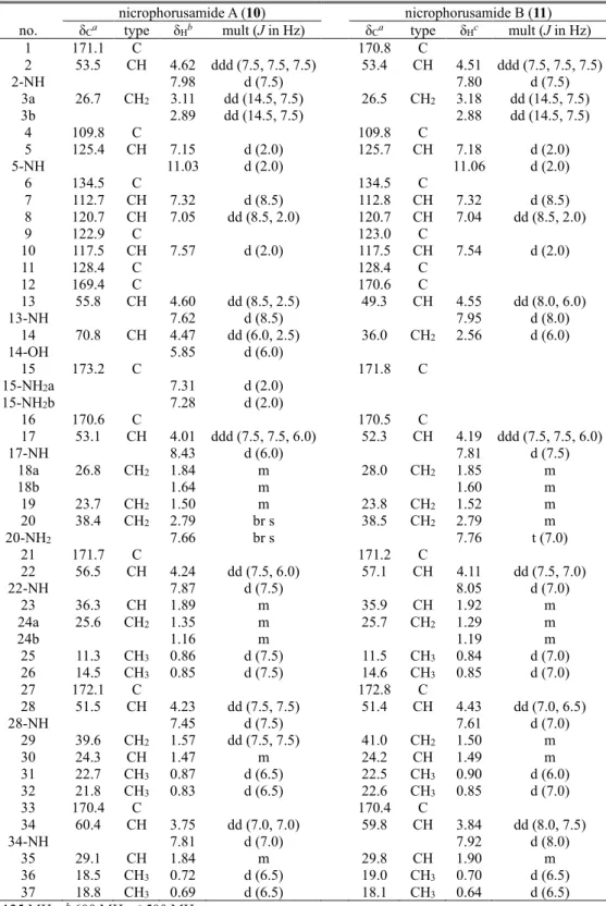
Nicrophorusamides A and B (10 and 11) were obtained during cultivation of a rare actinomycete Microbacterium sp. UTG9, which was isolated from the intestine of the carrion beetle Nicrophorus concolor. Based on the combinational analysis of their 1D & 2D NMR spectroscopic data, the structures of nicrophorusamides A and B (10 and 11) were established as a series of new chlorinated cyclic hexapeptides incorporating nonproteinogenic amino acid units such as 5-chloro-tryptophane and β-hydroxyasparagine. The absolute configurations of the amino acid residues in nicrophorusamide A (10) were determined by acid hydrolysis and L- and D- form of 1-fluoro-2,4-dinitrophenyl-5-alanine amide (FDAA) derivatizations followed by chromatographic analysis. In the case of nicrophorusamide B (11), the absolute configurations were deduced to be identical to those in nicrophorusamide A (10) based on identity between circular dichroism (CD) spectra and the common biosynthetic origin.
Nicrophorusamide A (10) displayed antibacterial activities against several human pathogenic bacterial strains.
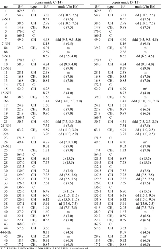
3. Coprisamides C and D, Cinnamic Acid Containing Cyclic Peptides from the Gut Micromonospora sp. of Carrion Beetle Silpha perforata
Coprisamides C and D (14 and 15), unreported cinnamic acid containing non- ribosomal peptides, were discovered from an intestinal Micromonospora sp. UTJ3, which was isolated from a gut of carrion beetle Silpha perforata. Based on the UV, MS, and NMR spectroscopic data, the planar structures of coprisamides C and D (14 and 15) were elucidated as heptacyclic depsipeptides bearing highly modified amino acid units such as β- methylaspartic acid (β-Me-Asp) and 2,3-diaminopropionic acid (2,3-Dap). The absolute configurations of coprisamides C and D (14 and 15) were identified by advanced Marfey’s method and phenylglycine methyl ester (PGME) derivatization followed by NMR spectroscopic analysis. The biosynthetic gene cluster and pathways were predicted based on
genomic analysis of the whole genome sequence data of the producing strain. Coprisamide C (14) exhibited mild growth inhibition activity against human tuberculosis causing bacteria Mycobacterium tuberculosis mc2 6230.
Keywords: Actinobacteria, Insect-associated bacteria, Secondary metabolites, Structure determination, Bioactivity, Biosynthesis
Student number: 2014-21975
V
List of Contents
Abstract ... I List of Contents ... V List of Figures ... VII List of Tables ... IX
Introduction ... 1 Introduction ... 2 General Experimental Procedures ... 6
Bombyxamycins A–C and Piceamycin, Cytotoxic Macrocyclic Lactams from the Gut Streptomyces sp. of the Silkworm Bombyx mori ... 7 Results and Discussion ... 8 Experimental Section ... 24
Nicrophorusamides A and B, Antibacterial Chlorinated Cyclic Peptides from the Gut Microbacterium sp. of the Carrion Beetle Nicrophorus concolor ... 41 Results and Discussion ... 42 Experimental Section ... 49
Coprisamides C and D, Antituberculosis Cinnamic Acid Containing Cyclic Peptides from the Gut Micromonospora sp. of the Carrion Beetle Silpha perforata ... 57 Results and Discussion ... 58 Experimental Section ... 67
Summary ... 73
References ... 76
Appendix: Supporting Information ... 82
List of Figures ... 83
List of Tables ... 91
Abstract in Korean ... 182
Publication List ... 194
Papers ... 195
Patents ... 197
Permissions for Republication of the Published Paper in Thesis ... 198
Acknowledgements ... 202
VII
List of Figures
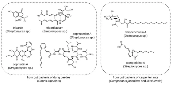
Figure 1. Previously reported secondary metabolites produced by intestinal bacteria of insects.
... 2
Figure 2. Key COSY and HMBC correlations for constructing the planar structures of bombyxamycins A–C (1–3) and piceamycin (4). ... 9
Figure 3. ΔδS-R values of S- and R-MTPA esters of bombyxamycins A–C (1–3). ... 10
Figure 4. Chemical reactions for the identification of the absolute configuration at C-24 of bombyxamycin A (1). ... 10
Figure 5. 13C NMR spectra of bombyxamycin B (2) in CD3OD and CD3OD:CD3OH = 1:1.
... 12
Figure 6. Preparing S- and R-PGME amides products from piceamycin (4). ... 15
Figure 7. Chromatographic analysis for the determination of the absolute configuration of the β-amino acid unit of piceamycin (4) by PGME derivatization. ... 17
Figure 8. Energy-minimized structures of S- and R-PGME amides of 3-amino-2- methylpropanoic acids (6–9). ... 18
Figure 9. Comparison of the experimental ECD data of piceamycin (4) with the calculated ECD data. ... 18
Figure 10. Biosynthetic gene cluster and pathways for bombyxamycins A–C (1–3) and piceamycin (4). ... 23
Figure 11. Chemical structures of nicrophorusamides A and B (10 and 11). ... 43
Figure 12. Structure determination of the uncommon amino acid units in nicrophorusamide A (10) based on COSY and HMBC correlations. ... 43
Figure 13. Identification of the amino acid sequence in nicrophorusamide A (10) based on HMBC and ROESY NMR spectra. ... 44
Figure 14. Energy minimized conformations of nicrophorusamides A and B (10 and 11).
... 48
Figure 15. Chemical structures of coprisamides C and D (14 and 15). ... 59
Figure 16. Key COSY and HMBC correlations for structural determining of coprisamide C (14).
... 60
Figure 17. Determining amino acid sequence of coprisamide C (14) based on HMBC and ROESY correlations. ... 62
Figure 18. Identification of the absolute configuration at C-3 based on the ΔδS-R values in ppm of the S- and R-PGME amides of coprisamide C (14a and 14b). ... 62
Figure 19. Dose response curve of coprisamides C and D (14 and 15) against Mycobacterium tuberculosis mc2 6230. ... 63
Figure 20. Biosynthetic gene cluster and pathways for coprisamides C and D (14 and 15).
... 66
IX
List of Tables
Table 1. 1H and 13C NMR data for bombyxamycins A and B (1 and 2). ... 38
Table 2. 1H and 13C NMR data for bombyxamycin C and piceamycin (3 and 4). ... 39
Table 3. Antibacterial activities of bombyxamycins A–C (1–3) and piceamycin (4) against human and silkworm pathogenic strains. ... 40
Table 4. Antiproliferative activities of bombyxamycins A–C (1–3) and piceamycin (4) against various human cancer cell lines. ... 40
Table 5. 1H and 13C NMR Data for nicrophorusamides A and B (10 and 11) in DMSO-d6. ... 55
Table 6. Inhibitory activities of nicrophorusamides A and B (10 and 11) against bacterial strains. ... 56
Table 7. 1H and 13C NMR spectra data for coprisamides C and D (14 and 15) in DMSO-d6. ... 72
Introduction
2
Introduction
Figure 1. Previously reported secondary metabolites produced by intestinal bacteria of insects.
Insects are the largest animal clade on Earth based on their diversity in species and entire biomass.1 The total number of their species are estimated to be around 1 million, which means that over half of all reported eukaryotes (~1.8 million) belong to insects.2 Insects are also ubiquitous because they are one of the oldest animals on our planet, so they have adapted to a number of different environments for survive. Based on these kinds of diversity, insects could provide exponentially various habitats for their gut bacteria communities increasing their genetic diversity. As a result, investigating secondary metabolites from insect’s gut bacteria lead me to discover unprecedent and diverse bioactive small molecules. For example, several kinds of secondary metabolites produced by dung beetles’ (Copris tripartitus) gut Streptomyces strains have been reported (Figure 1) such as tripartin,3 histone demethylase inhibiting dechlorinated indanone, tripartilactam,4 cytotoxic tricyclic macrolactam, coprisidins,5 naphthoquinone–oxindole alkaloids, and coprisamides,6 branched cyclic heptapeptides. Deinococcucins, which exhibited quinone reductase inducing activity, were
from gut bacteria of dung beetles (Copris tripartitus)
tripartilactam (Streptomycessp.)
coprisidin A (Streptomycessp.)
coprisamide A (Streptomycessp.) tripartin
(Streptomycessp.) deinococcucin A
(Deinococcussp.)
camporidine A (Streptomyces sp.)
from gut bacteria of carpenter ants (Camponotus japonicus and kiusiuensis)
also discovered from a gut Deinococcus strain of queen carpenter ants, Camponotus japonicus (Figure 1).7 Furthermore, another study on intestinal bacteria of other species of carpenter ants, Camponotus kiusiuensis, discovered two unreported alkaloids with anti- inflammatory activity, camporidines A and B (Figure 1).8
During my research, intestinal bacterial strains were isolated from Coleoptera and Lepidoptera, which are two of the hugest orders among insect. At first, intestinal bacteria of Lepidopteran insects were isolated and chemically investigated. Though Lepidoptera is the second largest order after Coleoptera,9 the chemistry of microbes associated with Lepidopteran insects was rarely studied. Only a proteinaceous toxin, identified as an antibiotic modulating the gut microbiota from a symbiotic bacterium, Enterococcus mundtii, in the cotton leafworm Spodoptera littoralis was discovered in 2017.10 The silkworm Bombyx mori, including its microbiota, is the most heavily studied Lepidopteran insect because of its industrial and medicinal importance.11 The paucity of chemical investigations of microbes in B. mori led me to focus on silkworm gut bacteria. In chemical examination of gut microorganisms of B. mori larvae, a bacterial strain, Streptomyces sp. SD53, was thought to produce a new series of polyene compounds based on their ultraviolet (UV) spectroscopic data and mass spectrometry (MS) data. Large-scale culture of the Streptomyces strain and subsequent chromatographic purification allowed the discovery of previously unreported macrocyclic lactams, bombyxamycins A–C, as the first small molecules from Lepidopteran insect-associated microbes. In addition, further investigations based on chemical profiling of the bacterial strain during cultivation in various culture media allowed me to detect additional macrocyclic lactams from the strain SD53. Large-scale cultivation of the strain in a specific medium enabled the production of piceamycin, which was discovered from Streptomyces sp.
GB4-2 isolated from the mycorrhizosphere of Picea abies.12 Although the planar structure of
4
originates from a secondary cyclization after the biosynthesis of the 26-membered macrocycle. In this work, a convenient chromatography-based method to determine the chirality of a carbon possessing a branching methyl group to the amide nitrogen, which is a motif commonly found in macrolactams prepared from a -amino acid, was developed and electronic circular dichroism (ECD) calculations was used to elucidate the stereogenic center in the cyclopentenone of piceamycin. The biological evaluation of piceamycin and bombyxamycins A–C in antibacterial and antiproliferative assays were also reported in this paper as well as the predicted post-PKS modification biosynthetic steps leading to bombyxamycin B and piceamycin from bombyxamycins A and C, respectively.
Gut bacteria isolated from Coleopteran insects were also chemically investigated. Especially, carrion beetles (Coleoptera, Silphidae) are ecologically interesting because they utilize vertebrate carrion to rear their offspring.13 Carrion beetles are, thus, exposed to carrion-borne bacteria, which may be pathogenic to them during development.14 Carrion beetles could be classified into two subfamilies, Nicrophorinae and Siphinae.15 A recent analysis of the gut microbiome of six carrion beetle species belonging to the genus Nicrophorus (subfamily Nicrophorinae) indicated that these carrion-feeding beetles harbor gut microbial communities that are distinctively different from those of herbivorous, xylophagous, humivorous, omnivorous, and predatory beetles.13 Although that study proposed hypothetical roles of carrion beetles’ gut symbionts, such as carcass degradation, detoxification, and defense,13 the mechanisms by which carrion beetles defend themselves against entomopathogenic bacteria originating from carrion have not yet been clearly elucidated. In this context, I assumed that the gut symbiotic bacteria of the carrion beetle Nicrophorus concolor may be a potential source of antimicrobial compounds. Therefore, the intestinal parts were extracted from a N.
concolor specimen and the bacterial strains were isolated, targeting chemically prolific actinobacteria. The isolated actinobacterial strains were cultivated and chemically analyzed.
During the chemical analysis, a rare actinomycete strain (UTG9) belonging to the genus Microbacterium was found to produce a series of previously unreported compounds based
on UV and MS data with a characteristic isotopic pattern corresponding to chlorination. This initial chemical evaluation prompted a large-scale cultivation and deeper chemical investigation by chromatographic purification and spectroscopic analysis of the two major compounds, nicrophorusamides A and B.
On the other hand, gut bacteria of Silpha perforata, a flightless roving carrion beetle species classified as a species of the other subfamily Siphinae16 were also subjected because microbes associated even in the genus Silpha have not been chemically investigated yet as only chemical reports about the genus were defensive steroids produced from rectal glands in S.
novaboracensis and S. americana.17, 18 Chemically profiling of gut bacterial isolates of S.
perforata based on their LC/MS chromatogram data detected the production of unidentified secondary metabolites in the culture of Micromonospora sp. UTJ3 strain. Based on the UV spectra of these two compounds and the mass spectrometric data along with accumulated UV library (~2,000 compounds) indicated that they were recognized as undiscovered bacterial small molecules, leading me to further investigation. Finally, large-scale culture, chromatographic purification, and spectroscopic analysis revealed the planar structures of the unknown compounds, coprisamides C and D, which structurally related to coprisamides A and B, which were isolated from the gut bacterium Streptomyces sp. SNU533 of the dung beetle Copris tripartitus.6 Coprisamides bear 2-alkenyl-cinnamic acid unit as an acyl chain and modified amino acids with a distinct branched amino acid chain, indicating an interesting but unreported biosynthetic pathway.
6
General Experimental Procedures
Optical rotations were measured on a JASCO P-2000 polarimeter with a 1-cm cell at 20 °C and 25 °C. Ultraviolet (UV) spectral data and circular dichroism (CD) data were recorded on an Applied Photophysics ChirascanTM plus circular dichroism detector at 25 °C using a 1-cm quartz cell. Infrared (IR) spectral data were collected on a JASCO FT/IR-4200 FT-IR spectrometer. 1H, 13C, and 2D NMR experiments were conducted by using Bruker Avance III 500 MHz, 600 MHz, 800 MHz, and 850 MHz spectrometers. Low-resolution electrospray ionization mass spectrometry (LR-ESI-MS) and UV chromatogram data were acquired using an Agilent Technologies 6130 quadrupole mass spectrometer coupled with an Agilent Technologies 1200-series HPLC using a reversed-phase C18(2) column (Phenomenex Luna, 5 μm, 100 4.6 mm). HR-FAB (fast atom bombardment)-MS data and HR-ESI-MS data were acquired by using a JEOL JMS-700 HR-MS and AB Sciex 5600 QTOF HR-MS, respectively. The NMR spectrometers (500 MHz, 600 MHz, and 850 MHz) and HR-FAB- MS were located at the National Center for Interuniversity Research Facilities (NCIRF) in Seoul National University. The HR-ESI-MS was located at the National Instrumentation Center for Environmental Management (NICEM) in Seoul National University.
1. Bombyxamycins A–C and Piceamycin, Cytotoxic Macrocyclic Lactams from the Gut Streptomyces sp.
of the Silkworm Bombyx mori
8
1.1. Results and Discussion
Bombyxamycin A (1) was isolated as yellow powder. A molecular formula of C27H33NO3
was identified by high-resolution fast atom bombardment mass spectrometry (HR-FAB-MS).
Based on the molecular formula, the unsaturation number of 1 was deduced as 12. Combined analysis of 1H and HSQC NMR data of 1 showed the existence of one amide proton (δH 7.97) and 17 olefinic methine protons in the downfield region (δH 7.45–5.23) (Table 1). One oxygen-bound methine proton (δH 4.04), one hydroxy proton (δH 4.77), six protons (δH 3.46, 2.86, 2.46, 2.31 [2H], and 2.30) belonging to three methylene groups, one aliphatic methine proton (δH 3.12), and two methyl group protons (δH 1.64 and 0.94) were also identified by 1H and HSQC NMR data. The 13C NMR spectral data (Table 1) revealed that 1 bears two carbonyl carbons (δC 199.8 and 166.3), eighteen olefinic carbons (δC 140–120), one oxygen- bound carbon (δC 68.5), and six aliphatic carbons, including two methyl groups (δC 17.9 and 12.5). Based on the NMR and UV spectroscopic data, 1 was deduced to possess a couple of conjugated chromophores with double bonds in its structure.
By interpretation of COSY NMR correlations of 1, three discrete spin systems were identified. One of the spin systems from C-2 could be successfully traced to C-7 by a series of COSY correlations from H-2 (δH 5.55) to H-7 (δH 6.41). Consecutive 1H–1H couplings from H-9 (δH 5.52) to H2-12 (δH 2.86 and 2.46) through H-10 (δH 2.31) and H-11 (δH 4.04) established the C-9–C-10–C-11–C-12 connectivity. The last spin system was assigned based on an array of COSY signals from H-14 (δH 6.15) to 25-NH (δH 7.97). H3-2′ (δH 0.94) was connected to C-24 by H3-2′/H-24 (δH 3.12) COSY correlations.
An HMBC correlation from H-2 to C-1 (δC 166.3) established the connectivity between the olefinic carbon C-2 (δC 121.3) and the carbonyl carbon C-1. Analysis of HMBC correlations from the methyl protons H3-1′ (δH 1.64) to C-7 (δC 140.1), C-8 (δC 135.1), and C-9 (δC 131.4) connected the first and the second spin systems through the fully substituted olefinic carbon C-8. Furthermore, the 2-bond HMBC signals from H2-12 and H-14 to C-13 (δC 199.8) indicated that the carbonyl carbon C-13 was between the second and the third spin systems,
resulting in a pentaenone-bearing structure. Because two carbonyl groups and nine double bonds explained 11 unsaturation equivalents out of 12, 1 was deduced to possess a ring.
Finally, a 25-NH/C-1 HMBC correlation established the 26-membered monocyclic structure of 1 (Figure 2a). The double bond geometries in 1 were clarified as 2Z, 4Z, 6E, 14E, 16Z, 18Z, 20E, and 22Z by measuring 3JHH values of the olefinic protons (Table 1 and Figure S2).
The 8Z geometry was established by H-9/H3-1′ NOESY correlation.
Figure 2. Key COSY and HMBC correlations for constructing the planar structures of (a) bombyxamycin A (1), (b) bombyxamycin B (2), (c) bombyxamycin C (3), and (d) piceamycin (4).
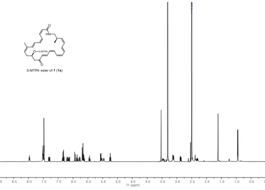
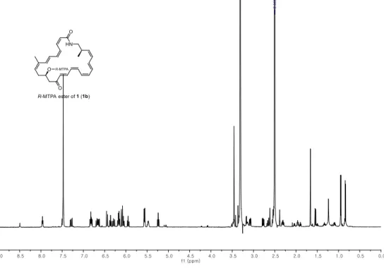
The absolute configuration of C-11 of 1 was established by the modified Mosher’s method.19 The secondary alcohol at C-11 was derivatized with R- and S-α-methoxy-(trifluoromethyl) phenyl acetyl chloride (MTPA-Cl) to yield S- and R-MTPA esters (1a and 1b) (Figure 3a).
ΔδS-R values were calculated by analysis of 1H and COSY NMR spectra of 1a and 1b. The signs of ΔδS-R values were consistently distributed, assigning the 11R configuration (Figure 3a).
To determine the absolute configuration of C-24 in 1, ozonolysis and acid hydrolysis of 1 were conducted (Figure 4).4 After the sequential chemical reactions, the desired β-amino acid (3-amino-2-methyl-propanoic acid) was derivatized with Sanger’s reagent (1-fluoro-2,4-
COSY HMBC (a)
bombyxamycin A (1) bombyxamycin B (2) bombyxamycin C (3) piceamycin (4)
(b) (c) (d)
10
products (5a and 5b) enabled the determination of the absolute configuration of 5 as 2S based on ΔδS-R values, thus establishing the 24R configuration of 1 (Figure 4).
Figure 3. ΔδS-R values of S- and R-MTPA esters of (a) bombyxamycin A (1), (b) bombyxamycin B (2), and (c) bombyxamycin C (3).
Figure 4. Chemical reactions for the identification of the absolute configuration at C-24 of bombyxamycin A (1). ΔδS-R values in ppm are noted for 5a and 5b.
Bombyxamycin B (2) was obtained as pale-yellow powder by consecutive chromatographic isolation. The molecular formula of 2 could be assigned as C27H31NO5 with an unsaturation number of 13 by using HR-FAB-MS data. Analysis of 1D and 2D NMR data of 2 (Table 1) revealed that it possesses two carbonyl carbons, nine double bonds, three oxygen-bearing sp3 carbons, and four aliphatic carbons, including two methyl groups. Because 2 has one more unsaturation equivalent than and the same numbers of carbonyl carbons and double bonds as
(a) (b) (c)
1, it was deduced that 2 must bear an additional ring. Examination of the COSY NMR spectrum elucidated the two polyene spin systems (C-1 to C-7 and C-14 to 25-NH) that were consistently found in 1 (Figure 2b). The other spin system was composed of two oxygen- bearing methine groups, and two hydroxyl groups were identified by H-9/H-10, H-9/9-OH, and H-10/10-OH COSY correlations. The HMBC correlation signals from H3-1′ to C-7, C-8, and C-9 demonstrated C-7–C-8–C-9 connectivity. In addition, 1H–13C correlations from H- 10 to C-11 and from H-12 to C-11, C-13, and C-14 allowed for the construction of a long polyketide chain from C-1 to 25-NH. As shown in 1, a 26-membered ring structure of 2 could be secured by HMBC correlation between 25-NH and C-1 (Figure 2b).
The planar structure of 2 was then proposed by connecting the last oxygen-bearing sp3 carbon, C-8 (δC 89.1), and the fully substituted, highly deshielded olefinic carbon C-11 (δC
167.6) through an ether linkage to make one more ring based on the molecular formula even though HMBC correlations indicating the five-membered ether ring could not be observed because C-8 and C-11 do not have a proton (Figure 2b). However, the ether bond was further supported by 13C NMR experiments for 2 in CD3OD and a 1:1 mixture of CD3OD–CD3OH (Figure 5). Carbon signals that are located at the β-position of exchangeable protons are doubled or broadened in a 1:1 mixture of CD3OD and CD3OH by a β-isotope effect.21 The carbon peaks of C-9 and C-10 of 2 were broadened, whereas C-8 and C-11 peaks were not affected by the mixed solvent, indicating that C-8 and C-11 are oxygen bearing but not bound to –OH (Figure 5). This result strongly supported the elucidated structure of 2.
The relative configuration of the tetrahydrofuran (THF) moiety was assigned by analysis of NOESY NMR data. H-7/H-9, and H3-1′/H-10 NOESY correlations indicated that H-7 and H-9 are in the same phase. In contrast, H3-1′ displayed a NOESY correlation with H-10, indicating that these protons are on the other side of the molecule. Therefore, the relative
12
Figure 5. 13C NMR spectra of bombyxamycin B (2) in (a) CD3OD and (b) CD3OD:CD3OH
= 1:1.
The absolute configurations at C-8, C-9, and C-10 of 2 were determined by the application of the modified Mosher’s method.19 With a short derivatization time (5 min) and using R- and S-MTPA-Cl, mono-S- and R-MTPA esters of 2 (2a and 2b) were predominantly obtained rather than bis-MTPA esters. Analysis of 1H and COSY NMR spectral data of 2a and 2b revealed that 10-OH in 2 was esterified. Calculation of ΔδS-R values enabled the determination of the 10S configuration. Based on the relative configurations, the absolute configurations of C-8 and C-9 were assigned as 8R and 9S (Figure 3b). The absolute configuration of C-24 was determined as R by the same procedure mentioned above for 1.
Bombyxamycin C (3), purified as a dark yellow powder, was found to have a formula of C27H33NO3, corresponding to 12 degrees of unsaturation, by HR-FAB-MS. Analysis of its 1H and HSQC NMR spectroscopic data (Table 2) indicated that 3 possessed one amide proton (δH 7.71), 17 olefinic protons (δH 7.00–5.19), and one carbinol proton (δH 4.09) in the deshielded region. In addition, six methylene protons (δH 3.36, 3.08, 2.74, 2.19 (2H), and 2.18), one aliphatic methine proton (δH 2.71), and two methyl groups (δH 1.49 and 0.96) were identified in the shielded region. Interpretation of the 13C and HSQC NMR spectra of 3 indicated the presence of two carbonyl carbons (δC 199.1 and 165.9) and 18 olefinic methine carbons (δC 144.3, 140.3, 137.8, 136.6, 135.7, 134.6, 133.3, 132.3, 132.3, 131.2, 130.6, 130.5, 129.7, 127.5, 127.0, 124.4, 123.3, and 120.9) in the structure. In addition, one oxygen-bound carbon (C 67.3) and six aliphatic carbons (δC 44.0, 43.0, 34.4, 33.7, 17.7, and 12.3) were identified (Table 2). Initial analysis of its 1D and HSQC NMR spectra suggested that
(a) CD3OD (b) CD3OD:CD3OH = 1:1
C-11 C-8 C-9 C-10 C-11 C-8 C-9 C-10
bombyxamycin C (3) should possess two carbonyl groups and nine double bonds, explaining 11 out of the 12 unsaturations inherent in the molecular formula. Therefore, 3 must have one ring.
The discrete spin systems were elucidated by analysis of the COSY NMR correlations.
Consecutive COSY correlations of the olefinic methine protons, from H-2 (δH 5.58) to H-7 (δH 6.50), revealed the connectivity from C-2 (δC 123.3) to C-7 (δC 140.3). Another spin system (C-9–C-12 linkage) was constructed via three-bond 1H–1H couplings among H-9 (δH
5.84), H2-10 (δH 2.19), H-11 (δH 4.09), and H2-12 (δH 3.08 and 2.18). A long spin system from H-14 (δH 5.99) to 25-NH (δH 7.71) was established based on the COSY signals of the protons belonging to this system. CH3-2′ could be assigned at C-24 by the 1H–1H COSY correlation between H3-2′ (δH 0.96) and H-24 (δH 2.71). An HMBC correlation between H-2 and the amide carbon C-1 (δC 165.9) indicated that C-1 is adjacent to C-2. 1H–13C HMBC signals from H-1′ (δH 1.49) to C-7, C-8 (δC 135.7), and C-9 (δC 129.7) connected the first and the second spin systems, forming the tetraenone chromophore of 3. This partial structure could be combined with the long spin system bearing the pentaene through the C-13 ketone carbon based on the HMBC correlations from H2-12 and H-14 to C-13 (δC 199.1). Furthermore, the 25-NH/C-1 1H–13C correlation connected 25-NH to C-1, generating the 26-membered macrocyclic structure of 3 (Figure 2c). The geometries of the double bonds in bombyxamycin C (3) could be assigned as 2Z, 4Z, 6E, 14E, 16E, 18Z, 20E, and 22Z from the 3JHH values between olefinic protons (Table 2). In addition, the NOESY correlation between H3-1′ and H-9 enabled the identification of the 8Z geometry.
Bombyxamycin C (3) has two stereogenic centers: C-11 bearing a secondary alcohol and C-24 at the β-position of the amide nitrogen. The secondary hydroxy group at C-11 was esterified with R- and S-MTPA-Cl, yielding S- and R-MTPA esters of 3 (3a and 3b),
14
During the cultivation of Streptomyces sp. SD53 in YPM medium, a metabolite with a polyene UV signature (max values of 296 and 410 nm), which is unlike those of bombyxamycins A–C (1–3), was detected by LC/MS analysis. Subsequent chromatographic separation yielded a pure orange powder, enabling spectroscopic analysis of the metabolite (4). The molecular formula of 4 was identified as C27H29NO4, corresponding to 14 double bond equivalents, by HR-FAB-MS data. The planar structure of 4 was deduced as that of piceamycin, which was previously reported from Streptomyces sp. GB 4-2 isolated from the mycorrhizosphere of Picea abies, by comparing its 1H and 13C NMR chemical shifts in DMSO-d6 (Table 2) with those in the literature.12 Its was further confirmed to be piceamycin by COSY, HSQC, and HMBC spectroscopic analyses (Figure 2d). Careful comparison of the NMR data of 4 with the literature data of piceamycin revealed that the chemical shifts and coupling constants of the methylene protons at C-9 must be revised. In the original report, H2-9 was assigned as δH 2.42 (d, J = 5.53 Hz). However, if the methylene protons are magnetically equivalent, these protons should appear as a singlet because this methylene group is flanked by a ketone group and an aliphatic quaternary carbon. In fact, these two protons are not magnetically equivalent and should be considered separate signals at δH 2.43 and 2.39 with 19.0 Hz geminal coupling (Table 2), which is characteristic of a five-membered ring.
The two stereogenic centers of piceamycin (4) remain unassigned, prompting me to take on this challenge. Indeed, the stereogenic center at the -position of the amide nitrogen is derived from the -amino acid starting material (3-amino-2-methylpropanoic acid) and is a common motif to all macrocyclic lactams in this class, as shown in cyclamenol A,22 macrotermycins A–D,23 sceliphrolactam,24 niizalactam C,25 and vicenistatin.26 However, the absolute configurations of the chiral carbons at this position can be either R or S depending on the natural product, indicating that L-glutamic acid is being converted to 3-amino-2- methylpropanoic acid stereo-selectively by an unknown mechanism in the biosynthesis of the starting material. The unambiguous determination of this -amino acid starting material-
derived stereogenic center has been time consuming and inefficient as a total synthesis or at least a 4-step degradation process of a macrocyclic lactam followed by product purification and 1H NMR spectroscopic analysis were required.4
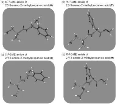
Figure 6. Preparing S- and R-PGME amides products from piceamycin (4).
Therefore, a new method for elucidating this common but problematic stereogenic center by combining simple chemical derivatization and chromatographic analysis without preparative purification and 1H NMR spectroscopic analysis of the products (Figure 6) was developed. First, to obtain a β-amino acid unit bearing the C-24 stereogenic center, piceamycin (4) was sequentially subjected to ozonolysis and acid hydrolysis. Then, portions of the hydrolysate were separately derivatized with S- or R-phenylglycine methyl ester (PGME).20 The S- and R-PGME products were analyzed via LC/MS, and their retention times were compared with those of the PGME amides (6–9) prepared with authentic 2S- and 2R-3- amino-2-methylpropanoic acids (Figure 7). The S-PGME adduct (6) of 2S-3-amino-2- methylpropanoic acid eluted later (8.3 min) than the R-PGME derivative (7) (6.4 min) (Figure 7). LC/MS analysis of the PGME adducts of 2R-3-amino-2-methylpropanoic acid showed
16
methyl group from the -amino acid and the phenyl group of PGME on the same side, which allows the formation of more hydrophobic interaction with the reversed-phase stationary phase, resulting in a higher retention time (Figures 7a and 7b). On the other hand, these hydrophobic functional groups are oriented in opposite directions in the S–R (or R–S) PGME amide, resulting in weaker interactions with the reversed-phase stationary phase and thus faster elution (Figures 7a and 7b). The PGME derivatives originating from 4 showed retention times consistent with those of the PGME amides of 2S-3-amino-2-methylpropanoic acid (Figure 7c), confirming the absolute configuration of 3-amino-2-methylpropanoic acid derived from piceamycin (4) as 2S and thus 24R in 4. This convenient procedure provides an empirical rule: if the S-PGME amide of the 3-amino-2-methylpropanoic acid unit derived from a macrocyclic lactam elutes slower than its R-PGME amide, this -amino acid must possess a 2S-configuration. Conversely, the 2R configuration can be assigned when the R- PGME amide elutes later than its S-PGME adduct. This method was also conducted with a small quantity of piceamycin (4, 0.2 mg), which provided the identical experimental results, indicating its validity on the 0.2 mg scale (Figure S65). The absolute configuration of the asymmetric carbon at C-24 of 3 was also determined as R by the same three-step reaction followed by LC/MS analysis as used for 4, and this process did not require NMR experiments.
As a proof of the versatility of the method developed in this work, this chiral center was successfully established using only 1 mg of 3.
Figure 7. Chromatographic analysis for the determination of the absolute configuration of the β-amino acid unit (3-amino-2-methylpropanoic acid) of 4 by PGME derivatization.
LC/MS chromatograms of the S- and R-PGME derivatives of (a) 2S-3-amino-2- methylpropanoic acid (6 and 8), (b) 2R-3-amino-2-methylpropanoic acid (7 and 9), and (c) 3-amino-2-methylpropanoic acid from 4 (Phenomenex, Luna, C18(2), 100 × 4.6 mm; 10%−25%
CH3CN−H2O over 30 min with 0.1% formic acid; flow rate: 0.7 mL/min; ion extraction for [M + H]+ m/z at 251).
18
Figure 8. Energy-minimized structures of S- and R-PGME amides of 3-amino-2- methylpropanoic acids (6–9).
Figure 9. Comparison of the experimental ECD data of piceamycin (4) with the calculated ECD data; solid line: experimental ECD data; dotted line: calculated ECD data (8S, 24R);
dashed line: calculated ECD data (8R, 24R).
The other asymmetric carbon in piceamycin (4) is the unprotonated C-8 moiety of the cyclopentenone. Because chemical derivatization of this chiral center for stereochemical analysis is not possible, electronic circular dichroism (ECD) calculations were utilized.27 Three-dimensional models of two possible diastereomers of 4 (8S/24R and 8R/24R) were
Experimental ECD of piceamycin (4) Calculated ECD of 4(8S, 24R) Calculated ECD of 4(8R, 24R)
250 300 350 400 450 500
-8 -6 -4 -2 0 2 4
Δε
wavelength (nm)
constructed, and ECD calculations were conducted for each diastereomer. The experimental ECD spectrum of 4 showed a positive Cotton effect at 300 nm and a negative Cotton effect at 455 nm. The calculated spectrum of the 8S,24R diastereomer was similar to the ECD spectrum calculated for that diastereomer (positive Cotton effect at 305 nm and negative Cotton effect at 460 nm) (Figure 9), indicating that the configuration of C-8 of 4 is 8S.
Compounds 1–4 were biologically evaluated for antibacterial activity (Table 3) against several human pathogenic bacterial strains (Staphylococcus aureus ATCC 25923, Enterococcus faecalis ATCC 19433, Enterococcus faecium ATCC 19434, Proteus hauseri NBRC 3851, Klebsiella pneumoniae ATCC 10031, Salmonella enterica ATCC 14028, and Escherichia coli ATCC 25922) and silkworm pathogenic bacterial strain Bacillus thuringiensis KACC 10168. Bombyxamycin A (1) showed significant inhibitory activity against P. hauseri (MIC = 0.5 μg/mL) and S. enterica (MIC = 1 μg/mL) and S. aureus (MIC
= 8 μg/mL), whereas bombyxamycin B (2) moderately inhibited only S. aureus (MIC = 64 μg/mL). Bombyxamycin C (3) exhibited only weak activity against Gram-negative pathogens P. hauseri and S. enterica with an MIC value of 128 μg/mL, and Gram-positive pathogen E. faecium (MIC = 128 g/mL) but did not suppress the growth of S. aureus and E.
faecalis. Piceamycin (4), which was previously reported inactive against Gram-negative bacteria,12 displayed potent activity against Gram-negative pathogens P. hauseri NBRC 3851 (MIC = 0.025 μg/mL) and S. enterica ATCC 14028 (MIC = 0.083 μg/mL). Piceamycin also inhibited Gram-positive pathogenic bacteria S. aureus ATCC 25923, E. faecalis ATCC 19433, and E. faecium ATCC 19434 (MIC = 0.025–32 μg/mL). In addition, piceamycin (4) strongly inhibited B. thuringiensis KACC 10168 with an MIC value of 3.2 g/mL, whereas bombyxamycin C (3) displayed weak activity (MIC = 380 g/mL). Bombyxamycin A (1) showed even weaker inhibitory activity (MIC = 2.1 mg/mL). These results indicated the
20
The antiproliferation activities of 1–4 were also evaluated in a panel of human cancer cell lines representing lung cancer (A549), colon cancer (HCT116), stomach cancer (SNU638), liver cancer (SK-HEP-1), breast cancer (MDA-MB-231), and leukemia (K562) (Table 4).
Bombyxamycin A (1) displayed remarkable antiproliferation activities against most of the tested cancer cell lines (IC50 = 0.93–4.20 M). However, 2 exhibited weak activity against only K562 cells, with an IC50 value of 82.93 M. Bombyxamycin C (3) also inhibited the proliferation of the tested cancer cells (IC50 = 0.78–1.87 M). Piceamycin (4) exhibited potent cytotoxicity comparable to that of the positive control (etoposide) against these cancer cell lines, with IC50 values between 0.22 and 0.74 M.
Bombyxamycins A–C and piceamycin exhibited no significant inhibitory effect (MIC 128 g/mL) against the tested pathogenic fungi (Candida albicans ATCC 10231, Aspergillus fumigatus HIC 6094, Trichophyton rubrum NBRC 9185, and Trichophyton mentagrophytes IFM 40996).
Approximately 8.1 Mb of draft genome sequence was obtained from the bombyxamycin producer strain Streptomyces sp. SD53. Analysis of the sequences with antiSMASH 4.028 revealed that the bombyxamycin biosynthetic gene clusters (GenBank accession No.
MK433001) contained six genes encoding polyketide synthases (PKSs), eight characteristic enzymes involved in β-amino acid synthesis, and several putative post-PKS tailoring enzymes (Figure 10a and Table S8). The core macrolactam ring of bombyxamycin is biosynthesized by a type I modular PKS, BomP1–BomP6, which comprises a loading and 11 elongation modules. These modules are consistent with the 26-membered structure of 1 except for the presence of the ketoreductase (KR) domain in module 6. However, a detailed sequence analysis of the modules revealed that the ketoreductase domain in module 6 is nonfunctional due to the absence of the highly conserved KR active site motif and an NADP(H) binding motif, which explains the ketone group at C-13 (Figure S67).29 In addition, the KR domain of module 7 is an A-type KR, which contains a conserved tryptophan residue (“W” motif) instead of the LDD motif in B-type KRs (Figure S67).30 This sequence analysis
is consistent with the chemically determined 11R configuration of 1. A -amino acid starter unit (3-amino-2-methyl-propanoic acid) is predicted to be synthesized from glutamic acid by glutamate mutase BomG/BomH, decarboxylase BomI, acyl carrier protein (ACP) BomD, two ATP-dependent ligases (BomE and BomJ), and acyltransferase BomM in a similar way as the starter unit biosynthesis of vicenistatin.31 After the polyketide chain is fully processed, I propose that the terminal acyl group (alanine) is removed by the L-amino acid amidase BomC before macrolactam formation by the thioesterase domain of BomP1 (Figure 10b).32
Compound 1 appeared to be modified by post-PKS modification enzymes to produce 2.
Although the detailed post-PKS modification route is unresolved, I propose a pathway for 2 biosynthesis based on sequence analysis and the deletion of one of the genes involved (Figure 10c). First, a cytochrome P450 hydroxylase (BomK, BomN, or BomO) catalyzes the hydroxylation at C-10 of 1. With the action of a monooxygenase (BomA and BomB), the intermediate is hydrated, and nonbonding electrons of C-11 hydroxyl group could attack C- 8, forming a THF ring structure. To identify the gene responsible for the C-10 hydroxylation step, each gene encoding bomK, bomN and bomO was inactivated by in-frame deletion (Figures S68–S70). Compound 2 production was not changed in the knockout mutants ΔbomN and ΔbomO (Figures S71 and S72). However, the ΔbomK mutant led to the selective loss of 2 production and the contrasting accumulation of 1, confirming that this gene is involved in 2 biosynthesis (Figure S73).
Piceamycin (4) appear to be biosynthesized through the bombyxamycin biosynthetic pathway. The post-PKS modification pathways involved in the transformation of bombyxamycin C (3) into piceamycin (4) seem to be similar to the proposed post-PKS tailoring steps in the synthesis of hitachimycin, which also possesses a penta-carbocyclic moiety in its structure.33 Although the mechanism of the formation of the cyclopentenone
22
the hydroxy group at C-11 of bombyxamycin C is likely converted to a ketone by BomP (GenBank accession No. MN095224), a putative short-chain dehydrogenase/reductase. Then, BomK (putative cytochrome P450) catalyzes a hydroxylation at C-10 and further oxidation to a carbonyl. With the catalytic action of sugar phosphate isomerase/epimerase (BomL), this α, β-unsaturated ketone moiety in the intermediate could be transformed into a cyclopentenone by a Michael addition (Figure 10d), as proposed in the biosynthesis of hitachimycin.33
Figure 10. (a) Biosynthetic gene cluster for bombyxamycins A−C (1–3) and piceamycin (4).
(b) Schematic representation of predicted biosynthetic procedure for bombyxamycin A (1).
Domains within each module are represented by circles. The black circle indicates a domain that is predicted from the final structure and deletions in the active sites to not be active. KS, ketoacyl synthase; AT, acyl transferase; DH, dehydratase; KR, keto reductase; ACP, acyl carrier protein: TE, thioesterase. (c) The proposed biosynthetic pathway for producing bombyxamycin B (2) from bombyxamycin A (1). (d) Predicted post-PKS modification pathway for piceamycin (4) from bombyxamycin C (3).
24
1.2. Experimental Section
Bacterial isolation. Silkworm specimens (Bombyx mori, 4th–5th instar) used for bacterial isolation were collected from Boeun-eup, Boeun-gun, Chungcheongbuk-do, Republic of Korea, in 2014. Suspensions of intestinal bacteria of Bombyx mori were obtained from the samples and spread onto the surfaces of diverse solid media for bacterial isolation. The plates were incubated at 25 °C for 3 weeks. An actinobacterial strain was isolated from A1 + C medium (10 g of starch, 4 g of yeast extract, 2 g of peptone, 1 g of CaCO3, 18 g of agar powder, and 100 mg of cycloheximide per 1 L of sterilized water) and inoculated onto fresh YEME solid medium (4 g of yeast extract, 10 g of malt extract, 4 g of glucose, and 18 g of agar powder per 1 L of sterilized water) to obtain a single strain. The bacterial strain SD53 was identified as a Streptomyces sp. (99% identity with Streptomyces drozdowiczii) by comparative analysis of the 16S rDNA sequence (GenBank accession No. KY249557).
Cultivation and extraction. For preparing 1 and 3, spores of Streptomyces sp. SD53 were inoculated into 50 mL of YPM liquid medium (2 g of yeast extract, 4 g of mannitol, and 2 g of peptone per 1 L of sterilized water) and cultured with 200 rpm shaking at 30 °C for 2 days.
A portion of the seed culture (5 mL) was transferred to 125 mL of K liquid medium (2 g of yeast extract, 15 g of soybean peptone, and 25 g of starch per 1 L of sterilized water) and cultivated for 5 days under the same conditions as the seed culture. The whole culture (24 L) was extracted with an equal volume of ethyl acetate and concentrated in vacuo, finally yielding 5 g of K medium extract. The production of 2 and 4 was achieved through the following procedure. Five milliliters of the SD53 YPM seed culture was transferred to 180 mL of YPM liquid media and cultivated (180 rpm, 30 °C). After 2 days, 25 mL of the bacterial culture was inoculated into 1 L of YPM liquid medium and shaken at 160 rpm and 30 °C for 5 days. The entire large-scale culture (24 L in total) was extracted with ethyl acetate by using a separation funnel. The whole extract was concentrated under low pressure. As a result, 3.5 g of dry YPM media extract was acquired.
Purification. The bacterial crude extract was directly injected into HPLC for purification to minimize the degradation of the polyene metabolites 1–4 by light exposure. The dried K medium extract was dissolved in HPLC-grade methanol (MeOH) and strained through a hydrophilic syringe filter (ADVANTEC, 25HP045AN) to remove small particles. The filtered extract was directly injected into semipreparative reversed-phase HPLC. The purification was conducted under an isocratic solvent system (58% CH3CN–H2O, flow rate:
2 mL/min, detection: UV 280 nm) using a YMC column (C18(2), 5 μm, 250 10 mm).
Compound 1 and 3 eluted at 24 min and 14 min, respectively. Bombyxamycin C (3) was further purified by HPLC with a gradient solvent system (YMC C18(2) column, 5 μm, 250 10 mm, 65%–90% CH3OH–H2O over 60 min with 0.1% formic acid, UV 280 nm, flow rate:
2 mL/min) (retention time: 21 min).
Compound 2 and 4 were also directly purified without other fractionation steps. The YPM medium extract was filtered as mentioned above and directly purified under isocratic solvent conditions using semipreparative HPLC (reversed-phase C18(2) YMC column, 5 μm, 250 10 mm, 58% CH3CN–H2O, flow rate: 2 mL/min, detection: UV 280 nm). Compound 2 and 4 eluted at 11 min and 24 min, respectively, and compound 2 was furtherly purified under gradient solvent conditions (35%–60% CH3CN–H2O over 50 min, flow rate: 2 mL/min, detection: UV 280 nm). Pure 2 was obtained 28 min after injection.
Bombyxamycin A (1): yellow powder; UV (MeOH) λmax (log ε) 323 (2.38), 383 (1.93) nm;
ECD (c 0.2 10-4 M, MeOH) λmax (Δε) 215 (-1.26), 268 (1.63), 320 (-2.26), 380 (2.26) nm;
IR (neat) νmax 2922, 2864, 1641, 1053, 1032, 1010 cm-1; For NMR spectral data, see Table 1;
HR-FAB-MS m/z at 420.2540 [M+H]+ (calcd for C27H34NO3, 420.2539). [𝛼]D20 was not exactly measurable (+73 ~ +167).
26
Table 1; HR-FAB-MS m/z at 450.2284 [M+H]+ (calcd for C27H32NO5, 450.2280). [𝛼]D20 was not exactly measurable (-826 ~ -410).
Bombyxamycin C (3): dark yellow powder; UV (MeOH) λmax (log ε) 320 (2.42), 396 (1.95) nm; ECD (c 0.1 × 10⁻⁴ M, MeOH) λmax (Δε) 216 (-1.01), 244 (2.72), 290 (0.44), 320 (1.74), 376 (-1.50) nm; IR (neat) νmax 2920, 2860, 1639, 1052, 1032, 1010 cm-1; For NMR spectral data, see Table 2; HR-FAB-MS m/z at 420.2534 [M+H]+ (calcd for C27H34NO3, 420.2539).
[𝛼]D20 was not exactly measurable (+86 ~ +140).
Piceamycin (4): orange powder; UV (MeOH) λmax (log ε) 296 (2.52), 410 (1.89) nm; ECD (c 0.7 × 10⁻⁴ M, MeOH) λmax (Δε) 228 (-1.25), 300 (1.31), 455 (-2.99) nm; IR (neat) νmax
2970, 2870, 1349, 1051, 1022, 1010 cm-1; For NMR spectral data, see Table 2; HR-FAB-MS m/z at 432.2174 [M+H]+ (calcd for C27H30NO4, 432.2175). [𝛼]D20 was not exactly measurable (-619 ~ -487).
Preparing MTPA esters. Four milligrams of 1 was split into two amber vials and dried under high vacuum to eliminate residual water. The compound in each vial was dissolved in 1 mL of distilled anhydrous pyridine. Then, 15 μL of R-MTPA-Cl reactant was added to one of the vials, and the same volume of an S-MTPA-Cl mixture was added to the other reaction vial. The reactions were maintained at rt for 1 h, and 30 μL of MeOH was added to each reaction vial to quench the reaction. The mixtures were concentrated under low pressure. To prepare pure state S- and R-MTPA ester products of 1, the reaction products were purified by using HPLC (YMC, 5 μm, C18(2), 250 10 mm) with gradient solvent conditions (0–20 min:
40%–100% CH3OH–H2O; 20–40 min: 100% CH3OH–H2O). The molecular formulas of S- and R-MTPA esters of 1 (1a and 1b) were determined to be C37H40F3NO5 by LR-ESI-MS ([M+H]+ m/z at 636, [M+Na]+ m/z at 658). S- and R-MTPA esters of 2 (2a and 2b) were prepared in a similar manner. The MTPA esters 2a and 2b were purified under gradient solvent conditions (YMC, 5 μm, C18(2), 250 10 mm, 0–20 min: 20%–85% CH3CN–H2O, 20–40 min: 85% CH3CN–H2O, flow rate: 2 mL/min, detection: UV 280 nm) at retention
times of 37 min (2a) and 36 min (2b), respectively. The molecular formula of 2a and 2b was confirmed as C37H38F3NO7 with LR-ESI-MS ([M+H]+ m/z at 666, [M+Na]+ m/z at 688).
Careful analysis of 1H and COSY NMR spectral data indicated that 2a and 2b have an MTPA ester at C-10. Bombyxamycin C (3, 1 mg) was also transferred into an amber vial and completely dried under high vacuum. Then, the compound was dissolved in 1 mL of distilled pyridine, and 15 μL of R-MTPA-Cl was added into the vial. The reaction mixture was stirred at rt for 30 min and then quenched with 30 μL of MeOH. The mixture was dried in vacuo and resuspended in MeOH for purification by reversed phase HPLC (Kromasil, C18(2), 5 μm, 250
10 mm). The desired product, S-MTPA-ester of 3 (3a, 0.5 mg), was observed at tR 26.3 min under gradient solvent conditions (0–20 min: 40%–100% CH3OH–H2O; 20–40 min: 100%
CH3OH; detection: UV 280 nm; flow rate: 2 mL/min). The R-MTPA-ester of 3 (3b) was also prepared through the same procedure used for 3a. R-MTPA-ester (3b, 0.4 mg) eluted at 26.7 min under the same gradient conditions.
S-MTPA ester of 1 (1a): 1H NMR (DMSO-d6, 500 MHz) δH 7.974 (1H, dd, J = 6.0, 6.0 Hz), 7.487 (1H, m), 7.481–7.450 (5H, m), 7.286 (1H, dd, J = 15.0, 11.5 Hz), 6.827 (1H, dd, J = 11.5, 11.5 Hz), 6.816 (1H, dd, J = 11.5, 11.5 Hz), 6.681 (1H, dd, J = 13.5, 11.5 Hz), 6.647 (1H, dd, J = 15.0, 11.0 Hz), 6.444 (1H, d, J = 15.0 Hz), 6.363 (1H, dd, J = 11.0, 11.0 Hz), 6.288 (1H, dd, J = 13.5, 11.0 Hz), 6.207–6.135 (2H, m), 6.093 (1H, d, J = 15.0 Hz), 6.060 (1H, dd, J = 11.5, 11.5 Hz), 5.954 (1H, dd, J = 11.5, 11.0 Hz), 5.564 (1H, d, J = 11.5 Hz), 5.556 (1H, m), 5.475 (1H, m), 5.241 (1H, dd, J = 10.5, 10.5 Hz), 3.476 (1H, m), 3.456 (3H, s), 3.143 (1H, m), 3.077 (1H, dd, J = 15.5, 5.0 Hz), 2.771 (1H, dd, J = 15.5, 7.5 Hz), 2.695–
2.611 (2H, m), 2.310 (1H, m), 1.658 (3H, s), 0.945 (3H, d, J = 6.5 Hz)
R-MTPA ester of 1 (1b): 1H NMR (DMSO-d6, 500 MHz) δH 7.970 (1H, dd, J = 6.0, 6.0 Hz),
28
11.5, 11.5 Hz), 6.281 (1H, dd, J = 14.0, 11.5 Hz), 6.176 (1H, d, J = 15.5 Hz), 6.175–6.143 (2H, m), 6.133 (1H, dd, J = 11.5, 11.5 Hz), 5.944 (1H, dd, J = 11.5, 11.5 Hz), 5.565 (1H, d, J = 11.5 Hz), 5.528 (1H, dd, J = 10.5, 5.0 Hz), 5.460 (1H, m), 5.244 (1H, dd, J = 10.5, 10.5 Hz), 3.531 (3H, s), 3.468 (1H, ddd, J = 13.0, 6.0, 4.0 Hz), 3.143 (1H, m), 3.121 (1H, dd, J = 15.5, 5.0 Hz), 2.872 (1H, dd, J = 15.5, 6.5 Hz), 2.500 (1H, m), 2.374 (1H, m), 2.311 (1H, ddd, J = 13.0, 11.5, 6.0 Hz), 1.612 (3H, s), 0.947 (3H, d, J = 6.5 Hz)
mono-S-MTPA ester of 2 (2a): 1H NMR (DMSO-d6, 500 MHz) δH 8.005 (1H, dd, J = 6.0, 6.0 Hz), 7.551–7.496 (5H, m), 7.438 (1H, dd, J = 11.5, 11.5 Hz), 7.348 (1H, dd, J = 15.5, 11.5 Hz), 6.818 (1H, dd, J = 15.0, 11.5 Hz), 6.794 (1H, dd, J = 11.5, 11.5 Hz), 6.788 (1H, dd, J = 11.5, 11.5 Hz), 6.685 (1H, dd, J = 14.5, 11.5 Hz), 6.342 (1H, dd, J = 14.5, 11.5 Hz), 6.247 (1H, d, J = 5.0 Hz), 6.214 (1H, d, J = 15.5 Hz), 6.134 (1H, dd, J = 11.5, 11.5 Hz), 6.132 (1H, dd, J = 11.5, 11.5 Hz), 6.032 (1H, dd, J = 11.5, 11.5 Hz), 5.974 (1H, dd, J = 11.5, 11.5 Hz), 5.852 (1H, d, J = 1.5 Hz), 5.740 (1H, dd, J = 11.5, 11.5 Hz), 5.708 (1H, d, J = 15.0 Hz), 5.626 (1H, d, J = 11.5 Hz), 5.263 (1H, s), 5.232 (1H, dd, J = 11.5, 11.5 Hz), 3.963 (1H, dd, J = 5.0, 1.5 Hz), 3.542 (3H, s), 3.397 (1H, ddd, J = 13.0, 6.0, 4.0 Hz), 3.139 (1H, m), 2.425 (1H, ddd, J = 13.0, 11.5, 6.0 Hz), 1.424 (3H, s), 0.945 (3H, d, J = 6.5 Hz)
mono-R-MTPA ester of 2 (2b): 1H NMR (DMSO-d6, 500 MHz) δH 7.997 (1H, dd, J = 6.0, 6.0 Hz), 7.540–7.463 (5H, m), 7.436 (1H, dd, J = 11.5, 11.5 Hz), 7.193 (1H, dd, J = 15.5, 11.5 Hz), 6.808 (1H, dd, J = 15.0, 11.5 Hz), 6.791 (1H, dd, J = 11.5, 11.5 Hz), 6.775 (1H, dd, J = 11.5, 11.5 Hz), 6.687 (1H, dd, J = 14.5, 11.5 Hz), 6.370 (1H, dd, J = 14.5, 11.5 Hz), 6.207 (1H, br s), 6.182 (1H, d, J = 15.5 Hz), 6.138 (1H, dd, J = 11.5, 11.5 Hz), 6.102 (1H, dd, J = 11.5, 11.5 Hz), 5.996 (1H, dd, J = 11.5, 11.5 Hz), 5.921 (1H, dd, J = 11.5, 11.5 Hz), 5.882 (1H, s), 5.860 (1H, dd, J = 11.5, 11.5 Hz), 5.755 (1H, d, J = 15.0 Hz), 5.635 (1H, d, J
= 11.5 Hz), 5.230 (1H, dd, J = 11.5, 11.5 Hz), 5.103 (1H, s), 4.090 (1H, br s), 3.598 (3H, s), 3.393 (1H, m), 3.154 (1H, m), 2.438 (1H, m), 1.418 (3H, s), 0.949 (3H, d, J = 6.5 Hz) S-MTPA ester of 3 (3a): 1H NMR (CD3OD, 850 MHz) δH 7.4821–7.4370 (5H, m), 7.1267 (1H, dd, J = 15.5, 11.0 Hz), 6.9389 (1H, dd, J = 14.5, 12.0 Hz), 6.8294 (1H, dd, J = 12.0,