저작자표시-비영리-변경금지 2.0 대한민국 이용자는 아래의 조건을 따르는 경우에 한하여 자유롭게
l 이 저작물을 복제, 배포, 전송, 전시, 공연 및 방송할 수 있습니다. 다음과 같은 조건을 따라야 합니다:
l 귀하는, 이 저작물의 재이용이나 배포의 경우, 이 저작물에 적용된 이용허락조건 을 명확하게 나타내어야 합니다.
l 저작권자로부터 별도의 허가를 받으면 이러한 조건들은 적용되지 않습니다.
저작권법에 따른 이용자의 권리는 위의 내용에 의하여 영향을 받지 않습니다. 이것은 이용허락규약(Legal Code)을 이해하기 쉽게 요약한 것입니다.
Disclaimer
저작자표시. 귀하는 원저작자를 표시하여야 합니다.
비영리. 귀하는 이 저작물을 영리 목적으로 이용할 수 없습니다.
변경금지. 귀하는 이 저작물을 개작, 변형 또는 가공할 수 없습니다.
A Thesis for the Degree of Doctor of Philosophy in Pharmacology
Structure Determination of Bioactive Secondary Metabolites from Streptomyces spp. from a Saltern
and the Dung Beetle, Copris tripartitus
February 2016
by
Seong-Hwan Kim
Natural Products Science Major, College of Pharmacy Doctoral Course in the Graduate School
Seoul National University
Abstract
Structure Determination of Bioactive Secondary Metabolites from Streptomyces spp. from a Saltern
and the Dung Beetle, Copris tripartitus
Seong-Hwan Kim Natural Products Science Major College of Pharmacy Doctoral Course in the Graduate School Seoul National University
Part A. Structure Determination of the New Bioactive Metabolites from Streptomyces spp. in a Korea Saltern
Microbial natural products have been proven to be a prolific source of novel therapeutic agents that have demonstrated significant activities in various pathological conditions including cancer, viral infection, inflammation, analgesia, and immune modulation. Halophilic Streptomyces have not been studied in the search of new secondary metabolites produced by bacteria adapted to hyper-saline environments.
The secondary metabolites produced by adaptive Streptomyces have been expected to have new carbon skeleton for bioactivity of metabolites.
Chemical studies of selected two halophilic strains lead to isolation of 12 novel compounds and 2 known compounds. These compounds have been structurally elucidated by combined spectroscopic methods and chemical analysis. The structures of 12 new compounds are belonged to various structural classes and have been derived from various biogenetic origins.
Salternamides A-E, the first secondary metabolites discovered from saltern-derived actinomycetes, were isolated from a halophilic Streptomyces strain isolated from a saltern on Shinui Island in the Republic of Korea. Salternamide A, which is the first chlorinated compound in the manumycin family, exhibited potent cytotoxicity against a human colon cancer cell line (HCT116) and a gastric cancer cell line (SNU638) with submicromolar IC50 values. Xiamycins C-E, were isolated from a Streptomyces sp. (#HK18) culture inhabiting the topsoil in a Korean solar saltern. Xiamycin D shows the potential of indolo- sesquiterpenoids as new and promising chemical skeletons against Porcine Epidemic Diarrhea Virus-related viruses. Starkmycins A-D, the secondary metabolites discovered from saltern-derived actinomycetes, were isolated from a halophilic Streptomyces strain isolated from a saltern on Shinui Island in Korea.
Part B. Structure Determination of the New Bioactive Metabolites from Streptomyces sp. from the Dung Beetle, Copris tripartitus
Investigation of the bacterial symbionts of eukaryotic hosts has become a powerful approach for the discovery of new chemical entities. In particular, insects, the phylogenetic group with the most biodiversity on Earth, host numerous chemically prolific bacteria. The bacteria associated with the dung beetle Copris tripartitus Waterhouse have been investigated in the search for new chemotypes because its system comprises extensive microbial communities that possibly originated from the feces of herbivores, which the beetle utilizes for feeding and making brood balls.
The purpose of this work is the investigation of new bioactive metabolites from a larva of Dung beetle, Copris tripartitus. Based upon chemical analysis and bioassay, novel substances from a larva of Dung beetle, Copris tripartitus have been isolated and demonstrated the biomedical potential as new drug candidates. Chemical studies of selected Streptomyces sp. lead to isolation of novel compound. This compound have been structurally elucidated by combined spectroscopic methods and crystallization analysis. These results strongly suggest that novel compound can specifically inhibit histone H3 lysine 9 demethylase (KDM4).
Key words: saltern, halophilic bacteria, structure determination, Copris tripartitus, dung beetle, symbiotic bacteria, cytotoxic activity, antiviral activity, enzyme inhibition activity
Student number: 2012-30456
List of Contents
Abstract in English ... I List of Contents ... V List of Tables ... VII List of Figures ... VIII
Part A. Structure Determination of the New Bioactive Metabolites from
Streptomyces spp. in a Korea Saltern ... 1
I. Introduction ... 2
II. Salternamides A-E from a Halophilic Streptomyces sp. HK10 ... 5
III. Xiamycins C-E from a Halophilic Streptomyces sp. HK18 ... 48
IV. Starkmycins A-D from a Halophilic Streptomyces sp. HK10 ... 86
V. Conclusion ... 105
Part B. Structure Determination of the New Bioactive Metabolites from Streptomyces sp. from the Dung Beetle, Copris tripartitus ... 106
I. Introduction ... 107
II. Tripartin from Streptomyces sp. SNC023 Associated with the Dung Beetle Larva ... 110
III. Conclusion ... 127
Summary ... 128
References ... 132
Appendix A: NMR Spectroscopic Data ... 141
Appendix B: Supporting Information ... 156
Abstract in Korean ... 165
Permissions for republication of the published papers in thesis ... 167
Acknowledgements ... 171
List of Tables
Table 1. Inhibitory effects of Salternamides A-E (1-5) on the proliferation
of human cancer cells in IC50 (mM) ... 28
Table 2. 13C and 1H NMR assignment for compound 1 ... 43
Table 3. 13C and 1H NMR assignment for compound 2 ... 44
Table 4. 13C and 1H NMR assignment for compound 3 ... 45
Table 5. 13C and 1H NMR assignment for compound 4 ... 46
Table 6. 13C and 1H NMR assignment for compound 5 ... 47
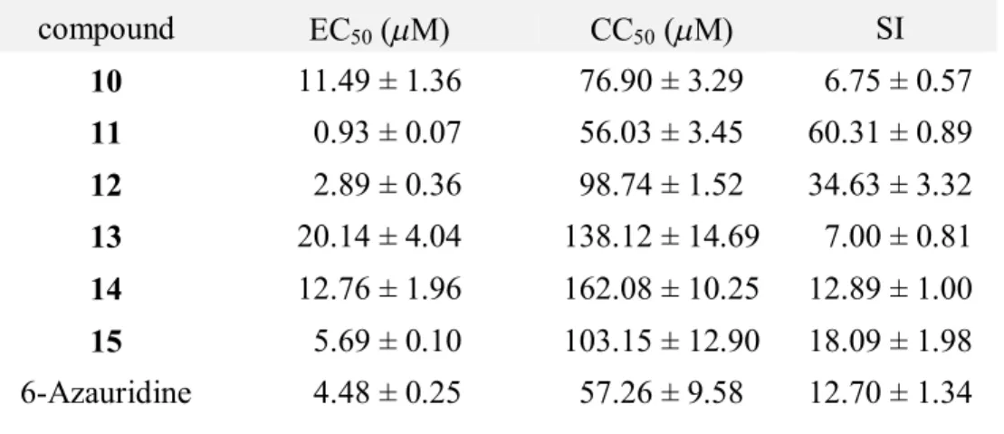
Table 7. The inhibitory effect of compounds 10-15 on PEDV induced CPE ... 61
Table 8. 13C and 1H NMR assignment for compound 10 ... 83
Table 9. 13C and 1H NMR assignment for compound 11 ... 84
Table 10. 13C and 1H NMR assignment for compound 12 ... 85
Table 11. 13C and 1H NMR assignment for compound 16 ... 101
Table 12. 13C and 1H NMR assignment for compound 17 ... 102
Table 13. 13C and 1H NMR assignment for compound 18 ... 103
Table 14. 13C and 1H NMR assignment for compound 19 ... 104
Table 15. 13C and 1H NMR assignment for compound 20 ... 126
List of Figures
Figure 1. Chemical structures of isolated compounds (1-5)... 9 Figure 2. Key COSY and HMBC correlations of salternamides A and C
(1 and 3) ... 16 Figure 3. Key NOESY correlations of the cyclohexenone ring of
salternamide C (3) ... 20 Figure 4. DdS-R values (ppm) obtained for the S- and R-MTPA esters (7a
and 7b) ... 21 Figure 5. J-based configuration analysis of salternamide C (3) at (a) C-4¢
and C-5¢ and (b) C-5¢ and C-6¢ ... 23 Figure 6. Experimental ECD spectrum (black) of (E)-4,6,8-
trimethylnona-2,7-dienamide (8) and calculated ECD spectra of (E)-4(S),6(R),8-trimethylnona-2,7-dienamide (red) and its enantiomer (blue)... 24 Figure 7. Chemical structures of isolated compounds (10-15) ... 52 Figure 8. The COSY (bold line) and HMBC (arrows) correlations of
xiamycin C (10) ... 55 Figure 9. NOESY correlations of the decaline of xiamycin C (10) ... 57 Figure 10. Comparison of the experimental ECD spectrum of 10 with the
calculated ECD spectra of two possible enantiomers ... 58
Figure 11. Inhibitory effects of xiamycin D (11) and E (12) against the PEDV virus ... 63
Figure 12. Inhibitory effects of compound (11) on PEDV RNA expression and protein synthesis in a dose-dependent manner ... 66
Figure 13. Immunofluorescence images show that xiamycin D (11) inhibited PEDV replication in a dose-dependent manner ... 68
Figure 14. Chemical structures of isolated compounds (16-19) ... 88
Figure 15. Key HMBC (→) and COSY (─) correlations of starkmycin A (16) and HMBC corrlations of methylation derivative (16a) 91
Figure 16. Key HMBC (→) correlations of starkmycin C (18) ... 93
Figure 17. Chemical structure of Tripatin (20) ... 114
Figure 18. Key HMBC correlations of tripartin (20) ... 115
Figure 19. ORTEP diagram for tripartin (20) ... 116
Figure 20. Effect of tripartin (20) on various histone H3 lysine methylation and acetylation ... 118
Part A.
Structure Determination of the New Bioactive
Metabolites from Streptomyces spp. in a Korea
Saltern
I. Introduction
Examining the natural product profiles of underexploited microorganisms has become a research hotspot in drug discovery.1 Especially, actinobacteria have been recognized as talented chemical synthesizers that produce structurally diverse bioactive compounds, many of which have been developed as drugs since the early years of antibiotic discovery in the 1940s.2 Thus, investigation of unique niches harboring chemically new actinomycetes, rather than typical terrestrial environments, is required to explore the full chemical diversity of this chemically prolific bacterial group.3
Halophilc microorganism are broadly defined as those that require at least 0.2 M NaCl for their survival. So extereme halophiles have specific character through adaptive physiologies.4 Halophilc Streptomyces have not been studied in the search of new secondary metabolites produced by Streptomyces adapted to hyper-saline environments. The secondary metabolites produced by adaptive streptomyces are expected to have new carbon skeleton for bioactivity of metabolites.
The purpose of this thesis is to investigate new secondary metabolites produced from halophilc actinomycetes isolated from hyper-
saline environments, such as a solar saltern. The halophilic eniveronment spot was established in a saltern of Shinui Island at Jollanam-Do in Republic of Korea. Numerous halophic bacteria strains, which required salinity for growth, were isolated.
Two strains (#HK10, #HK18) produced interesting small molecule through single LC-MS chemical screening. 12 new compounds were purely isolated from the culture extract of #HK10 and #HK18.
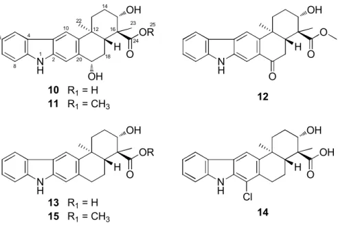
Secondary metabolites from halophilic actinomycetes were strucually determined using integrated spectroscopic analysis, chemical reactions, and computer modeling approaches. These compounds belonged to diverse structural classes with various biosynthetic origins: manumycin family, indolosesquiterpenoids, and benzo-oxazinone amines.
The biological activities of the separated secondary metabolites were determined various bioactivity tests: cytotoxicity, antimicrobial activities, enzyme inhibition activity aginst Na+/K+-ATPase, and antiviral activity aginst porcine epidemic diarrhea virus (PEDV). The cyototoxicity of the compounds was examined on A549, HCT116, SNU638, SK-HEP1, MDA-MB231, and K562 human cancer cell lines. The inhibitory activitiy against Na+/K+-ATPase, which plays a crucial role in cellular function, was tested. In addition, the aniti-viral inhibition activity against PEDV
virus, a kind of coronavirus occurs in upper respiratory and enteric diseases of animals, was also evaluated.
Salternamides displayed significant anticancer activity and xiamycins showed a strong inhibition against PEDV virus. Structure- activity relationship were also deduced.
II. Salternamides A-E from a Halophilic Streptomyces sp.
HK10
“Reproduced from [Kim, S.-H.; Shin, Y.; Lee, S.-H.; Oh, K.-B.; Lee, S.
K.; Shin, J.; Oh, D.-C. Jouranl of Natural Products, 2015, 78, 836-843.].”
“Reproduced from [Kim, S.-H.; Shin, Y.; Lee, S. K.; Shin, J.; Oh, D.-C.
Natural Product Sciences, 2015, 21, 273-277.].”
Salternamides A-E (1-5), the first secondary metabolites discovered from saltern-derived actinomycetes, were isolated from a halophilic Streptomyces strain isolated from a saltern on Shinui Island in the Republic of Korea. The planar structures of the salternamides, which are new members of the manumycin family, were elucidated by a combination of spectroscopic analyses. The absolute configurations of the salternamides were determined by chemical and spectroscopic methods, including the modified Mosher’s method, J-based configuration analysis, and circular dichroism spectroscopy. Salternamide A (1), which is the first chlorinated compound in the manumycin family, exhibited potent cytotoxicity against a human colon cancer cell line (HCT116) and a gastric cancer cell line (SNU638) with submicromolar IC50 values. Salternamides
A and D were also determined to be weak Na+/K+ ATPase inhibitors.
2-1. Introduction
Actinobacteria have been recognized as talented chemical synthesizers that produce structurally diverse bioactive compounds, many of which have been developed as drugs since the early years of antibiotic discovery in the 1940s.2 Although actinomycetes are a major source of microbial compounds, the proportion of the total number of known microbial metabolites that are derived from actinomycetes has decreased from 62% during the period from 1940 to 1974 to 28.5% in the first decade of the 21st century.5 Thus, investigation of unique niches harboring chemically new actinomycetes, rather than typical terrestrial environments, is required to explore the full chemical diversity of this chemically prolific bacterial group.3
Halophilic and halotolerant actinomycetes from saline environments are distinct from their terrestrial counterparts in terms of their physiology and secondary metabolites and thus were expected to be a promising source of novel bioactive compounds that may be of value in drug discovery.6 Since the discovery of a novel anticancer drug candidate, salinosporamide A, from an obligate marine actinomycete, studies of halophilic and halotolerant actinomycete-derived secondary metabolites have demonstrated the chemical potential of actinomycetes from saline
environments.7 However, salterns, which are representative of possibly the most saline environments, have not been investigated in the search for new bioactive compounds produced by actinomycetes adapted to such extreme habitats. A comprehensive literature search revealed that only a few studies have examined the phylogenetic diversity of actinomycetes from salterns; however, their chemical diversity has not been explored.8 It is hypothesized that saltern actinomycetes may have developed unique biosynthetic machinery in the course of their adaptation to extremely saline substrates and that they therefore may produce novel bioactive secondary metabolites.
Thus, halophilic and halotolerant actinomycete strains were selectively isolated from saltern topsoil samples collected on Shinui Island in the Republic of Korea and chemically screened their secondary metabolites by LC/MS. Notably, through the screening process, a chlorinated compound was observed, salternamide A (1), that was produced by one of the halophilic actinomycete strains (#HK10), which required seawater for growth. This initial analysis prompted the scale-up of the culture of this strain and the isolation of a series of metabolites from these bacteria. Here, the structures and biological activities of salternamides A-E (1-5) were reported, new members of the manumycin
class (Figure 1).
Figure 1. Chemical structures of isolated compounds (1-5).
2-2. Results and discussion
Salternamide A (1) was obtained as an amorphous oil that was determined to have the molecular formula C23H32ClNO5 by HRESIMS in combination with 1H and 13C NMR spectroscopic data. The 1H NMR spectrum of 1 in CD3OD clearly showed five olefinic resonances in the deshielded region (dH 7.68, 7.14, 6.79, 6.16, and 4.87), 12 aliphatic protons between dH 2.43 and 1.28, and four methyl groups in the shielded region (dH 1.66, 1.61, 1.04, and 0.91). The 13C and HSQC NMR spectra of 1 revealed three carbonyl carbons at dC 177.7, 176.0, and 167.5; eight olefinic carbons at dC 153.9 ~ 122.6, including four methines and four non-protonated carbons; an oxygen-bearing, fully substituted carbon at dC
72.1; two aliphatic methines (dC 35.8, and 31.5); five aliphatic methylenes (dC 45.5, 41.7, 35.1, 26.4, and 24.5); and four methyl carbons (dC 25.9, 21.7, 19.8, and 18.1). Because the eight olefinic carbons (four double bonds) and three carbonyl resonances accounted for seven out of eight unsaturation equivalents contained in the molecular formula of 1, salternamide A must be a monocyclic compound.
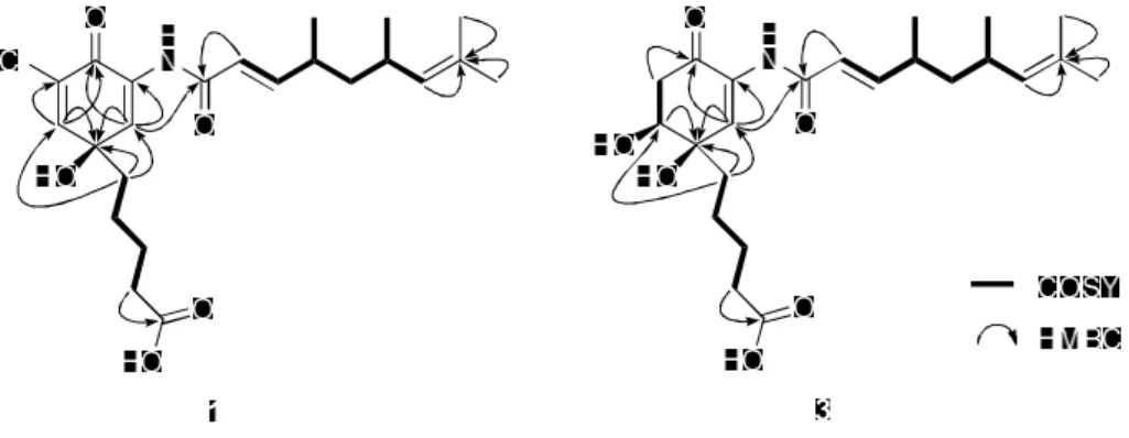
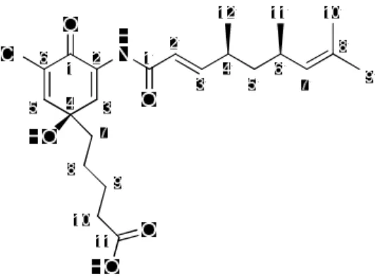
An analysis of COSY and NMR spectra of 1 resulted in the construction of three partial structures. The COSY correlations of H2-7,
H2-8, H2-9, and H2-10 connected these four consecutive aliphatic methylenes from C-7 to C-10. This chain was connected to a carboxylic acid carbon, C-11 at dC 177.7, by the HMBC correlation from H2-10 to C- 11, thus elucidating the structure of the pentanoic acid moiety. The second partial structure, the trimethyl nonadienamide chain, was also established with the COSY correlations connecting the backbone core from C-2′ to C- 7′. The doublet methyl groups C-11′ and C-12′ were connected to C-6′ and C-4′ by the COSY correlations of H3-11′/H-6′ and H3-12′/H-4′. The long- range heteronuclear couplings from H3-9′ and H3-10′ to the olefinic double-bond carbons C-7′ and C-8′ secured the connectivity from C-7′ to C-8′ bearing these dimethyl groups. Strong HMBC correlations from H-2′
and H-3′ to the carbonyl carbon C-1′ at dC 167.5 extended the chain to C- 1′. Both the molecular formula, which contained a nitrogen atom, and the chemical shift of C-1′ (dC 167.5) implied that this carbonyl carbon was part of an amide functional group. The geometry of the C-2′ double bond was determined to be E by observation of the trans 1H-1H coupling constant (J2′3′ = 15.5 Hz) between H-2′ and H-3′.
The HMBC correlations of the protons H-3 (dH 7.68) and H-5 (dH
7.14) were utilized to establish the third partial structure, which contained a ring system. First, H-3 exhibited correlations with C-1 and C-2, while H-
5 displayed heteronuclear couplings with C-1 and C-6. Due to the common HMBC correlations of these olefinic protons to the carbonyl carbon C-1 (dC 176.0), a cross-conjugated ketone system was deduced.
Further sharing of HMBC couplings with C-4, a fully-substituted oxygenated carbon, led to the construction of a cyclohexadienone moiety as the third partial structure.
These three partial structures, pentanoic acid, the trimethyl nonadienamide chain, and cyclohexadienone, were assembled by the analysis of long-range 1H-13C correlations. The HMBC correlation from H-7 of pentanoic acid to C-4 connected the chain to C-4. Also the HMBC correlations of H-7 with C-3 and C-5 supported the connectivity between pentanoic acid and cyclohexadienone ring. The trimethyl nonadienamide moiety was adjacent to C-3 based on a 4-bond correlation from H-3 to C- 1′. This assignment was further supported by the chemical shifts of C-2 (dC 131.8) and C-3 (dC 134.7), because the location of the amide chain at C-2 was able to reduce the polarization of the double bond by the conjugated ketone system and thus resulted in the similar chemical shifts of C-2 and C-3. Based on the molecular formula and the 13C chemical shifts, a hydroxy group was located at C-4 (dC 72.1), and a chlorine atom was assigned at C-6, thus completing the planar structure of salternamide
A (1) (Figure 2). The relative and absolute configurations in this compound are discussed below, along with those of the analogs.
Salternamide B (2) was isolated as an amorphous oil, of which the molecular formula was deduced to be C23H33NO5, based on HRESIMS data in combination with 1H and 13C NMR spectroscopic data. The 1H NMR spectrum of 2 in CD3OD exhibited a pattern analogous to that of 1.
However, salternamide B exhibited substantially different 1H resonances in the olefinic region, with a doublet proton at dH 6.21 (J=10.0 Hz) and a double-doublet proton at dH 6.94 (J=10.0, 3.0 Hz). The 13C NMR spectrum of 2 also exhibited features similar to those of 1. The HSQC NMR spectrum of 2 assigned all of the carbon-bound protons to the corresponding carbons. In particular, the proton at dH 6.21, which is different from that of 1, directly coupled with an olefinic carbon at dC
126.6, indicating that one more methine was present in 2 than 1. This correlation suggested that the olefinic carbon bearing a chlorine atom in 1 was replaced by this additional methine in 2.
The spectroscopic differences were readily accounted for by the replacement of the C-6 chlorine in 1 with a proton in 2, as was demonstrated by a combination of COSY and HMBC analyses, in which
key correlations were obtained from the H-5/H-6, H-5/C-1 and H-6/C-1 correlations. Thus, the structure of salternamide B (2) was defined as the dechlorinated derivative of salternamide A (1).
Salternamide C (3) was purified as an amorphous oil. Its molecular formula was determined to be C23H35NO6, based on HRESIMS data, together with 1H and 13C NMR spectroscopic data. The 1H NMR spectrum of 3 in CD3OD clearly showed four olefinic protons (dH 7.57, 6.76, 6.11, and 4.85), 15 aliphatic protons (dH 3.96~1.28), and four methyl groups (dH 1.65, 1.60, 1.02, and 0.91). An analysis of 13C and HSQC NMR spectra of 3 revealed the existence of three carbonyl carbons at dC 194.1, 177.6, and 167.2; six olefinic carbons, consisting of four methines (dC
153.5, 133.0, 132.1, and 122.7) and two fully substituted carbons (dC
133.7 and 131.2); a fully-substituted oxygenated carbon at dC 73.4; an oxymethine at dC 70.9; six aliphatic methylenes (dC 45.4, 42.4, 39.3, 34.9, 26.6, and 24.5); two aliphatic methines (dC 34.9 and 31.4); and four methyl carbons (dC 25.9, 21.6, 19.8, and 18.0). Once the protons and their associated carbons were matched using the HSQC NMR spectroscopic data, three main partial structures of salternamide C were further elucidated using COSY and HMBC NMR spectroscopic data (Figure 2).
Similar to salternamides A and B, salternamide C possessed pentanoic acid
and trimethyl nonadienamide moieties. Careful analysis of 13C chemical shifts and of the correlations in the COSY and HMBC NMR spectra revealed that the cyclohexadienone in 1 and 2 had been modified in 3. The carbonyl carbon of C-1 resonated at dC 194.1, indicating that this carbonyl carbon is a part of an a,b-unsaturated ketone rather than a cross- conjugated ketone. The three bond HMBC correlations from the olefinic proton H-3 to C-1 confirmed this a,b-unsaturated ketone moiety. The COSY correlation between H-5 and H2-6 connected C-5 and C-6. The methylene group of H2-6 also exhibited an HMBC correlation with C-1, which demonstrated the connectivity between C-6 and C-1. The H-3/C-4 and H-5/C-4 HMBC correlations closed the cyclohexenone ring structure.
The three partial structures, pentanoic acid, trimethyl nonadienamide, and cyclohexenone, were confidently connected based on long-range 1H-13C couplings. The H-3/C-1′ HMBC correlation assigned the trimethyl nonadienamide to C-2 of cyclohexenone, whereas pentanoic acid was connected to C-4 of cyclohexenone by the H-7/C-4, H-7/C-3, and H-7/C-5 HMBC correlations.
Figure 2. Key COSY and HMBC correlations of salternamides A and C (1 and 3).
Salternamide D (4) was obtained as an amorphous oil that was determined to have the molecular formula C24H37NO6, based on HRESIMS observations, together with 1H and 13C NMR spectroscopic data. Although the 1H and 13C NMR spectra of 4 exhibited a pattern analogous to that of 3, salternamide D (4) was shown to possess three olefinic protons, in contrast with salternamide C (3), which bears four olefinic protons. In particular, the H-2′ doublet proton (dH 6.11) associated with the trimethyl nonadienamide moiety in 3 disappeared in 4, and instead, an additional allylic methyl group (dH 1.88) was observed. Thus, the structure of salternamide D (4) was deduced to be a methyl-substituted analog of 3. This structure was confirmed by comprehensive NMR analyses, including HMBC data, in which important correlations were
observed between the allylic methyl protons (2′-CH3) and the amide carbonyl carbon C-1′ and the double bond carbons C-2′ and C-3′.
Salternamide E (5) was obtained as a colorless powder. The molecular formula was determined to be C23H37NO6 by the molecular ion peak [M+Na]+ at m/z 446.2511 (calcd for C23H38NO6Na, 446.2519) in the positive ion mode of HR-FABMS. The IR spectrum of 5 indicated the presence of hydroxy (3377 cm-1) and carbonyl functional groups (1672 and 1640 cm-1). The 1H NMR spectrum of 5 in CD3OD displayed signals for four methyl groups at dH 1.05 (d, J = 6.5 Hz), 0.87 (d, J = 6.5 Hz), 0.86 (d, J = 6.5 Hz), and 0.85 (d, J = 6.5 Hz), an oxymethine proton signal at dH
3.98 (dd, J = 8.0, 4.0 Hz), three olefinic proton signals at dH 7.58 (s), 6.71 (dd, J = 15.5, 8.0 Hz), and 6.15 (dd, J = 15.5, 1.0 Hz), ten aliphatic protons between dH 2.82 ~ 1.01 (Table 1). In the 13C NMR spectrum, 23 carbon signals appeared, including four methyl carbons at dC 23.7, 22.7, 20.9, and 20.0, an oxygenated methine carbon at dC 70.9, four olefinic carbons at dC 153.2, 133.8, 133.1, and 123.5, a quaternary carbon at dC
73.5, and three carbonyl carbons at dC 194.2, 178.3, and 167.2.
The interpretation of 2D HSQC and HMBC NMR experiments allowed all of the protons and carbons to be assigned. An analysis of COSY correlations revealed that salternamide E possesses three spin
systems: one composed of the protons at C-5 and C-6, another built with the protons of the linear chain group from C-7 to C-10, and the last assembled with the protons of the C-2¢ to C-12¢ chain involving four methyl groups. These three spin systems are extended to three partial structures based on HMBC correlation. First, the HMBC correlations of [H-3/C-1, C-2, C-5], [H-5/C-3, C-4, C-6], and [H2-6/C-1, C-2, C-4, C-5]
elucidated a 6-membered ring system bearing an α,β-unsaturated ketone group, thus providing a cyclohexenone moiety bearing two hydroxy groups at C-4 and C-5. Second, an HMBC correlation from H2-10 to C-11 (dC 178.3) assigned a pentanoic acid substructure from C-7 to C-11. Lastly, the third spin system from C-2¢ to C-12¢ was extended to C-1¢ by the HMBC correlation from H-2¢ to the amide carbonyl carbon C-1¢ (dC
167.2), completing a 4,6,8-trimethylnon-2-enamide chain. The geometry of the C-2¢ double bond was assigned as E by a trans 1H-1H coupling constant between H-2¢ and H-3¢ (JH2¢H3¢ = 15.5 Hz).
These three substructures were connected by the interpretation of the HMBC NMR spectrum. In particular, 2- and 3-bond 1H-13C couplings from H2-7 to C-4, C-3, and C-5 assigned the pentanoic acid at C-4. Finally, the 4,6,8-trimethylnon-2-enamide chain was assembled to C-2 of the cyclohexenone moiety based on the molecular formula, thus elucidating
the planar structure of salternamide E (5).
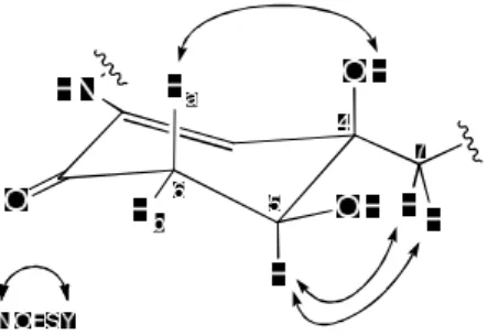
Salternamides A-E (1-5) have asymmetric carbon centers at the six-membered ring and amide chain moieties. For the determination of the absolute configurations, stereochemical analysis focused on salternamide C (3) because this major compound has a secondary hydroxy group at C-5, to which the modified Mosher’s method can be applied. First, salternamide C was derivatized using 2 M TMS diazomethane to yield a methyl ester (6), which is more soluble and thus more readily undergoes further chemical transformation. The 1D- and 2D-NMR spectra of 6 were acquired in pyridine-d5 to observe exchangeable protons. An analysis of the NOESY NMR spectrum and of the 1H-1H coupling constants (J values) determined the relative configuration of the cyclohexenone system of salternamide C (3). The carbinol proton H-5 (dH 3.96) showed a double- doublet with J values of 8.0 and 4.0 Hz. The large coupling constant (8.0 Hz) indicated that H-5 is in a pseudoaxial position. H-6a (dH 2.81) showed a double-doublet splitting pattern with geminal coupling of 17.0 Hz and vicinal coupling of 8.0 Hz, which assigned H-6a in a pseudoaxial position;
H-6b (dH 2.72) was situated in a pseudoequatorial position, based on the small vicinal coupling constant (4.0 Hz; Figure 2). In the NOESY spectrum of the methyl ester (6) of salternamide C, 4-OH (dH 6.72 in
pyridine-d5) exhibited a NOESY correlation with H-6a, thereby establishing a pseudoaxial position for 4-OH. The pentanoic acid chain was assigned a pseudoequatorial position by the H-5/H2-7 NOESY correlation, thus establishing the relative configuration of the ring (Figure 3).
Figure 3. Key NOESY correlations of the cyclohexenone ring of salternamide C (3).
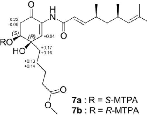
The absolute configuration of the stereogenic center at C-5, which bears a secondary alcohol, was determined using the modified Mosher’s method.9 The hydroxy group 5-OH was derivatized with R- and S-α- methoxy-α-(trifluoromethyl)phenylacetyl chloride (MTPA-Cl) to yield S- and R-MTPA esters (7a and 7b), respectively. The 1H chemical shifts of 7a and 7b were precisely assigned based on their 1H and COSY NMR spectra.
The calculation of the ΔδS-R values for 7a and 7b established the absolute configuration of 5S (Figure 4). Based on the relative configurations, the
absolute configuration of C-4 was simultaneously determined to be R.
(S) (R)
HN O
HO
O
O O RO
7a: R =S-MTPA 7b: R =R-MTPA
+0.04 +0.17 +0.16 -0.22
-0.09
+0.13 +0.14
Figure 4. DdS-R values (ppm) obtained for the S- and R-MTPA esters (7a and 7b).
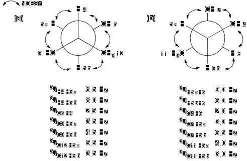
To determine the relative configuration of the stereogenic centers at C-4′ and C-6′ in the trimethyl nonadienamide moiety, J-based configuration analysis10 was utilized with vicinal 1H-1H and 3-bond 1H-
13C coupling constants and NOESY correlations. The 1H-1H coupling constants were measured in the 1H and 1H homo-decoupling NMR spectra, and long-range heteronuclear coupling constants were acquired via a hetero half-filtered TOCSY (HETLOC) NMR experiment11 and a heteronuclear scalar coupling HSQC-HECADE (heteronuclear couplings from aSSCI-domain experiments with e.COSY-type cross peaks) NMR experiment.12 The 3JH4′/H5′b value (8.0 Hz) suggested an anti relationship between H-4′ and H-5′b. The large value of 3JC12′/H5′a (5.5 Hz) indicated a
relative anti relationship between C-12′ and H-5′a. Further analysis of the
1H-1H and 1H-13C coupling constants strongly suggested the rotamer depicted in Figure 5a. The NOESY correlations of H3-12′ (dH 1.02) to H- 5′b (dH 1.28) and H-6′, of H-4′ to H-5′a and H-6′, and of H-3′ to H-5′a and H-5′b completely supported the configuration shown in Figure 4a. The large vicinal 3JH5′a/H6′ coupling constant (9.0 Hz) assigned their anti relationship. Another anti relationship between H-5′b and C-7′ was deduced based on the large 3JC7′/H5′b value (6.5 Hz). Additional HETLOC and HECADE analyses of the small values of 3JC4′/H6′ (2.0 Hz), 3JC7′/H5′a
(3.0 Hz), 3JC11′/H5′a (4.0 Hz), and 3JC11′/H5′b (3.0 Hz) fully supported the rotamer structure shown in Figure 5b. Careful analysis of the NOESY spectrum resulted in further confidence in the rotamer shown in Figure 4b, based on the correlations of H-5′a to H-11′ and H-7′, of H-4′ to H-7′and H-6′, and of H-5′b to H-11′ and H-6′. Thus, the relative configurations of the asymmetric carbons in the trimethyl nonadienamide moiety were determined to be 4′S* and 6′R*, which compose 1,3-syn dimethyl groups.
Further analysis of the conformation of the chain using TD-DFT (time dependent-density functional theory) calculations and long-range NOESY correlations also supported the 4′S* and 6′R* configurations. Particularly, a strong NOESY correlation was observed between H-4′ and H-7′, which
was reasonably rationalized based on the distances between the protons measured in the DFT-calculated conformation with 4′S* and 6′R*
configurations.
Figure 5. J-based configuration analysis of salternamide C (3) at (a) C-4¢
and C-5¢ and (b) C-5¢ and C-6¢.
To determine the absolute configuration of the trimethyl nonadienamide chain, I attempted to hydrolyze the amide bond of salternamide C (3) to obtain the part of the chain bearing the C-4′ and C-6′
stereogenic centers. However, both acidic and basic hydrolysis reactions decomposed salternamide C (3). Alternatively, I planned to introduce hydroxy groups into the double bonds in 3 by OsO4 oxidation,13 the
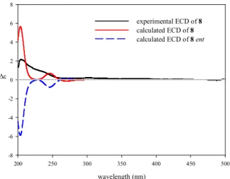
product of which would be suitable for analysis using the modified Mosher’s method. Fortunately, the (E)-4,6,8-trimethylnona-2,7-dienamide moiety was cleaved during the oxidation reaction, along with the oxidation product with a 1,2-diol at C-2′ and C-3′. I acquired an ECD spectrum of the intact (E)-4,6,8-trimethylnona-2,7-dienamide chain (8) and compared it with the calculated ECD spectra of two possible enantiomers, (4′S, 6′R)- 8 and (4′R, 6′S)-8. The ECD spectrum of (4′S, 6′R)-8 was consistent with the experimental ECD spectrum of 8 and exhibited a positive Cotton effect at 220~250 nm, whereas the calculated ECD spectrum of (4′R, 6′S)-8 exhibited the opposite profile. Consequently, the absolute configurations of the stereogenic centers in the chain were proposed to be 4′S and 6′R (Figure 6).
wavelength (nm)
200 250 300 350 400 450 500
De
-8 -6 -4 -2 0 2 4 6 8
experimental ECD of 8 calculated ECD of 8 calculated ECD of 8 ent
Figure 6. Experimental ECD spectrum (black) of (E)-4,6,8-trimethylnona-
2,7-dienamide (8) and calculated ECD spectra of (E)-4(S),6(R),8- trimethylnona-2,7-dienamide (red) and its enantiomer (blue).
Once the absolute configurations of 3 were fully established as 4R, 5S, 4′S, and 6′R, the absolute configurations of salternamide D (4), which is the 2′-methyl analog of 3, were easily deduced to be identical to those of 3, based on their virtually identical ECD spectra. However, the experimental ECD spectra of salternamides A and B (1-2) were significantly different from that of 3 because of an additional double bond at C-5 that comprised a cross-conjugated ketone system, thus prohibiting the assignment of their absolute configurations by CD spectroscopic comparison. Thus, I decided to eliminate the 5-OH in salternamide C methyl ester (6) to generate a double bond. This hydroxy group was successfully eliminated by treating 6 with methane sulfonyl chloride (MsCl) and triethylamine (TEA) to yield salternamide B methyl ester (9) (Scheme 1). The identical product (9) was also generated by methylation of 2 with TMS diazomethane (Scheme 1). The two products (9) from 2 and 6 had identical 1H NMR and CD spectra, suggesting that salternamide B (2) possesses 4S, 4′S, and 6′R configurations. Salternamide A (1) clearly exhibited a CD spectrum consistent with that of 2, thus indicating that the same arrangement of atoms was present in both compounds. However, the
absolute configurations of 1 were assigned as 4R, 4′S, and 6′R because the substitution of chlorine at C-6 changes the priorities of the atoms around C-4.
Scheme 1. Formation of salternamide B methyl ester (9) from salternamide C methyl ester (6) and salternamide B (2).
The relative configuration of the cyclohexenone system in 5 was determined by the analysis of the NOESY NMR spectrum and the 1H-1H coupling constants (J values). The carbinol proton H-5 (dH 3.98) showed a double-doublet with J values of 8.0 and 4.0 Hz. The large coupling constant (8.0 Hz) indicated that H-5 is located in a pseudoaxial position.
H-6a (dH 2.82) showed a double-doublet splitting pattern with geminal coupling of 16.5 Hz and vicinal coupling of 8.0 Hz, which established the pseudoaxial position of H-6a. H-6b (dH 2.73) was assigned in a pseudoequatorial position based on the small vicinal coupling constant (4.0 Hz). In the NOESY spectrum of the salternamide E (5), the pentanoic acid chain was assigned in a pseudoequatorial position by the H-5/H2-7
NOESY correlation, thus establishing the relative configuration of the ring.
The relative configuration of the two methyl groups at C-11¢ and C-12¢ in the 4,6,8-trimethylnon-2-enamide chain was deduced as syn based on the common biogenic origin of 5 with previously reported salternamides A-D.
The structure of salternamide E (5) is similar to that of salternamide C. The difference is at the sp3 bond between C-7¢ and C-8¢, whereas salternamide C has a sp2 bond between these carbons and thus possesses a trimethyl nonadienamide chain. To determine the absolute configuration of salternamide E (5), I compared experimental ECD spectra of salternamide E and salternamide C, of which the absolute configuration was unequivocally determined to be 4R, 5S, 4S¢, and 6S¢ by chemical derivatizations. Based on the high similarity between the experimental ECD spectra of these compounds, the absolute configuration of salternamide E (5) was fully established to be 4R, 5S, 4S¢, and 6S¢. Thus, the configurations of 1-5 were unambiguously determined by combined chemical and spectroscopic analyses.
The biological activities of salternamides A-E (1-5) were primarily measured using assays that evaluated their abilities to inhibit the growth and survival of a variety of human cancer cells. As summarized in
Table 2, salternamide A (1) exhibited potent anti-proliferative effects against HCT-116 (colon cancer) and SNU638 (gastric cancer) cells, with an IC50 value of 0.96 mM in each case. Salternamide A (1) also exhibited significant cytotoxicity against K562 (leukemia), SK-HEP1 (liver cancer), and MDA-MB231 (breast cancer) cells, with micromolar IC50 values of 8.9, 2.7, and 8.9 mM, respectively. This compound did not significantly inhibit the lung cancer cell line A549 (IC50>10 mM). Salternamides B-E (2-5) did not exhibit significant cytotoxic effects against the cancer cell lines tested (IC50>10 mM), indicating that the chlorine substitution at C-6 is critical for the cytotoxic effects of salternamides against cancer cells.
Furthermore, salternamides A and D (1 and 4) were moderate inhibitors of Na+/K+ ATPase,14 with IC50 values of 62 and 30 mM, respectively.
Table 1. Inhibitory Effects of Salternamides A-E (1-5) on the Proliferation of Human Cancer Cells in IC50 (mM)
compound A549 HCT116 SNU638 K562 SK-
HEP1
MDA- MB231
1 36 0.96 0.96 8.9 2.7 8.9
2 82 55 27 >100 56 27
3 >100 >100 >100 >100 >100 >100
4 >100 34 15 59 32 92
5 83 85 75 70 91 54
Etoposide 0.80 1.9 0.50 3.1 1.1 11
Salternamides A-E (1-5) are related to the manumycin group of metabolites from Streptomyces spp., in that salternamides A-E (1-5) are composed of a six-membered ring bearing a carbonyl carbon at C-1, a hydroxy group at C-4, an amide-linked chain connected to C-2, and a hydrocarbon chain directly attached to C-4 of the ring.15 However, salternamide A is the first chlorinated member to be identified in the manumycin family. Chlorination at the a-position of the C-1 carbonyl group may be occur via electrophilic halogenation by a haloperoxidase in a chlorine-rich environment.16 The upper chain, (E)-4,6,8-trimethylnona- 2,7-dienamide, is identical to that of U-62162, a member of the manumycin group from the soil-derived Streptomyces verdensis.17 The absolute configuration of the chain in U-62162 has not been determined previously. Only the relative configuration (4′R* and 6′S*) of the chain was reported via an analysis of the racemic total synthesis of this chain.18 Our CD analysis of the cleaved chain allowed the assignment of the absolute configuration of these challenging stereogenic centers. The cross- conjugated ketone system in salternamides A and B has been found previously in the manumycin family only in daryamides B and C from a marine-derived Streptomyces.19 However, because this cyclohexadienone moiety does not incorporate a secondary alcohol, the absolute
configuration of the C-4 stereogenic center has not been established for the daryamides. Our dehydration of the methyl ester of 3 using MsCl successfully generated the cross-conjugated ketone system in 2 and allowed the determination of the absolute configuration of the cyclohexadienone moiety in a rigorous manner.
To date, only a handful of secondary metabolites have been reported from solar saltern-derived microorganisms, such as fungi20 and cyanobacteria.21 No secondary metabolite from a saltern-derived actinomycete has been reported. The discovery of the salternamides from a saltern actinomycete isolate suggests that there may be chemically prolific actinomycete communities in salterns; thus, the chemical investigation of these microorganisms could potentially lead to the discovery of more natural chemical diversity and pharmaceutically valuable compounds.
2-3. Experimental section
General Experimental Procedures. Optical rotations were measured on a Jasco P-1020 polarimeter using a 1-cm cell. UV spectra were acquired using a PerkinElmer Lambda 35 UV/VIS spectrophotometer. CD spectra were recorded using an Applied Photophysics Chirascan-plus circular dichroism detector. IR spectra were obtained on a Thermo N1COLET iS10 spectrometer. 1H, 13C, and 2D NMR spectra were acquired on a Bruker Avance 600-MHz and 500-MHz spectrometers at the NCIRF (National Center for Interuniversity Research Facilities at Seoul National University) and on a Bruker Avance II 900- MHz NMR spectrometer at the KBSI (Korea Basic Science Institute at Ochang). Electrospray ionization (ESI) low-resolution LC/MS data were acquired on an Agilent Technologies 6130 quadrupole mass spectrometer coupled with an Agilent Technologies 1200-series HPLC using a reversed- phase C18 column (Phenomenex Luna, 100×4.6 mm). High-resolution electrospray ionization (HRESI) mass spectra were obtained using a Thermo Scientific Q high-resolution mass spectrometer at the NICEM (National Instrumentation Center for Environmental Management at Seoul National University). High-resolution fast atom bombardment (HRFAB) mass spectra were obtained using a JMS-700 mass spectrometer at the
NCIRF (National Center for Interuniversity Research Facilities at Seoul National University). Instant Ocean was utilized to generate artificial seawater.
Bacterial Isolation. A saltern sediment sample was collected on Shin-eui Island in the Republic of Korea. The sample (1 g) was diluted in 10 mL of sterilized artificial saline water (3´ strength of seawater) and vortexed. The mixture was spread onto actinomycete isolation agar, A4 medium (1 L of artificial seawater, 18 g of agar, and 100 mg/L of cycloheximide), A5 medium (750 mL of artificial seawater, 250 mL of distilled H2O, 18 g of agar, and 100 mg/L of cycloheximide), A6 medium (1 L of artificial seawater, 18 g of agar, and 5 mg/L of polymyxin B sulfate), A7 medium (1 L of artificial seawater, 18 g of agar, and 5 mg/L of kanamycin) and chitin-based agar (1 L of artificial seawater, chitin 4 g, K2HPO4 0.75 g, MgSO4∙7H2O 0.5 g, KH2PO4 3.5 g, FeSO4∙7H2O 10 mg, MnCl2∙4H2O 10 mg, ZnSO4∙7H2O 10 mg, 18 g of agar, and 100 mg/L of cycloheximide). The strain HK10 was isolated on actinomycete isolation agar medium. Colonies were repeatedly inoculated onto fresh agar plates to obtain single strains. HK10 16S rDNA sequence analysis data obtained from COSMO Co., Ltd. revealed that HK10 is most closely related to Streptomyces radiopugnans (99% identity), identifying the strain as a
Streptomyces sp. (GenBank accession number: LC013479).
Cultivation and Extraction. The HK10 strain was cultivated in 50 mL of YEME medium (4 g of yeast extract, 10 g of malt extract, and 4 g of glucose in 1 L of artificial seawater) in a 125-mL Erlenmeyer flask.
After the strain was cultivated for 3 days on a rotary shaker at 200 rpm at 30 °C, 10 mL of the culture was inoculated in 1 L of YPM + starch medium (6 g of yeast extract, 4 g of peptone, 10 g of starch, and 4 g of mannitol in 1 L of artificial seawater) in 2.8-L Fernbach flasks. These large cultures (60 L) were incubated at 180 rpm at 30 °C. After 5 days, the entire culture volume was extracted twice using EtOAc. The EtOAc layer was separated and dried over anhydrous sodium sulfate. The EtOAc extract was enriched in vacuo to yield 5 g of dried material.
Isolation of Salternamides A-E (1-5). The dried extract was absorbed on celite, loaded onto a 20 g C18 resin, and fractionated with 200 mL each of 20%, 40%, 60%, 80% and 100% MeOH in H2O and 1:1 MeOH/CH2Cl2. Salternamides A-E (1-5) were detected in the 80% MeOH/
H2O fraction. To purify 1-4, the 80% fraction was chromatographed using reversed-phase HPLC on a C18column (Kromasil, 5 μm, 250×10 mm) under isocratic conditions (55:45 CH3CN/H2O, UV detection at 254 nm,
flow rate of 2 mL/min). Finally, salternamide A (1) (5 mg), salternamide B (2) (8 mg), salternamide C (3) (20 mg), salternamide D (4) (3 mg), and salternamide E (5) (2 mg) were isolated as pure compounds at retention times of 28, 25, 21, 29 and 31 min, respectively.
Salternamide A (1): pale yellow amorphous oil; [a]20D +120 (c 0.1, MeOH); UV (MeOH) lmax (log e) 253 (2.97) nm; CD (c 2.2´10-4 M, MeOH) lmax (De) 211 (3.87), 236 (-6.84), 259 (7.72), 311 (-2.98), 354 (0.86) nm; IR (ZnSe) nmax 3329, 2957, 2925, 1711, 1662, 1522 cm-1; for
1H and 13C NMR data, Table 2; HRESIMS m/z 438.2043 [M+H]+ (calcd for C23H33ClNO5, 438.2047).
Salternamide B (2): pale yellow amorphous oil; [a]20D +110 (c 0.1, MeOH); UV (MeOH) lmax (log e) 238 (3.04) nm; CD (c 2.4´10-4 M, MeOH) lmax (De) 202 (1.45), 227 (-6.94), 249 (7.45), 299 (-1.83), 353 (0.74) nm; IR (ZnSe) nmax 3328, 2957, 2925, 1711, 1662, 1522 cm-1; for
1H and 13C NMR data, Table 3; HRESIMS m/z 404.2435 [M+H]+ (calcd for C23H34NO5, 404.2437).
Salternamide C (3): pale yellow amorphous oil; [a]20D +170 (c 0.1, MeOH); UV (MeOH) lmax (log e) 238 (3.69), 273 (3.41) nm; CD (c
2.3´10-4 M, MeOH) lmax (De) 203 (5.34), 262 (-2.51), 320 (2.26) nm; IR (ZnSe) nmax 3371, 2959, 2926, 1671, 1520 cm-1; for 1H and 13C NMR data, Table 4; HRESIMS m/z 422.2546 [M+H]+ (calcd for C23H36NO6, 422.2543).
Salternamide D (4): pale yellow amorphous oil; [a]20D +140 (c 0.1, MeOH); UV (MeOH) lmax (log e) 236 (4.06), 274 (3.96) nm; CD (c 2.2´10-4 M, MeOH) lmax (De) 220 (-1.75), 242 (3.40), 270 (-0.95), 311 (3.20) nm; IR (ZnSe) nmax 3372, 2958, 2928, 1673, 1518 cm-1; for 1H and
13C NMR data, Table 5; HRESIMS m/z 436.2701 [M+H]+ (calcd for C24H38NO6, 436.2699).
Salternamide E (5): colorless powder; [a]20D +30.1 (c 0.1, MeOH);
UV (MeOH) λmax (log ε) 227 (3.75), 272 (3.57) nm; CD (c 2.4 ´ 10-4 M, MeOH)λmax(De) 206 (2.36), 258 (-2.92), 319 (2.00); IR (ZnSe) νmax cm-1: 3701, 3377, 2926, 1672, 1640, 1516, 1404, 1052, 7521H, 13C NMR: Table 6.; ESIMS m/z 446 [M+Na]+; HRFABMS m/z 446.2511 [M+Na]+; (calcd for C23H37NO6Na, 446.2519).
Methylation of Salternamide C. Salternamide C (3) (10 mg) was dissolved in 2 mL of anhydrous MeOH. Then, 200 μL of 2 M TMS
diazomethane in diethyl ether was added to the salternamide C (3) solution, and the reaction mixture was stirred at 25 °C for 2 h. After the reaction mixture dried, salternamide C methyl ester (6) was purified by reversed- phase HPLC using a C18 column (Kromasil,5 μm, 250×10 mm) with an acetonitrile gradient (from 50% to 100% CH3CN/H2O in 60 min, UV detection at 254 nm, flow rate of 2 mL/min, tR=27 min).
Salternamide C methyl ester (6): 1H NMR (500 MHz, pyridine-d5) d 9.19 (s, NH), 8.43 (s, 1H), 7.13 (dd, J=16.0, 7.0 Hz, 1H), 6.89 (br s, OH), 6.72 (br s, OH), 6.38 (d, J=16.0 Hz, 1H), 4.88 (d, J=10.0 Hz, 1H), 4.37 (dd, J=8.0, 4.0 Hz, 1H), 3.59 (s, 3H), 3.33 (dd, J=16.0, 7.0 Hz, 1H), 3.09 (dd, J=16.0, 4.0 Hz, 1H), 2.41 (m, 1H), 2.32 (t, J=7.0 Hz, 2H), 2.30 (m, 1H), 2.12 (dt, J=13.0, 5.0 Hz, 1H), 1.99 (dt, J=13.0, 5.0 Hz, 1H), 1.81 (m, 1H), 1.78 (m, 1H), 1.69 (m, 1H), 1.66 (m, 1H), 1.65 (s, 3H), 1.55 (s, 3H), 1.29 (ddd, J=13.5, 9.0, 6.5 Hz, 1H), 1.16 (ddd, J=13.5, 8.0, 6.0 Hz, 1H), 0.94 (d, J=6.0 Hz, 3H), 0.89 (d, J=6.0 Hz, 3H). For 1H and 13C NMR assignments, ESIMS m/z 436 [M+H]+, 458 [M+Na]+.
MTPA Esterification of Salternamide C Methyl Ester.
Salternamide C methyl ester (6) was prepared in two 40-mL vials (two 1- mg samples), which were dried completely under high vacuum for 6 h.
Then, freshly distilled anhydrous pyridine (1 mL) was added under argon gas. The reaction mixtures were stirred at room temperature for 10 min.
After 10 min, R- and S-a-methoxy-(trifluoromethyl)phenylacetic acid (MTPA) chloride (30 μL) were added separately. The reaction mixtures were stirred at 25 °C for 2 h. The reactions were quenched by adding 50 mL of MeOH. The products were then purified using a reversed-phase C18
column (Kromasil, 5 μm, 250×10.0 mm) with an CH3CN gradient ranging from 40% to 100% aqueous CH3CN over 50 min. The S-MTPA ester (7a) and the R-MTPA ester (7b) eluted at 48 and 49 min, respectively. The 1H NMR chemical shifts of 6a and 6b were assigned by 1H and 1H-1H COSY NMR experiments. ΔδS-R values around the C-5 stereogenic center were calculated.
S-MTPA ester (7a) of salternamide C methyl ester (6): 1H NMR (500 MHz, pyridine-d5) d 9.28 (s, NH), 8.45 (s, 1H), 7.86 (d, J=7.5 Hz, 2H), 7.41-7.37 (m, 3H), 7.11 (dd, J=15.0, 7.5 Hz, 1H), 6.37 (d, J=15.5 Hz, 1H), 5.98 (dd, J=5.0, 5.0 Hz, 1H), 4.87 (d, J=9.5 Hz, 1H), 3.70 (s, 3H), 3.59 (s, 3H), 3.30 (d, J=5.0 Hz, 2H), 2.40 (m, 1H), 2.33 (t, J=7.5 Hz, 2H), 2.28 (m, 1H), 2.10 (dt, J=14.0, 5.5 Hz, 1H), 2.01 (dt, J=14.0, 4.5 Hz, 1H), 1.83 (m, 1H), 1.80 (m, 1H), 1.69 (m, 1H), 1.64 (m, 1H), 1.64 (s, 3H), 1.54 (s, 3H), 1.27 (ddd, J=13.5, 9.0, 6.5, 1H), 1.15 (ddd, J=13.5, 8.0, 6.0 Hz,
1H), 0.93 (d, J=6.5 Hz, 3H), 0.88 (d, J=6.5 Hz, 3H); ESIMS m/z 674 [M+Na]+.
R-MTPA ester (7b) of salternamide C methyl ester (6): 1H NMR (500 MHz, pyridine-d5) d 9.37 (s, NH), 8.42 (s, 1H), 7.81 (d, J=7.5 Hz, 2H), 7.42-7.38 (m, 3H), 7.15 (dd, J=15.0, 7.5 Hz, 1H), 6.43 (d, J=15.5 Hz, 1H), 5.92 (dd, J=8.5, 4.0 Hz, 1H), 4.88 (d, J=9.5 Hz, 1H), 3.64 (s, 3H), 3.61 (s, 3H), 3.53 (dd, J=16.5, 8.5 Hz, 1H), 3.39 (dd, J=17.0, 4.0 Hz, 1H), 2.41 (m, 1H), 2.31 (t, J=7.0 Hz, 2H), 2.30 (m, 1H), 1.92 (m, 1H), 1.89 (m, 1H), 1.67-1.64 (m, 4H), 1.65 (s, 3H), 1.55 (s, 3H), 1.29 (m, 1H), 1.16 (m, 1H), 0.94 (d, J=6.5 Hz, 3H), 0.89 (d, J=6.5 Hz, 3H); ESIMS m/z 674 [M+Na]+.
Cleavage of the Trimethyl Nonadienamide Chain (8).
Salternamide C (3) (10 mg) was dissolved in acetone (2 mL), and 4- methylmorpholine N-oxide (NMO, 3 mg) and a solution of OsO4 in H2O (30 μL) were added. The reaction mixture was stirred at 25 °C for 10 h.
The reaction was quenched by the removal of acetone, and the residue was eluted using a 1 g Sep-pak C18 column with 100% MeOH. After removal of the solvent under vacuum, the products were purified by reversed-phase HPLC using a C18 column (Kromasil,5 μm, 250×10 mm) with an CH3CN
gradient (from 50% to 100% CH3CN/H2O in 60 min, UV detection at 254 nm, flow rate of 2 mL/min). Trimethyl nonadienamide (8) was obtained at 30 min.
(E)-4,6,8-trimethylnona-2,7-dienamide (8): white amorphous solid;
[a]20D -11 (c 0.1, MeOH); UV (MeOH) lmax (log e) 204 (4.47) nm; CD (c 5.1´10-4 M, MeOH) lmax (De) 207 (6.52), 224 (3.20) nm; IR (ZnSe) nmax
3342, 3197, 2959, 2925, 2854, 1674, 1604, 1512 cm-1, 1H NMR (600 MHz, CD3OD) d 6.69 (dd, J=16.0, 7.5 Hz, 1H), 5.89 (dd, J=16.0, 1.0 Hz, 1H), 4.86 (d, J=10.0 Hz, 1H), 2.43 (m, 1H), 2.29 (m, 1H), 1.67 (d, J=1.0 Hz, 3H), 1.61 (d, J=1.0 Hz, 3H), 1.30 (ddd, J=13.5, 9.0, 6.5, 1H), 1.27 (ddd, J=13.5, 8.0, 6.0, 1H), 1.02 (d, J=6.0 Hz, 3H), 0.91 (d, J=6.0 Hz, 3H);
HRESIMS m/z 196.1701 [M+H]+ (calcd for C12H22NO, 196.1701).
Formation of Salternamide B Methyl Ester (9). The methyl ester (6) of 4.8 mg of salternamide C was prepared in a 4-mL vial and was dried completely under high vacuum for 12 h. First, freshly distilled anhydrous CH2Cl2 (0.5 mL) was added under argon gas. The reaction mixture was stirred at 0 °C for 5 min. After 5 min, methane sulfonyl chloride (MsCl, 1.0 μL, 1.2 eq) and triethylamine (2.0 μL, 1.2 eq) were added separately. The reaction was performed at 0 °C for 3 h. The reaction
was quenched by adding 1 mL of saturated aqueous NaHCO3 over a 15- min period. The reaction mixture was dried for purification through a 1-g silica Sep-Pak column. The reaction product was eluted by 100% EtOAc.
The product was then purified using a reversed-phase C18 column (Kromasil, 5 μm, 250×10.0 mm) with an CH3CN gradient ranging from 50% to 100% aqueous CH3CN over 50 min. Salternamide B methyl ester (9) eluted at 35 min. Independent of the formation of 8 by treating 5 with MsCl under basic conditions, salternamide B methyl ester (9) was obtained by methylation of 2 with TMS diazomethane. The methylation method used was the same as that described above for the methylation of 3.
Salternamide B methyl ester (9): 1H NMR (600 MHz, CD3OD) d 7.64 (d, J=3.0 Hz, 1H), 6.94 (dd, J=10.0, 3.0 Hz, 1H), 6.79 (dd, J=15.0, 8.0 Hz, 1H), 6.22 (d, J=10.0 Hz, 1H), 6.15 (dd, J=15.0, 1.0 Hz, 1H), 4.87 (d, J=9.5 Hz, 1H), 3.63 (s, 3H), 2.44 (m, 1H), 2.35 (m, 1H), 2.30 (m, 2H), 1.78 (m, 2H), 1.67 (d, J=1.0 Hz, 3H), 1.62 (d, J=1.0 Hz, 3H), 1.61 (m, 2H), 1.33-1.29 (m, 4H), 1.04 (d, J=6.5 Hz, 3H), 0.92 (d, J=6.5 Hz, 3H);
ESIMS m/z 418 [M+H]+.
Computational Analysis. The ground-state geometries were optimized with density functional theory (DFT) calculations using