저작자표시-비영리-변경금지 2.0 대한민국 이용자는 아래의 조건을 따르는 경우에 한하여 자유롭게
l 이 저작물을 복제, 배포, 전송, 전시, 공연 및 방송할 수 있습니다. 다음과 같은 조건을 따라야 합니다:
l 귀하는, 이 저작물의 재이용이나 배포의 경우, 이 저작물에 적용된 이용허락조건 을 명확하게 나타내어야 합니다.
l 저작권자로부터 별도의 허가를 받으면 이러한 조건들은 적용되지 않습니다.
저작권법에 따른 이용자의 권리는 위의 내용에 의하여 영향을 받지 않습니다. 이것은 이용허락규약(Legal Code)을 이해하기 쉽게 요약한 것입니다.
Disclaimer
저작자표시. 귀하는 원저작자를 표시하여야 합니다.
비영리. 귀하는 이 저작물을 영리 목적으로 이용할 수 없습니다.
변경금지. 귀하는 이 저작물을 개작, 변형 또는 가공할 수 없습니다.
약학석사 학위논문
AKAP12 is downregulated in liver fibrosis and its deficiency promotes
thioacetamide-induced hepatocarcinogenesis
간섬유증에서 AKAP12의 발현 및 간암화 과정에서의 역할에 관한 연구
2 0 1 6 년 8 월
서울대학교 융합과학기술대학원
분자의학 및 바이오제약학과
최 진 혁
ABSTRACT
AKAP12 is downregulated in liver fibrosis and its deficiency promotes
thioacetamide-induced hepatocarcinogenesis
Jinhyeok Choi Department of Molecular Medicine and Biopharmaceutical Sciences Graduate School of Convergence Science and Technology Seoul National University
Liver fibrosis is originated from wound healing response to chronic liver injury from infectious (mostly HBV and HCV), toxic-induced, drug-induced, metabolic and autoimmune causes. Furthermore, liver fibrosis can lead to liver cirrhosis by reiterated liver damages. It is difficult for cirrhotic liver to function normally, threatening our lives.
Moreover, liver cirrhosis is strongly associated with liver cancer, and
curative therapy is only liver transplant. Therefore, liver fibrosis has been considered appropriate for therapy, with its reversibility.
Nevertheless, there are no approved agents as anti-fibrotic drugs to date. Accordingly, development of therapeutic modalities for treatment of liver fibrosis is thought to be necessary at present.
A-Kinase Anchoring Protein 12 (AKAP12) has been reported as tumor suppressor gene in various cancers such as prostate, ovarian, breast, lung and hepatocellular carcinoma (HCC). However, the role of AKAP12 in liver fibrosis still remains largely unknown.
In this study, I report that AKAP12 is expressed in liver sinusoidal endothelial cells (LSECs) and portal fibroblasts (PFs) in normal mouse liver. I analyzed AKAP12 expression in mice liver with portal fibrosis induced by 3,5-diethoxycarbonyl-1,4-dihydrocollidine (DDC) diet, which represented reduction of AKAP12 expression in activated PFs and capillarized LSECs around fibrotic regions. However, there is no difference between WT and AKAP12-/- mice liver damaged by DDC diet.
AKAP12 expression was also examined in mice liver with centrilobular fibrosis by TAA injection in the same manner. I found that AKAP12 expression decreased only in capillarized LSECs along fibrotic region. As in portal fibrosis, there is no difference in AKAP12-/- mice liver with centrilobular fibrosis by TAA, compared to WT.
Interestingly, liver progenitor cells (EpCAM+) were more highly induced in AKAP12-/- mice liver by TAA than WT, suggesting correlation of AKAP12 with cholangiocarcinoma resulting from long-term TAA administration. Indeed, It was found that AKAP12-/- mice were more susceptible for cholangiocarcinoma induced by TAA compared to WT, representing high incidence of large tumors (>2mm).
Taken together, AKAP12 was downregulated in animal models of liver fibrosis, suggesting that may function as the mediator of liver fibrosis, in particular, involved in fibrosis resolution, expressed in LSECs and PFs. In addition, the study for tumor implies that AKAP12 may be the tumor suppressor gene against cholangiocarcinoma with other various cancers.
Keywords : Liver fibrosis, AKAP12, PFs, LSECs, Cholangiocaricnoma Student Number : 2014-24872
TABLE OF CONTENTS
ABSTRACT ··· ⅰ LIST OF FIGURES ··· ⅵ
INTRODUCTION ··· 1
1. Liver fibrosis ··· 1
2. Animal models of liver fibrosis ··· 5
3. Cholangiocarcinoma ··· 6
4. A-kinase anchoring protein 12 (AKAP12) ··· 9
MATERIALS AND METHODS ··· 12
1. Animals ··· 12
2. Thioacetamide (TAA) administration ··· 12
3. 3,5-diethoxycarbonyl-1,4-dihydrocollidine (DDC) diet ··· 13
4. Sirius Red staining ··· 13
5. Immunohistochemistry ··· 13
6. Quantitative analysis ··· 14
7. Statistics ··· 14
RESULTS ··· 15
1. AKAP12 is expressed in normal mouse liver ··· 15
2. AKAP12 is expressed in portal fibroblasts and liver sinusoidal endothelial cells in mouse liver ··· 17
3. AKAP12 expression is reduced in portal fibrosis induced by DDC diet in mouse liver ··· 21
4. There is no difference between WT and AKAP12
-/-mice in portal fibrosis induced by DDC diet ··· 27
5. AKAP12 expression decreases in liver sinusoidal endothelial cells of mouse with centrilobular fibrosis by TAA administration ··· 30
6. Liver progenitor cells are more induced in AKAP12
-/-mice than WT in centrilobular fibrosis ··· 36
7. Hepatocarcinogenesis more severely occurs in AKAP12
-/-mouse than WT under long-term TAA administration ··· 39
DISCUSSION ··· 42
REFERENCES ··· 46
ABSTRACT IN KOREAN (국문초록) ··· 50
LIST OF FIGURES
Figure 1. Development and progression of liver fibrosis ··· 3
Figure 2. Schematic representation of liver ··· 4
Figure 3. The subtypes of cholangiocarcinoma ··· 8
Figure 4. The gene structures of AKAP12 ··· 11
Figure 5. AKAP12 is expressed in sinusoid and around portal vein ··· 16
Figure 6. AKAP12 is expressed in portal fibroblasts and liver sinusoidal endothelial cells ··· 19
Figure 7. Portal fibrosis is induced by DDC diet in mouse ··· 23
Figure 8. Expression of portal fibrosis marker protein increases in mouse liver injured by DDC diet ··· 24
Figure 9. AKAP12 expression decreases in portal fibrosis induced by DDC diet ··· 26
Figure 10. There is no difference in AKAP12
-/-mouse liver compared to WT with portal fibrosis by DDC diet ··· 28
Figure 11. Centrilobular fibrosis is induced by TAA injection in mouse ··· 32
Figure 12. AKAP12 expression is reduced unlike fibrosis marker
proteins in centrilobular fibrosis induced by TAA ··· 34
Figure 13. Liver progenitor cells are more highly induced in AKAP12
-/-mice compared to WT after TAA injection ··· 37
Figure 14. Hepatocarcinogenesis occurs more severely in AKAP12
-/-mouse liver than WT subjected to long-term TAA
administration ··· 40
INTRODUCTION
1. Liver fibrosis
Liver is known as a regenerative organ. The liver has remarkable capacity to endure liver injury via tissue repair (Pellicoro et al. 2014).
Tissue damages from infective (mostly HBV and HCV), toxic-induced, drug-induced, metabolic and autoimmune causes, provokes wound-healing response characterized by dynamic process balancing collagen accumulation and degradation. Liver fibrosis is mainly caused by chronic wound healing response by chronic liver injury, representing continuous accumulation of fibrillar extracellular matrix (ECM) components with insufficient degradation of them (Pinzani et el. 2015).
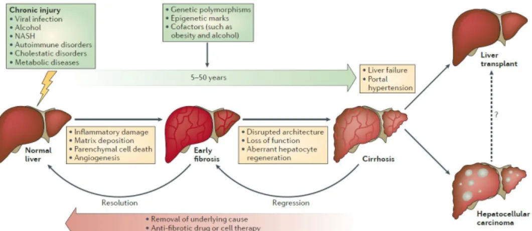
It is evident that liver fibrosis is reversible to normal state according to previous many evidences. Despite its reversibility, chronic and continuous liver injury promotes progression of liver fibrosis, leading to liver cirrhosis. The cirrhotic liver is not able to function normally, which results in many complications such as hepatic failure, diabetes and obesity, even liver cancer (Fig. 1). In this context, liver fibrosis as previous stage of cirrhosis is a prominent therapeutic target due to its reversibility (Pellicoro et al. 2012). Despite this fact, there are not many drugs available for liver fibrosis, and, in particular, no single compound approved for anti-fibrogenic therapy in chronic liver disease (Weiskirchen 2016). Therefore, the study for liver fibrosis is necessary.
There are several cell types contributing to liver fibrosis (Xu et al.
2014) (Fig. 2). Among them, hepatic stellate cells (HSCs) are known as main provider of fibrosis (Mederacke et al. 2013). HSCs are
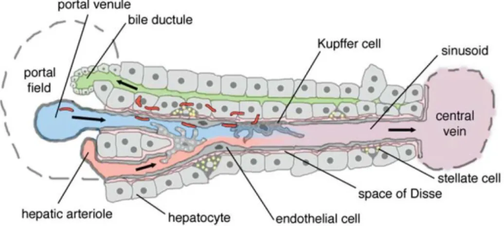
mesenchymal cells and reside in space of disse between liver sinusoidal endothelium and hepatocytes. HSCs can be activated to myofibroblasts-like cell types expressing Smooth Muscle Actin. Once activated, it presents contractile properties and produce extracellular matrix (ECM) components, remodeling them. As the injury disappears, the activated HSCs go through apoptosis or are inactivated (Elpek 2014). Although this HSCs have been believed as a key player for liver fibrosis, recently, portal fibroblasts (PFs) have also been reported that contribute to liver fibrosis. In homeostatic state, PFs are located around bile duct epithelial cells and can be also activated to myofibroblasts-like cell types presenting contractile characteristic (Yovchev et al. 2009, Wells 2014). When activated, the PFs proliferate expanding from portal tract, producing ECM components such as Elastin, Collagen. Furthermore, it was reported that PFs play a major role especially in portal fibrosis, expressed around portal tract (Dudas et al. 2009, Wells 2014). In addition, liver sinusoidal endothelial cells (LSECs) are also important cell types involved in liver fibrosis (Iwakiri et al. 2014). It produces many cytokines involved in inflammation and activation of fibroblasts-like cells. Moreover, LSECs become capillarized via interaction with neighboring cells under liver injury, constructing basal lamina and promoting angiogenesis (Lemoinne et al. 2015, Xie et al. 2012). In contrast, it was reported that LSECs are participated in fibrosis resolution recruiting macrophages which can degrade ECM components (Yang et al. 2014, Kantari-Mimoun et al. 2014).
Figure 1. Development and progression of liver fibrosis (Pellicoro
et al. 2014)
Figure 2. Schematic representation of liver (Frevert et al. 2005)
2. Animal models of liver fibrosis
Liver fibrosis is classified into two subtypes. One is centrilobular fibrosis, the other is portal fibrosis.
The centrilobular fibrosis is that collagen accumulation expands from central vein. This fibrosis is caused by the death of hepatocytes, inducing inflammatory response. In this process, hepatic stellate cells (HSCs) mainly produce ECM components as wound-healing response for tissue repair. Finally, chronic liver injury induces liver fibrosis via activated HSCs, which can lead to liver cirrhosis. There are some animal models for centrilobular fibrosis (Marques et al. 2012). One of them is thioacetamide (TAA) administration model, used in this study.
TAA compound does not induce liver fibrosis itself. However, its metabolites by Cyt-p450 in hepatocytes are hepatotoxin, which damages to parenchymal cells, promoting centrilobular fibrosis (Akhtar et al. 2013). After intraperitoneal injection of TAA for 6-8 weeks, centrilobular fibrosis is induced in mice, elevating the activity of ALT and AST.
The portal fibrosis is that collagen accumulates around portal tracts.
Portal fibrosis is induced by cholestasis. Blockade of biliary tree disrupts the flow of bile acids, damaging to cholangiocytes, bile duct epithelial cells. Subsequently, it induces inflammatory signals, leading to liver fibrosis in periportal region (Hirschfield et al. 2010). Some animal models have been established to study portal fibrosis such as bile duct ligation (BDL), 3,5-diethoxycarbonyl-1,4-dihydrocollidine (DDC) diet. DDC diet was used in this study. DDC compound produces porphyrin crystals in liver, which block biliary tree, inducing portal fibrosis together with elevation of ALP activity (Fickert et al. 2007).
3. Cholangiocarcinoma
Cholangiocarcinoma (CCA) is malignant tumor arising from the epithelium of bile duct. CCA is the second most common primary hepatic malignancy after hepatocellular carcinoma. Many epidemiological studies suggest that the incidence and mortality are increasing worldwide (Blechacz et al. 2008). There are many risk factors for CCA including primary sclerosing cholangitis, parasitic infections, choledochal cysts, carcinogen exposure (Goodman 2007).
Advanced CCA has a devastating prognosis, presenting the median survival (<24 months) because the only curative therapy is surgical extirpation, or liver transplantation. Moreover, many of patients present with unresectable CCA because of the late clinical presentation. CCA progresses insidiously, and is difficult to be diagnosed. In addition, non-surgical therapy also has not yet been developed sufficiently.
Therefore, it is essential to develop new diagnostic and therapeutic modalities for early detection and non-surgical treatment for CCA (Khan et al. 2005).
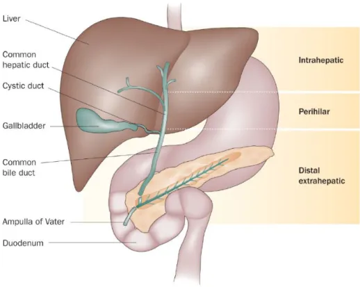
CCA is classified into extrahepatic and intrahepatic cholangiocarcinoma. Extrahepatic cholangiocarcinoma (EH-CCA) is more common, accounting for 80% to 90% of CCA. EH-CCA is further divided into proximal (peri-hilar), middle and distal subsets, depending on the location of the cancer within extrahepatic bile duct (Fig. 3).
Intrahepatic cholangiocarcinoma (IH-CCA) is relatively rare cancer than EH-CCA. IH-CCA appears as a mass-forming lesion within liver. It is confused with tumor metastasis because of its invasive feature from bile duct (Gatto et al. 2010). In this study, I was focused on this IH-CCA.
To investigate IH-CCA, I selected a mouse model established by orally administered TAA (Sekiya et al. 2012). Long-term oral administration of TAA results in early and severe dysplastic changes in the biliary epithelium, which subsequently progresses cytokeratin (CK-19)-expressing invasive IH-CCA. In addition to this sequence, this model is also associated with the upregulation of proto-oncogene such as c-met and c-erbB2, presenting one of the most common genetic changes appearing in human CCAs (Yeh et al. 2004).
Figure 3. The subtypes of cholangiocarcinoma (Blechacz et al.
2011)
4. A-kinase anchoring protein 12 (AKAP12)
A-kinase anchoring protein 12 is abbreviated to AKAP12. The AKAP12 is also known as Gravin in Human and SSeCKS in Mice.
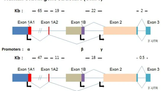
AKAP12 gene is regulated independently by three promoters; α, β and γ (Fig. 4). As three different transcripts are transcribed, AKAP12 protein also has three isoforms. AKAP12α and β isoform are expressed in all tissues, whereas AKAP12γ is only expressed in testis.
Predicted molecular size of AKAP12α and AKAP12β are 305 kDa and 287 kDa respectively. In addition, proteolytic fragments are produced by AKAP12α and AKAP12β by cleavage, showing following molecular sizes; 250 kDa and 43 kDa (Gelman 2002)
AKAP12 is a scaffolding protein, binding to various key signaling proteins, in particular, including mitogenesis related (G1->S) key proteins such as Calmodulin, PKA and PKC. By compartmentalization of key signaling protein via the ability to bind selectively, AKAP12 regulates diverse cellular signaling pathway related to mitogenesis, development, inflammatory response, differentiation and cytoskeletal architecture. In addition, AKAP12 is also known as tumor suppressor gene in various cancers such as prostate, ovarian, breast, lung and hepatocellular carcinoma (HCC). AKAP12 was reduced in metastastic progression of human prostate cancer, and AKAP12-deficient mice exhibited prostatic hyperplasia. Moreover, metastasis was suppressed by highly expressing AKAP12 protein in mice (Gelman 2012). In the liver tissues from HCC patients, AKAP12 was downregulated (Hayashi et al. 2012). Furthermore, as recent study, the micro RNA against 3`-UTR region of AKAP12 increased proliferation in HCC cell lines and promoted xenografted HCC tumor growth, whereas overexpression of
AKAP12 inhibited them (Xia et al. 2016).
Cholangiocarcinoma is the second most common primary hepatic malignancy following HCC, and its incidence and mortality rate are increasing worldwide according to several epidemiological studies.
Nevertheless, AKAP12 has never been studied in cholangiocarcinoma.
Furthermore, liver fibrosis is recently considered appropriate for therapy due to its reversibility. However, the role of AKAP12 is pooly studied in liver fibrosis. Therefore, the study for AKAP12 in cholangiocarcinoma and liver fibrosis, is necessary, so I researched it in this study.
Figure 4. The gene structures of AKAP12
MATERIALS AND METHODS
1. Animals
AKAP12-/- mice (C57BL/6 background) was generated and genotyped as previously reported (Akakura et al. 2008). The mice were inbred and maintained in the Specific-Pathogen-Free (SPF) room of the animal-housing facilities, Seoul National University (Seoul, Republic of Korea). All animal experiments in this study were approved by the Committee for Care and Use of Laboratory Animals at Seoul National University, according to the Guide for Animal Experiments edited by the Korean Academy for Medical Sciences.
2. Thioacetamide (TAA) administration
Male C57BL/6 mice, 8-10 weeks old, were subjected to TAA administration.
At first, WT mice were divided into two groups. The vehicle group (n=4) was treated with saline, and TAA group (n=10) was treated with TAA. AKAP12-/- mice also received TAA (n=6). TAA administration was done by intraperitoneal injection three times a week for a total duration of 8 weeks. Injection doses of TAA (Sigma-Aldrich) were 100 mg/kg at the first injection, 150 mg/kg at the rest of injections.
Next, WT and AKAP12-/- mice were subjected to long-term TAA administration by drinking water every day for a total duration of 26 weeks. The drinking water dose of dissolved TAA was 300 mg/l.
After TAA treatment, the mice were euthanized by deep anesthesia followed by cardiac perfusion with phosphate buffered saline (PBS).
3. 3,5-diethoxycarbonyl-1,4-dihydrocollidine (DDC) diet
Male C57BL/6 mice, weighing 25-27 g and 8-10 weeks old, were subjected to DDC diet. WT (n=5) and AKAP12-/- (n=5) mice were fed 0.1% DDC (Sigma-Aldrich)-containing diet every day for 3 weeks. After 3 weeks of DDC diet, the mice were euthanized via deep anesthesia followed by cardiac perfusion.
4. Sirius Red staining
The livers were dissected from the sacrificed mice, following cardiac perfusion with PBS. Lateral lobe was isolated from the whole liver, then fixed with 4% Paraformaldehyde (PFA) (Sigma-Aldrich) overnight at 4 ℃. The fixed lobe was embedded in paraffin, and sectioned.
Paraffin sections were de-paraffinized with xylene, followed by hydration with gradient ethanol and distilled water. The de-paraffinized paraffin sections were applied to Picro-Sirius Red staining (Abcam) according to manufacturer’s instructions. The Sirius Red staining images were taken under the microscope (Leica).
5. Immunohistochemistry
The livers were dissected from the euthanized mice after cardiac perfusion, and median lobe was frozen, embedded within O.C.T.
compound (Sakura) to make a frozen block. The frozen block was sectioned to 8 μm in width by using microtome (Thermo Scientific).
The frozen sections were fixed with 4% PFA for 10 min at room temperature, followed by washing in PBS. Then, the sections were permeabilized in PBS/0.1% Tween-20 (Amresco) solution for 25 min.
For the paraffin sections, there are additional steps for immunohistochemistry, including de-paraffinization and antigen retrieval.
Subsequent to de-paraffinization with xylene, antigen retrieval was carried out by incubating in Tris-EDTA buffer (10mM Tris Base, 1mM EDTA, 0.05% Tween-20, pH 9.0) for 40 min at 95 ℃. After blocking in 5% donkey serum (Sigma-Aldrich)/PBS solution for 1hr, primary antibodies for AKAP12 (1:300, I. Gelman, Roswell Park Cancer Institute), Thy-1 (1:50, BD), EpCAM (1:200, BD), CD31 (1:100, BD), Desmin (1:200, Abcam), α-SMA (1:200, Dako), Laminin (1:200, Sigma-Aldrich), CK-19 (1:100, Santa Cruz) and CollagenⅠ (1:200, Abcam) were incubated in humidity box overnight at 4 ℃. After intensive washing in PBS/0.1% Tween-20 solution, Alexa-488 and/or 546-conjugated secondary antibodies (1:750, Invitrogen) were treated for 1 hr at room temperature followed by counter staining with Hoechst 33342 (Sigma-Aldrich). Fluorescent images were taken under the confocal microscope (Carl Zeiss AG).
6. Quantitative analysis
Collagen-positive and immuno-positive areas in the images for Sirius Red staining and Immunohistochemistry respectively were quantified by using Image J software.
7. Statistics
All data were expressed as mean ± SEM by Prism 5 (GraphPad).
Unpaired two-tailed student's t-test was used for statistics. Differences were considered significant as a p-value < 0.05.
RESULTS
1. AKAP12 is expressed in normal mouse liver
In hepatocellular carcinoma (HCC), it has been already reported that AKAP12 is downregulated as tumor suppressor gene (Hayashi et al. 2012). However, AKAP12 is poorly studied in liver fibrosis, not even understood well that is expressed exactly where within the liver.
To unravel this, first, frozen and paraffin sections, prepared from normal mouse liver, were stained with AKAP12 antibody for Immunohistochemistry (Counter staining was done with Hoechst). Then, I found that AKAP12 is broadly expressed in mouse liver (Fig. 5A).
AKAP12 expression was observed in sinusoid and around portal vein.
However, there was no expression of AKAP12 in central vein. To confirm, normal AKAP12-/- mouse liver was subjected to staining with AKAP12 antibody, which demonstrated that the fluorescence for AKAP12 was not non-specific signal (Fig. 5B).
Figure 5. AKAP12 is expressed in sinusoid and around portal vein
(A) Immunofluorescent staining of AKAP12 in the normal mouse liver, showed AKAP12 expression in sinusoid and around portal vein. Scale bar, 200 μm. And, (B) It was confirmed by staining of AKAP12-/- mouse liver. DNA was stained with Hoechst. Scale bar, 50 μm. PV : portal vein, CV : central vein.
2. AKAP12 is expressed in portal fibroblasts and liver sinusoidal endothelial cells in mouse liver
Despite observation of the AKAP12 expression in mouse liver, It was still unknown which cell types expresses AKAP12. To investigate this, several markers were used for Immunofluorescence; EpCAM, Thy-1, Desmin, CD31. Portal tract consists of portal vein, bile duct and hepatic artery (Frevert et al. 2005). At first, strong expression of AKAP12 around portal vein was thought to come from bile duct epithelial cells based on nucleic pattern by counter staining. Therefore EpCAM, marker for bile duct epithelial cells, was double-stained with AKAP12 by Immunofluorescence. Unexpectedly, AKAP12 was expressed around bile duct epithelial cells, not merged with EpCAM expression (Fig. 6A). There is a cell type around bile duct epithelial cells; portal fibroblasts. Thy-1, marker for portal fibroblasts, was double-stained with AKAP12 by Immunofluorescence, showing co-expression in same region (Fig. 6B). This indicates portal fibroblats strongly expresses AKAP12 in normal mouse liver.
Next, to identify which cell types within sinusoid express AKAP12 in normal mouse liver, Desmin and CD31 antibody were used for Immunofluorescence. In sinusoid, there are mainly liver sinuoidal endothelial cells and hepatic stellate cells, closely attached to each other (Frevert et al. 2005). To specify which cell types expresses AKAP12 among these, first, Desmin, which is hepatic stellate cells marker, was double-stained with AKAP12 in the same manner as previously. As a result, Desmin expression seemed not to be merged with AKAP12 expression (Fig. 6C), successively CD31, endothelial cell marker, was stained with AKAP12 by Immunofluorescence (Fig. 6D).
In this staining, It was observed that AKAP12 and CD31 expression were merged each other, which demonstrated that AKAP12 is expressed in liver sinusoidal endothelial cells in normal mouse liver, not hepatic stellate cells.
Figure 6. AKAP12 is expressed in portal fibroblasts and liver sinusoidal endothelial cells
Normal mouse liver was analyzed for (A-F) AKAP12, (A) EpCAM, bile
duct epithelial cells marker, (B) Thy-1, portal fibroblasts marker, (C) Desmin, hepatic stellate cells marker, (D) CD31, endothelial cells marker, by Immunohistochemistry. (A) AKAP12 is expressed around bile duct epithelial cells stained with EpCAM. However, (B) AKAP12 expression was observed in portal fibroblasts. In sinusoid, (C,D) AKAP12 expression was only observed in liver sinusoidal endothelial cells, not hepatic stellate cells. DNA was stained with Hoechst. Scale bar, 50 μm. PV : portal vein
3. AKAP12 expression is reduced in portal fibrosis induced by DDC diet in mouse liver
Liver sinusoidal endothelial cells and portal fibroblasts have been reported that have crucial role in portal fibrosis (Wells 2014). As AKAP12 is expressed in these cells, I thought that AKAP12 might have certain roles in portal fibrosis. To investigate, 3,5-diethoxycarbonyl-1,4-dihydrocollidine (DDC) diet was used for portal fibrosis model in mouse. During DDC diet, the color of the mouse liver gradually became darkish due to porphyrin crystals caused by DDC diet (Fig. 7A). And also, the mouse livers treated with DDC were subjected to Sirius Red staining by which CollagenⅠ and Ⅲ fibers can be visualized. In the livers injured by DDC diet for 3 weeks, Collagen accumulation (Red) was observed around portal tract, demonstrating establishment of portal fibrosis model in mouse liver (Fig. 7B).
In addition, several portal fibrosis marker proteins were stained to confirm liver fibrosis model by Immunofluorescence; Desmin, a-SMA, Laminin, Thy-1 and CK-19.
As explained, Desmin is known as marker for hepatic stellate cells, and increased in portal fibrosis mouse liver sample. α-SMA, marker for myofibroblasts, also increased in DDC diet liver samples due to activation and proliferation of hepatic stellate cells and portal fibroblasts. Laminin is used for capillarized liver sinusoidal endothelial cells marker because liver sinusoidal endothelial cells do not express Laminin in normal state, and increased in portal fibrosis liver samples via DDC diet. Thy-1 expression was also elevated, indicating proliferation of portal fibroblasts. Finally, CK-19 is bile duct epithelial
cells marker like EpCAM, and was increased by proliferation of bile duct epithelial cells or liver progenitor cells (Fig. 8A). Then, positive areas of marker proteins in Immunofluorescene were quantified, all was significantly increased in portal fibrosis induced by DDC diet (Fig.
8B).
AKAP12 was also stained in portal fibrosis sample induced by DDC diet. Interestingly, unlike other marker proteins, only AKAP12 expression decreased in portal fibrosis by DDC diet, with significant reduction by quantitative analysis of Immunofluorescence (Fig. 9).
Previous report showed that liver sinusoidal endothelial cells around fibrotic region undergo capillarization and promotes angiogenesis (Lemoinne et al. 2015, Xie et al. 2012). As this report, in Immunofluorescent staining, AKAP12 expression was reduced in liver sinusoidal endothelial cells around portal fibrosis region damaged by DDC diet, suggesting that AKAP12 may have certain role during development of portal fibrosis. On the other hand, It has been previously reported that portal fibroblasts are transdifferentiated into myofibroblasts and proliferate (Wells 2014). In Immunofluorescence, the area of AKAP12 expression was broadened as portal fibroblasts proliferate, but the intensity of that was weakened in portal fibrosis mouse liver sample induced by DDC diet. Thus, although AKAP12 expression remain in the activated portal fibroblasts, decrease in those cells. It implies that AKAP12 may mediate the activation of portal fibroblasts in some degree.
Figure 7. Portal fibrosis is induced by DDC diet in mouse
DDC diet was treated to mice, (A) the color of whose liver gradually became darkish due to porphyrin crystals. (B) In Sirius Red staining, collagen deposition was observed in mouse liver damaged by DDC diet for 3 weeks. Scale bar, 400 μm.
Figure 8. Expression of portal fibrosis marker protein increases in mouse liver injured by DDC diet
(A) Representative images showed that Desmin, α-SMA, Laminin, Thy-1 and CK-19 expression were increased after 3 weeks DDC diet by Immunofluorescence. DNA (Blue) was stained with Hoechst. Scale
bar, 100 μm (upper three panels), 200 μm (lower two panels). Also, (B) quantitative analysis for immuno-positive area, showed that marker proteins were significantly increased in mouse liver treated with DDC.
Desmin, α-SMA, Thy-1, CK-19 (n=4). Laminin (n=3). Error bars indicate SEM. *p<0.01, ***p<0.001
Figure 9. AKAP12 expression decreases in portal fibrosis induced by DDC diet
Immunohistochemistry for DDC-fed mouse liver showed that AKAP12 expression was reduced in portal fibrosis, in both portal fibroblasts and liver sinusoidal endothelial cells. Quantitative analysis for AKAP12-positive area in Immunofluorescence, also indicates that AKAP12 significantly decreased in portal fibrosis liver induced by DDC diet (n=3). Scale bar, 100 μm. Error bars indicate SEM. **p<0.01 PV : portal vein, CV : central vein
4. There is no difference between WT and AKAP12
-/-mice in portal fibrosis induced by DDC diet
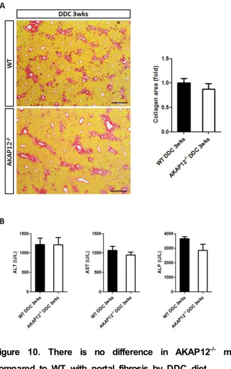
I preciously showed that AKAP12 is expressed in two major cell types involved in portal fibrosis, so I used AKAP12-/- mice to investigate difference between WT and AKAP12-/- mice. AKAP12-/- mice were fed on 0.1% DDC-containing diet with WT mice, these mice were sacrificed and subjected to Sirius Red staining. Contrary to expectations, there was no significant difference in collagen deposition between two groups (Fig. 10A). In addition, serum prepared from blood of WT and AKAP12-/- mice was used for liver function test, which confirmed no significant difference between them (Fig. 10B).
Figure 10. There is no difference in AKAP12
-/-mouse liver compared to WT with portal fibrosis by DDC diet
(A) Collagen-positive area was similar in WT and AKAP12-/- mouse liver treated with DDC in Sirius Red staining. Quantitative analysis for
collagen-positive area, also showed no significant difference between them (n=5). Scale bar, 400 μm. (B) Enzyme activity of ALT, AST and ALP was measured with serum, which represented no significant difference in AKAP12-/- compared with WT (each, n=5). Error bars indicate SEM.
5. AKAP12 expression decreases in liver sinusoidal endothelial cells of mouse with centrilobular fibrosis by TAA administration
To further study AKAP12 in centrilobular fibrosis, I injected thioacetamide (TAA) intraperitoneally three times a week for 8 weeks into mice to establish the centrilobular fibrosis model (Fig. 11A). Then, I performed Sirius Red staining with the mouse livers damaged by TAA, checked the establishment of centrilobular fibrosis via accumulation of collagen expanding from central veins (Fig. 11B).
According to previous report, liver sinusoidal endothelial cells become capillarized and promotes angiogenesis during development of centrilobular fibrosis (Lemoinne et al. 2015). To find out certain correlation of AKAP12 with liver sinusoidal endothelial cells, I stained vehicle liver samples and TAA 8 weeks liver samples with Laminin, α -SMA and AKAP12 antibody by Immunofluorescence. As previous reports, while Laminin+ liver sinusoidal endothelial cells and a-SMA+ myofibroblasts were not almost observed in vehicle samples, many of them were detected in fibrosis region in TAA 8 weeks samples.
However, AKAP12+ liver sinusoidal endothelial cells were decreased by centrilobular fibrosis induced by TAA along fibrosis region where Laminin and α-SMA expression were positive (Fig. 12A). Nevertheless, in non-fibrotic region where Laminin and α-SMA expression were lower, AKAP12 expression was shown to be maintained constantly.
Moreover, it is also understood well that hepatic stellate cells maintain tight attachment with liver sinusoidal endothelial cells in fibrotic environment, and the high magnification image of Immunofluorescent staining for AKAP12 and α-SMA, showed the loss of AKAP12
expression in liver sinusoidal endothelial cells in close proximity to α -SMA+ hepatic stellate cells (Fig. 12B). In other words, when liver sinusoidal endothelial cells are capillarized, AKAP12 is downregulated in those cells. It suggests that AKAP12 may mediate the vascular remodeling of liver sinusoidal endothelial cells in centrilobular fibrosis.
Figure 11. Centrilobular fibrosis is induced by TAA injection in mouse
(A) Thioacetamide (TAA) was intraperitoneally injected to mice every three days a week for 8 weeks. Injection dose for TAA was 150 mg/kg (Only 1st dose was 50 mg/kg). (B) Collagen accumulation was observed only in the mouse liver treated with TAA by Sirius Red staining. Vehicle (n=4), TAA 8wks (n=10). Scale bar, 400 μm.
Quantification of Sirius Red staining revealed significant induction of liver fibrosis. Error bars indicate SEM. ***p<0.001
Figure 12. AKAP12 expression is reduced unlike fibrosis marker protein in centrilobular fibrosis induced by TAA
(A) In Immunofluorescence, liver fibrosis markers, Laminin and α-SMA, were induced in centrilobular fibrosis liver by TAA. However, AKAP12 expression decreased, only along fibrotic region in centrilobular fibrosis injured by TAA. Scale bar, 200 μm. (B) As seen in high magnification image, AKAP12 expression diminished in liver sinusoidal endothelial
cells adjacent to myofibroblasts-like cells on fibrotic region. Scale bar, 50 μm. Arrows indicate fibrotic region (upper) and AKAP12-reduced region (lower).
6. Liver progenitor cells are more induced in AKAP12
-/-mice than WT in centrilobular fibrosis
Until now, I demonstrated that AKAP12 expression is reduced in capillarized liver sinusoidal endothelial cells in centrilobular fibrosis caused by chronic TAA injection, suggesting correlation with AKAP12 in terms of capillarization. To investigate this, I tried to inject TAA into AKAP12-/- mice as well as WT, expecting any difference between them. Then, these mice were sacrificed and subjected to Sirius Red staining, unexpectedly, which represented that there is no difference in collagen deposition between WT and AKAP12-/- mice with centrilobular fibrosis by TAA administration (Fig. 13A). Subsequently, I performed Immunofluorescent staining with paraffin sections from WT and AKAP12-/- mouse livers using CollagenⅠ, α-SMA, Laminin and EpCAM antibody. As the result of Sirius Red staining, CollagenⅠ, α-SMA and Laminin expression were similar between WT and AKAP12-/- mice, but interestingly, only EpCAM expression was significantly much higher in AKAP12-/- mice than WT in chronic TAA administration sample in quantitative analysis of Immunofluorescence (Fig. 13B, 13C). As described previously, chronic TAA administration induces EpCAM+ liver progenitor cells from hepatocytes. Therefore, although AKAP12 does not affect severity of TAA induced-centrilobular fibrosis, it could affect the formation of liver progenitor cells.
Figure 13. Liver progenitor cells are more highly induced in AKAP12
-/-mice compared to WT after TAA injection
(A) The degree of collagen deposition was similar in WT and AKAP12-/- mouse liver treated with TAA in Sirius Red staining. Scale bar, 400 μm. Quantitative analysis for collagen-positive area showed
no significant difference. WT (n=10), AKAP12-/- group(n=6). (B) While the other marker expression were similar in Immunofluorescence, EpCAM expression was highly induced in AKAP12-/- mouse liver with centrilobular fibrosis via TAA injection. Scale bar, 50 μm. (C) Quantification of immuno-positive area by Immunohistochemistry showed that only EpCAM+ area significantly increased in AKAP12-/- than WT (each, n=4). Error bars indicate SEM. **p<0.01
7. Hepatocarcinogenesis more severely occurs in AKAP12
-/-mouse than WT under long-term TAA administration
TAA administration induces not only liver progenitor cells from hepatocytes, but also induces cholangiocarcinoma caused by these liver progenitor cells when administration maintain for long-term (Sekiya et al. 2012). Since AKAP12-/- mice was shown to be more likely to induce liver progenitor cells from hepatocytes and AKAP12 is already known for tumor suppresor gene in several cancers including hepatocellular carcinoma, I hypothesized that AKAP12-/- mice are much more susceptible to cholangiocarcinoma. To verify this hypothesis, WT and AKAP12-/- mice were treated with TAA in their drinking water for 26 weeks. The mice were sacrificed and liver images were taken (Fig.
14A). Tumor occurred in both WT and AKAP12-/- mice liver, ranging from small to large size. However, tumor incidence was similar in WT and AKAP12-/- mice (Fig. 14B). Nonetheless, the large size tumor (>2mm) incidence was higher in AKAP12-/- mice than WT.
Figure 14. Hepatocarcinogenesis occurs more severely in AKAP12
-/-mouse liver than WT subjected to long-term TAA administration
(A) After treatment with TAA in drinking water for 26 weeks, tumor occurred in WT and AKAP12-/- mouse liver. Representative images showed that the number of tumors in each sample is much higher and the size of tumors is also larger in AKAP12-/- liver samples than WT. Arrows indicate tumors. (B) Total tumor incidence was similar each other, however the incidence of the tumors (>2mm) was higher in AKAP12-/- mice. WT and AKAP12-/- group (n=9). The numbers
above the bars mean (the number of samples including tumor) / (the number of total samples).
DISCUSSION
In this study, I demonstrated that AKAP12 is downregulated in liver fibrosis in portal fibroblasts (PFs) and liver sinusoidal endothelial cells (LSECs) I found that AKAP12 is expressed in PFs and LSECs by Immunofluorescence. PFs can be activated to myofibroblast-like cell types in response to fibrotic stimuli like hepatic stellate cells (HSCs).
As recent studies, PFs are considered as one of the contributable cells for portal fibrosis as well as HSCs, due to its ability to transdifferentiate into contractile cells. Once PFs are activated, they proliferate and expand from portal tracts, presenting contractile property, expressing α-SMA. Furthermore, the activated PFs can produce ECM components such as Collagen, Elastin. During this activation process induced by DDC diet, AKAP12 expression was reduced in the PFs. This result suggests that AKAP12 may play an important role for the activation of PFs. Besides PFs, LSECs are also important cells for induction of fibrosis including centrilobular and portal fibrosis. In homeostatic state, LSECs do not contain basal lamina (Laminin-). However, LSECs become capillarized charaterized by production of basal lamina (Laminin+) and disappearance of fenestrae in response to fibrotic stimulus, promoting angiogenesis. The LSECs interact with PFs, HSCs, kupffer cells (KCs) and hepatocytes via cell-cell interaction and soluble factors. In portal fibrosis, the LSECs near the injured portal tract, become capillarized, producing pro- and anti-fibrogenic signals to adjacent cells. As in the activation of PFs, AKAP12 was also reduced in capillarized LSECs located around damaged portal tract in portal fibrosis liver injured by DDC diet. This
result also implies that AKAP12 may have regulatory function involved in vascular remodeling in LSECs. To validate the assumptions for AKAP12 functions in PFs and LSECs, I compared WT mice liver with AKAP12-/- damaged by DDC diet for induction of potal fibrosis.
Unexpectedly, there is no significant difference between them in Sirius Red staining and liver function test for their serum. AKAP12 expressions in PFs and LSECs were already reduced at early time point during DDC diet by Immunofluorescence (Data is not shown), which could be the reason why there was no difference between WT and AKAP12-/- mice.
Next, to investigate the role of AKAP12 in centrilobular fibrosis, mice were subjected to TAA injection for 8 weeks. Collagen accumulation expanding from central vein was clearly observed in Sirius Red staining. As mentioned before, LSECs are able to be capillarized by fibrotic stimuli. In accordance with this, Immunofluorescent staining with Laminin and α-SMA antibody showed that LSECs became capillarized along fibrotic region represented by α -SMA. In this vascular remodeled LSECs, similarly to the case of portal fibrosis, AKAP12 expression significantly decreased. The activated HSCs producing ECM components more closely contact with LSECs strongly expressing Laminin than quiescent HSCs. In high-magnification images by Immunofluorescence, AKAP12 expression almost disappeared only in the LSECs closely proximal to α-SMA+ HSCs, implying that AKAP12 may act as a certain mediator during capillarization of LSECs.
AKAP12-/- mice was also subjected to chronic TAA administration to find any difference in centrilobular fibrosis compared with WT by Sirius Red staining. As a result, there was no difference between them in
centrilobular fibrosis as in portal fibrosis. They could not be different because there is already no AKAP12 expression in AKAP12-/- mice, even though AKAP12 expression is diminished in WT mice. In addition to Sirius Red staining, Immunofluorescent staining for CollagenⅠ, α -SMA and Laminin also showed no difference between WT and AKAP12-/- mice, but only for EpCAM represented clear difference. Liver progenitor cells (LPCs), which are EpCAM-immunoreactive, are known to be differentiated from hepatocytes and proliferate following TAA administration (Sekiya et al. 2012). Subsequently, the proliferating LPCs are transformed into cholangiocarcinoma by long-term TAA administration. I found that EpCAM+ LPCs were highly induced in AKAP12-/- mice liver, suggesting AKAP12-/- mice is more susceptible to cholangiocaricnoma. Therefore, to demonstrate, I tried to induce cholangiocaricnoma by long-term TAA administration for 26 weeks with WT and AKAP12-/- mice. After that, I observed various size of tumors on the surface of the livers. Tumor incidence was similar between WT and AKAP12-/- mice, accounting for about 60%. Nonetheless, the incidence of large tumors (>2mm) was higher in AKAP12-/- mice, presenting an evidence for the susceptibility to cholangiocarcinoma.
Taken together, AKAP12 is downregulated in LSECs and PFs during development of liver fibrosis including centrilobular and portal fibrosis in mice. It suggest that AKAP12 expression may function as a mediator for the capillarization of LSECs and the activation of PFs. In addition, although AKAP12 gene was deleted in mice, the severity of liver fibrosis was similar to WT, demonstrating that AKAP12 does not promote fibrogenic reaction. Recent experiment I did showed that AKAP12 expression increased again in the liver of the mice subjected to normal diet following DDC diet. Here I suggest that the role of
AKAP12 is involved in fibrosis resolution. My hypothesis for AKAP12 is as in the following. In normal state, LSECs and PFs maintain normal (quiescent) state, expressing AKAP12. However, once meet fibrotic stimuli, these cells get into the capillarization process and/or the activation process respectively, downregulating AKAP12. Then, if liver injury is ceased, Some of the activated PFs and the capillarized LSECs revert to normal state, re-expressing AKAP12. To demonstrate this hypothesis, overexpression of AKAP12 is necessary in vivo by using hydrodynamic injection of vector for AKAP12 and Tg-mice in liver fibrosis model (Liu et al. 1999), with in vitro experiments using primary cells or cell lines.
Moreover, while AKAP12 has already reported as tumor suppressor gene in several cancers including prostate cancer, cervical cancer, breast cancer and hepatocellular caricnoma (HCC), not reported in cholangiocarcinoma as tumor suppressor gene yet. Therefore, I’d like to suggest that AKAP12 may function as tumor suppressor gene in cholangiocarcinoma based on high incidence of large tumor in AKAP12-deficient liver. To confirm this, a longer administration of TAA for complete cholangiocarcinoma model and more mice for cholangiocarcinoma model are needed. And also, an analysis for correlation between AKAP12 gene and cholangiocarcinoma in human patients by bioinformatics, is thought to be essential to involve the results, which have been studied in mice, with human cholangiocarcinoma.
REFERENCES
Akakura S, Huang C, Nelson PJ, Foster B, Gelman IH. Loss of the SSeCKS/Gravin/AKAP12 gene results in prostatic hyperplasia. Cancer Res. 68(13): 5096-103 (2008)
Akhtar T, Sheikh N. An overview of thioacetamide-induced hepatotoxicity. Toxin Rev. 32(3): 43-46 (2013)
Dudas J, Mansuroglu T, Batusic D, Ramadori G. Thy-1 is expressed in myofibroblasts but not found in hepatic stellate cells following liver injury. Histochem Cell Biol. 131(1): 115-27 (2009)
Fickert P, Stoger U, Fuchsbichler A, Moustafa T, Marschall HU, Weiglein AH, Tsybrovskyy O, Jaeschke H, Zatloukal K, Denk H, Trauner M. A new xenobiotic-induced mouse model of sclerosing cholangitis and biliary fibrosis. Am J Pathol. 171(2): 525-36 (2007)
Gelman IH. Suppression of tumor and metastasis progression through the scaffolding functions of SSeCKS/Gravin/AKAP12. Cancer Metastasis Rev. 31(3-4): 493-500 (2012)
Gelman IH. The role of SSeCKS/gravin/AKAP12 scaffolding proteins in the spaciotemporal control of signaling pathways in oncogenesis and development. Front Biosci. 7: d1782-97 (2002)
Goodman ZD. Neoplasms of the liver. Mod Pathol. 20 Suppl 1:
S49-60 (2007)
Hirschfield GM, Heathcote EJ, Gershwin ME. Pathogenesis of cholestatic liver disease and therapeutic approaches. Gastroenterology.
139(5): 1481-96 (2010)
Iwakiri Y, Shah V, Rockey DC. Vascular pathobiology in chronic liver disease and cirrhosis - current status and future directions. J Hepatol.
61(4): 912-24 (2014)
Kantari-Mimoun C, Castells M, Klose R, Meinecke AK, Lemberger UJ3, Rautou PE, Pinot-Roussel H, Badoual C, Schrodter K, Osterreicher CH, Fandrey J, Stockmann C. Resolution of liver fibrosis requires myeloid cell-driven sinusoidal angiogenesis. Hepatology. 61(6): 2042-55 (2015)
Lemoinne S, Cadoret A, Rautou PE, El Mourabit H, Ratziu V, Corpechot C, Rey C, Bosselut N, Barbu V, Wendum D, Feldmann G, Boulanger C, Henegar C, Housset C, Thabut D. Portal myofibroblasts promote vascular remodeling underlying cirrhosis formation through the release of microparticles. Hepatology. 61(3): 1041-55 (2015)
Liu F, Song Y, Liu D. Hydrodynamics-based transfection in animals by systemic administration of plasmid DNA. Gene Ther. 6(7): 1258-66 (1999)
Pellicoro A, Ramachandran P, Iredale JP, Fallowfield JA. Liver fibrosis and repair: immune regulation of wound healing in a solid organ. Nat
Rev Immunol. 14(3): 181-94 (2014)
Pellicoro A, Ramachandran P, Iredale JP. Reversibility of liver fibrosis.
Fibrogenesis Tissue Repair. 5(Suppl 1): S26 (2012)
Pinzani M. Pathophysiology of Liver Fibrosis. Dig Dis. 33(4): 492-7 (2015)
Sekiya S, Suzuki A. Intrahepatic cholangiocarcinoma can arise from Notch-mediated conversion of hepatocytes. J Clin Invest. 122(11):
3914-8 (2012)
Wells RG. Portal Fibroblasts in Biliary Fibrosis. Curr Pathobiol Rep.
2(4): 185-190 (2014)
Wells RG. The portal fibroblast: not just a poor man's stellate cell.
Gastroenterology. 147(1): 41-7 (2014)
Xia W, Ni J, Zhuang J, Qian L, Wang P, Wang J. MiR-103 regulates hepatocellular carcinoma growth by targeting AKAP12. Int J Biochem Cell Biol. 71: 1-11 (2016)
Xie G, Wang X, Wang L, Wang L, Atkinson RD, Kanel GC, Gaarde WA, Deleve LD. Role of differentiation of liver sinusoidal endothelial cells in progression and regression of hepatic fibrosis in rats.
Gastroenterology. 142(4): 918-927 (2012)
Xu J, Liu X, Koyama Y, Wang P, Lan T, Kim IG, Kim IH, Ma HY,
Kisseleva T. The types of hepatic myofibroblasts contributing to liver fibrosis of different etiologies. Front Pharmacol. 5: 167 (2014)
Yang L, Kwon J, Popov Y, Gajdos GB, Ordog T, Brekken RA, Mukhopadhyay D, Schuppan D, Bi Y, Simonetto D, Shah VH. Vascular endothelial growth factor promotes fibrosis resolution and repair in mice. Gastroenterology. 146(5): 1339-50 (2014)
Yovchev MI, Zhang J, Neufeld DS, Grozdanov PN, Dabeva MD.
Thymus cell antigen-1-expressing cells in the oval cell compartment.
Hepatology. 50(2): 601-11 (2009)
국문초록
간섬유화는 감염 (주로 B형, C형 간염), 독성 물질, 대사 질환. 자가면 역에 의한 만성적 간 손상에 대한 복구 반응으로부터 야기된다. 게다가, 간섬유증은 반복적이고 지속적인 간 손상에 의해 간경화로 이어질 수 있 다. 간경화가 발생한 간 조직의 경우 정상 간 조직에 비해 간 기능이 현저 히 떨어지고, 이는 생명을 위협하게 된다. 더욱이 간경변증은 간암과 밀접 하게 연관되어 있고, 치료법도 오직 간 이식 뿐이다. 이러한 이유로, 가역 성을 띠는 간섬유증이 치료에 적절한 질병으로 여겨지고 있다. 그럼에도 불구하고, 지금까지도 항섬유성 약물로 승인된 물질이 아직 없다. 따라서 간섬유증의 치료를 위한 대상 물질을 개발하는 것이 현재로서 필요하다고 생각된다.
A-Kinase Anchoring Protein (AKAP12) 는 전립선암, 자궁암, 유방암, 폐암, 간암 등 다양한 암에서 종양 억제 유전자로 보고되어 왔다. 하지만, 간섬유증에서 AKAP12의 역할은 여전히 알려지지 않은 채로 남아있다.
이번 연구에서 AKAP12가 정상 생쥐의 간에서 동양혈관내피세포와 문 맥 섬유아세포에서 발현됨을 보였다. 간문맥 섬유증 유발을 위해, DDC 사 료를 먹인 생쥐의 간에서 AKAP12의 발현을 분석하였는데, 활성화된 문맥 섬유아세포와 섬유화가 발생한 영역 근처의 모세혈관화된 동양혈관내피세 포에서 AKAP12의 발현이 감소되는 것을 관찰하였다. 하지만 DDC 사료 로 간 손상을 준 야생형, AKAP12-/- 생쥐의 간 사이에는 차이가 없었다.
TAA 주사에 의해 소엽중심성 섬유증을 나타내는 생쥐의 간 또한 같은 방식으로 AKAP12의 발현이 조사되었고, 섬유화가 발생한 영역을 따라서 모세혈관화된 동양혈관내피세포에서 발현이 감소함을 발견하였다. 간문맥 섬유증에서처럼, TAA에 의해 소엽중심성 섬유증이 발생한 간에서도, 야생 형과 AKAP12-/- 생쥐 사이에 차이가 없었다.
흥미롭게도 EpCAM+ 간 전구세포가 야생형보다 AKAP12-/- 생쥐의 간에
서 TAA에 의해 더 많이 유도되었고, 이는 AKAP12와 장기간 TAA 투여에 의한 담관암 사이에 어떤 연관성이 있음을 암시한다. 실제로, TAA에 의해 유도된 큰 종양 (>2mm)의 발생 빈도가 더 높음을 나타내면서, AKAP12-/- 생쥐가 야생형에 비해 담관암에 더욱 걸리기 쉬울 것이라는 사실을 발견 했다.
종합해보면, AKAP12는 간섬유증 동물모델에서 하향조절되고, 이는 특 히 간섬유화 완화 과정과 관련하여, AKAP12가 동양혈관내피세포와 문맥 섬유아세포에서 발현되면서 간섬유화에 대한 조절자로 기능할 것임을 보 이고 있다. 또한 종양에 관한 연구에서는, AKAP12가 다른 다양한 암에서 처럼 담관암에 대해서도 종양 억제 유전자로 작용할 수도 있음을 나타내 고 있다.
주요어
: 간섬유증, AKAP12, 동양혈관내피세포, 문맥 섬유아세포, 담관암학번
: 2014-24872