저작자표시-비영리-변경금지 2.0 대한민국 이용자는 아래의 조건을 따르는 경우에 한하여 자유롭게
l 이 저작물을 복제, 배포, 전송, 전시, 공연 및 방송할 수 있습니다. 다음과 같은 조건을 따라야 합니다:
l 귀하는, 이 저작물의 재이용이나 배포의 경우, 이 저작물에 적용된 이용허락조건 을 명확하게 나타내어야 합니다.
l 저작권자로부터 별도의 허가를 받으면 이러한 조건들은 적용되지 않습니다.
저작권법에 따른 이용자의 권리는 위의 내용에 의하여 영향을 받지 않습니다. 이것은 이용허락규약(Legal Code)을 이해하기 쉽게 요약한 것입니다.
Disclaimer
저작자표시. 귀하는 원저작자를 표시하여야 합니다.
비영리. 귀하는 이 저작물을 영리 목적으로 이용할 수 없습니다.
변경금지. 귀하는 이 저작물을 개작, 변형 또는 가공할 수 없습니다.
A DISSERTATION
FOR THE DEGREE OF DOCTOR OF PHILOSOPHY
Control of Macrophage Phenotypic Polarization by TSG-6 Secreted from Human and Canine Adipose Tissue-derived Mesenchymal Stem Cells in Mouse Models of Inflammatory Bowel Disease
염증성 장질환 마우스 모델에서 사람과 개의 지방유래 중간엽줄기세포가 분비하는 TSG-6 에 의한
대식세포 표현형 조절
By Woojin Song
MAJOR IN VETERINARY CLINICAL SCIENCE (VETERINARY INTERNAL MEDICINE) COLLEGE OF VETERINARY MEDICINE
GRADUATE SCHOOL OF SEOUL NATIONAL UNIVESITY
February, 2019
염증성 장질환 마우스 모델에서 사람과 개의 지방유래 중간엽줄기세포가 분비하는
TSG-6 에 의한 대식세포 표현형 조절
지도교수 윤 화 영
이 논문을 수의학박사학위논문으로 제출함 2018 년 11 월
서울대학교 대학원
수의과대학 임상수의학 (수의내과학) 전공 송 우 진
송우진의 박사학위논문을 인준함 2018 년 12 월
위 원 장 김 용 백 (인)
부 위 원 장 윤 화 영 (인)
위 원 신 남 식 (인)
위 원 서 경 원 (인)
위 원 정 진 영 (인)
i
Control of Macrophage Phenotypic Polarization by TSG-6 Secreted from Human and Canine Adipose Tissue-derived Mesenchymal Stem Cells in Mouse Models of Inflammatory Bowel Disease
Woojin Song
(Supervised by Prof. Hwa-Young Youn)
Major in Veterinary Clinical Science (Veterinary Internal Medicine) College of Veterinary Medicine
Graduate School of Seoul National University
Abstract
Inflammatory bowel disease (IBD) is an intractable autoimmune disorder that markedly deteriorates quality of life. Mesenchymal stem cells (MSCs) alleviate inflammation by modulating inflammatory cytokines in inflamed tissues, and have been suggested as a promising alternative for IBD treatment in human and veterinary cases. Furthermore, tumor necrosis factor- α-induced gene/protein 6 (TSG-6) is a key factor influencing MSC immunomodulatory properties; however, the precise mechanism of TSG-6
ii
release from human and canine MSCs in IBD remains unclear. This study aimed to assess therapeutic effects of human adipose tissue-derived (hAT)- and canine adipose tissue-derived (cAT)-MSC-produced TSG-6 in an IBD mouse model and explore the mechanisms underlying the immunomodulatory properties.
This dissertation is composed of three parts. The first part of the study was performed to investigate to assess therapeutic effects of hAT-MSC and explore their mechanisms in dextran sulfate sodium (DSS)-induced colitis mice. DSS-induced colitis mice were infused with hAT-MSCs intraperitoneally and colon tissues were collected. hAT-MSCs were shown to induce the expression of M2 macrophage markers and to regulate the expression of pro- and anti-inflammatory cytokines in the colon. Quantitative real time-PCR analyses demonstrated that less than 20 hAT-MSCs, 0.001%
of all intraperitoneally injected hAT-MSCs, were detected in the inflamed colon. To investigate the effects of hAT-MSC-secreted factors in vitro, transwell co-culture system was used, demonstrating that tumor necrosis factor-α-induced gene/protein 6 (TSG-6) released by hAT-MSCs induces M2 macrophages. In vivo, hAT-MSCs transfected with TSG-6 small interfering RNA, administered intraperitoneally, were not able to induce M2 macrophage phenotype switch in the inflamed colon and had no significant effects on IBD severity.
iii
The second part of the dissertation was designed to evaluate whether cAT-MSC-secreted TSG-6 could ameliorate IBD and regulated colonic expression of pro- and anti-inflammatory cytokines such as tumor necrosis factor-α, interleukin-6, and interleukin-10. To investigate the effect of cAT- MSC-secreted TSG-6 on activated macrophages in vitro, a transwell co- culture system was used; TSG-6 released by cAT-MSCs induced macrophage phenotypic switch from M1 to M2. The cAT-MSC-secreted TSG-6 increased M2 macrophages in the inflamed colon in vivo.
The last part of the dissertation was designed to demonstrate whether cAT-MSCs stimulated with TNF-α could exert enhanced therapeutic effects in IBD mice models. DSS- or dinitrobenzene sulfonic acid (DNBS)-induced colitis mice were treated with cAT-MSCs pre-treated with TNF-α intraperitoneally. Colitis severity was assessed and colon tissues were collected for histopathological, enzyme-linked immunosorbent assay, and flow cytometry analysis cAT-MSCs stimulated with TNF-α secreted higher concentrations of immunomodulatory factors such as TSG-6 and PGE2, which play a key role in inducing macrophages phenotypic alteration. In consequence, TNF-α pre-treated cAT-MSCs further regulated colonic inflammatory cytokines such as interleukin (IL)-1β, IL-6, and IL-10, and ameliorated DSS- or DNBS-induced colitis in mice. Also, I demonstrated that M1 macrophages (F4/80+/iNOS+ cells) were more decreased and M2 macrophages (F4/80+/CD206+ cells) were more increased in colon tissues
iv
from mice treated with TNF-α pre-treated cAT-MSCs rather than those treated with naïve cAT-MSCs.
In conclusion, I demonstrated here that TSG-6 released from human or canine AT-MSCs can alleviate IBD in mice. Furthermore, pre-treatment with TNF-α enhanced anti-inflammatory effects of MSCs by secreting high concentrations of immunomodulatory factors (such as TSG-6 and PGE2) and inducing M2 macrophage phenotypic alteration. These findings might provide cell-based therapeutic options in IBD dogs.
Key words: / inflammatory bowel disease / auto-immune disease /
mesenchymal stem cells / TSG-6 / immunomodulation / anti-inflammation
Student number: 2013-21558
v
CONTENTS
ABSTRACT i
CONTENTS v
LIST OF FIGURES xi
LIST OF TABLES xiv
ABBREVIATIONS xv
LITERATURE REVIEW 1 1. Generalities of mesenchymal stem cells (MSCs) 2 2. Properties of canine MSCs 4 3. Immunomodulatory effects of canine MSCs 7 4. Preclinical and clinical application of canine MSCs in non-infectious inflammatory diseases 9 CHATER I. TSG-6 Secreted by Human Adipose Tissue-derived Mesenchymal Stem Cells Ameliorates Inflammatory Bowel Disease by Inducing M2 Macrophage Polarization in Mice 15
1. Introduction 16
2. Materials and Methods 19
2.1. Cell preparations 19
vi
2.2. Stimulation of hAT-MSCs with TNF-α 21
2.3. Transfection of hAT-MSCs with small interfering RNA 21
2.4. Animal experiments 21
2.5. Assessment of colitis severity 23
2.6. Histological analysis 23
2.7. Co-culturing of Raw 264.7 macrophages with hAT-MSCs 24
2.8. RNA extraction, cDNA synthesis, and qRT-PCR 24
2.9. Determination of TSG-6 expression by MSCs in the conditioned medium 25
2.10. Western blot analysis 25
2.11. Immunofluorescence analyses 26
2.12. Generation of GAPDH standard curve 27
2.13. Statistical analysis 27
3. Results 29
3.1. Intraperitoneally administered hAT-MSCs ameliorate IBD 29
3.2. hAT-MSCs reduce the inflammatory response by increasing the percentage of M2 macrophages in colon 30
3.3. Intraperitoneally injected hAT-MSCs do not migrate into the colon 31
3.4. hAT-MSC-produced TSG-6 induces the phenotypic switch to M2 macrophages in vitro 31
vii
3.5. Intraperitoneal administration of hAT-MSCs with TSG-6
knockdown cannot alleviate IBD 34
4. Discussion 36
5. Figures and Tables 43
CHAPTER II. TSG-6 Released from Intraperitoneally Injected Canine Adipose Tissue-derived Mesenchymal Stem Cells Ameliorate Inflammatory Bowel Disease by Inducing M2 Macrophage Switch in Mice 72
1. Introduction 73
2. Materials and Methods 76
2.1. Isolation and characterization of cAT-MSCs 76
2.2. Small interfering RNA transfection of cAT-MSCs 77
2.3. Animal experiments 78
2.4. Evaluating colitis severity 78
2.5. Histological analysis 79
2.6. Enzyme-linked immunosorbent assay 79
2.7. Obtaining canine peripheral blood mononuclear cell (cPBMC)- derived macrophages 80
2.8. Co-culture of canine PBMC-derived macrophages with cAT- MSCs 81
2.9. Flow cytometric analysis 81
viii
2.10. Immunofluorescence analysis 82
2.11. Annexin-V and propidium iodide staining 83
2.12. Generation of the GAPDH standard curve 84
2.13. Statistical analysis 84
3. Results 85
3.1. Intraperitoneally administered cAT-MSC-secreted TSG-6 played a crucial role in ameliorating IBD 85
3.2. cAT-MSC-secreted TSG-6 reduced inflammatory response and apoptosis in the colon 86
3.3 Intraperitoneally infused cAT-MSCs did not migrate to inflamed colon 87
3.4. TSG-6 produced by cAT-MSCs induced phenotypic switching from M1 to M2 macrophages in vitro 87
3.5. cAT-MSC-secreted TSG-6 increased M2 macrophages in inflamed colon 88
4. Discussion 90
5. Figures and Tables 95
CHAPTER III. Canine Adipose Tissue-derived Mesenchymal Stem Cells Pre-treated with TNF-alpha Enhanced Immunomodulatory Effects on Inflammatory Bowel Disease in Mice 116
1. Introduction 117
ix
2. Materials and Methods 119
2.1. Isolation, culture, and characterization of cAT-MSCs 119
2.2 TNF-α stimulation of cAT-MSCs 120
2.3. Animal experiments 121
2.4. Evaluation of colitis severity 121
2.5. Enzyme-linked immunosorbent assay 122
2.6. Flow cytometry analysis 123
2.7. Statistical analysis 123
3. Results 124
3.1. Phenotypic characterization of cAT-MSCs 124
3.2. Enhanced secretory abilities for immunomodulatory factors of TNF-α stimulated cAT-MSCs 124
3.3. Improved therapeutic effects of TNF-α stimulated cAT-MSCs on DSS- or DNBS-induced colitis mice 125
3.4. Enhanced anti-inflammatory effects of TNF-α primed cAT- MSCs on DSS-induced colitis mice 125
3.5. Increased alteration of macrophage phenotype in the inflamed colon of mice administered TNF-α primed cAT-MSCs 126
4. Discussion 127
5 Figures and Tables 132
GENERAL CONCLUSION 144
x
REFERENCES 148
ABSTRACT IN KOREAN (국문초록) 167
xi
LIST OF FIGURES
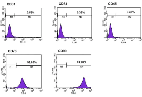
Figure 1. Immunophenotypic analysis of hAT-MSCs by flow cytometry.
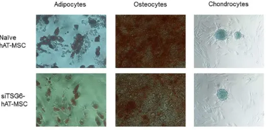
Figure 2. Differentiation of hAT-MSCs.
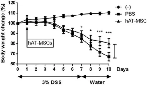
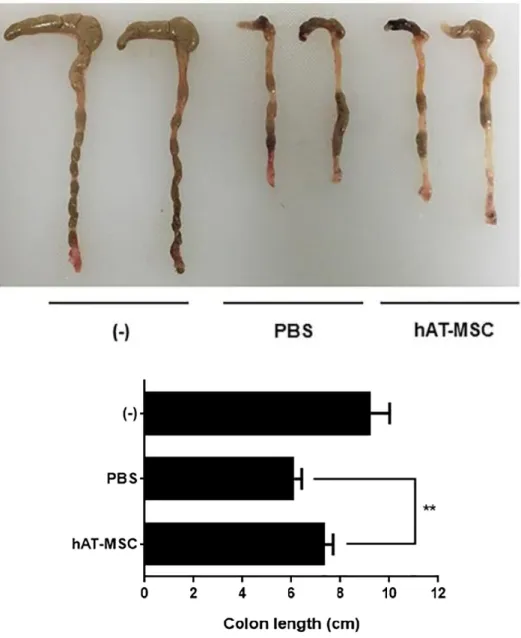
Figures 3-6. Intraperitoneally injected hAT-MSCs ameliorate IBD.
Figures 7-8. hAT-MSCs inhibit inflammatory response in the colon.
Figures 9-10. hAT-MSC administration leads to an increase in the percentage of M2 macrophages in the colon.
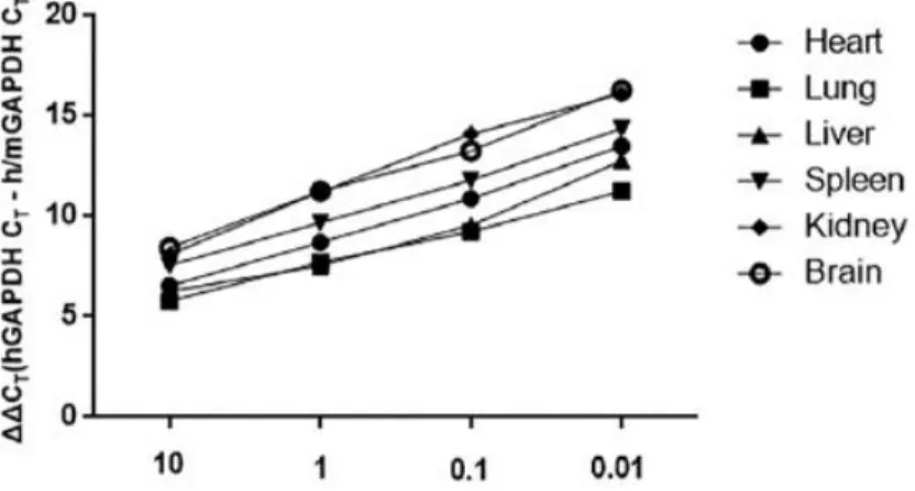
Figure 11. Standard curves for qRT-PCR assays of human mRNA for GAPDH.
Figure 12. Distribution of hAT-MSCs in colitis mice after intraperitoneally infusion.
Figure 13. Intraperitoneally administered hAT-MSCs do not migrate into the colon.
Figure 14. TSG-6 gene and protein expression levels in TNF-α-stimulated hAT-MSCs and naïve hAT-MSCs.
Figures 15-16. TNF-α-stimulated hAT-MSCs induce the expression of the M2 macrophage markers.
Figure 17. TSG-6 mRNA and protein expression levels in hAT-MSCs transfected with TSG-6 siRNA.
xii
Figures 18-19. Expression of M2 macrophage markers decreases in hAT- MSCs transfected with TSG-6 siRNA.
Figures 20-23. TSG-6 knockdown in hAT-MSCs inhibits their effect on IBD.
Figures 24-25. TSG-6 knockdown in hAT-MSCs inhibits their effect on M2 macrophage phenotypic switch.
Figure 26. Flow cytometric analysis of cAT-MSCs.
Figure 27. Differentiation of cAT-MSCs.
Figure 28. TSG-6 mRNA expression levels in cAT-MSCs transfected with TSG-6 siRNA.
Figures 29-32. Intraperitoneally infused cAT-MSC-secreted TSG-6 plays an essential role in alleviating IBD.
Figures 33-34. TSG-6 secreted by cAT-MSCs inhibits inflammatory response and apoptosis in the colon.
Figure 35. Standard curves for evaluating the migratory ability of intraperitoneally injected cAT-MSCs.
Figure 36. Intraperitoneally administered cAT-MSCs do not migrate into the colon.
Figures 37-38. cAT-MSC-secreted TSG-6 induce macrophage phenotypic switching from M1 to M2 in vitro.
Figures 39-40. TSG-6 secreted by cAT-MSCs induces M2 macrophage polarization in inflamed colon.
Figure 41. Characterization of cAT-MSCs.
xiii
Figure 42. cAT-MSCs stimulated with TNF-α released higher TSG-6 and PGE2.
Figure 43-46. cAT-MSCs stimulated with TNF-α showed enhanced therapeutic effects on DSS-induced colitis in mice.
Figure 47-50. cAT-MSCs stimulated with TNF-α showed enhanced therapeutic effects on DNBS-induced colitis in mice.
Figure 51. cAT-MSCs stimulated with TNF-α further regulated inflammation in the inflamed colon.
Figure 52. cAT-MSCs stimulated with TNF-α further decreased M1 macrophages and increased M2 macrophages in colons from IBD mice.
xiv
LIST OF TABLES
Table 1. Primers for qRT-PCR for the first part of this dissertation Table 2. Primers for qRT-PCR for the second part of this dissertation
xv
ABBREVIATIONS
APC Allophycocyanin
cAT-MSC Canine adipose tissue-derived mesenchymal stem cell cPBMC Canine peripheral blood mononuclear cell
DSS Dextran
ELISA Enzyme-linked immunosorbent assay FITC Fluorescein isothiocyante
GAPDH Glyceraldehyde 3-phosphate dehydrogenase IACUC Institutional Animal Care and Use Committee IBD Inflammatory bowel disease
IF Immunofluorescence
IL Interleukin
iNOS Inducible nitric oxide synthase
LPS Lipopolysaccharide
PE Phycoerythrin
PGE2 Prostaglandin E2
PI Propidium iodide
qRT-PCR Quantitative reverse transcription-polymerase chain reaction siRNA Small interfering ribonucleic acid
TNF Tumor necrosis factor
TSG-6 Tumor necrosis factor-α-induced gene/protein-6
1
LITERATURE REVIEW
2
1. Generalities of mesenchymal stem cells (MSCs)
Mesenchymal stem cells (MSCs) are a heterogeneous population of cells that proliferate in vitro as plastic-adherent cells, have fibroblast-like morphology, and can differentiate into adipocytes, osteocytes, and chondrocytes. MSCs have been successfully isolated from nearly every organ and many tissues including brain, liver, kidney, lung, bone marrow, muscle, thymus, pancreas, skin, aorta, vena cava, adipose tissue, fetal tissue, umbilical cord, Wharton’s jelly and placenta (da Silva Meirelles et al., 2006; N Momin et al., 2010; Romanov et al., 2003)
.
MSCs are regarded to be promising candidates for clinical application due to the ease of isolation from adult donors, thus obviating the ethical concerns concerning embryonic stem cell research.MSCs potently modulate immune responses and have paracrine effects through secretion of growth factors, cytokines and antifibrotic or angiogenic mediators (Uccelli et al., 2008). Thus, MSCs have a broad prospect of clinical application in regenerative medicine. Recent studies also have demonstrated that MSCs show anti-inflammatory effects and can alleviate the symptoms in various inflammatory disease models, including rheumatoid arthritis, peritonitis, and pancreatitis, as well as IBD(Gonzalez- Rey et al., 2009; Jung et al., 2011; Liu et al., 2010; Prockop and Oh, 2012).
Currently, the mechanisms underlying the anti-inflammatory effects are being
3
investigated, in order to establish the basis for effective clinical application of MSCs. In addition, MSCs derived from dogs, cats, or horses were shown to have immunomodulatory effects on activated immune cells and that cell- based therapy using MSCs is a potential treatment or intractable inflammatory diseases in veterinary medicine(Carrade Holt et al., 2014; Chae et al., 2017;
Kim et al., 2016).
4
2. Properties of canine MSCs
Canine MSCs could be derived from various stromal tissues such as adipose tissue, bone marrow, umbilical cord blood, and amniotic fluid.
Among them, canine adipose tissue or bone marrow derived MSCs have been used mainly for research and application. The samples can be obtained by appropriate general and local anesthesia. For collecting adipose tissue samples, our group use medetomidine (400-500 μg/m2, IV), tramadol (4 mg/kg, IV), and lidocaine (local injection). For bone marrow samples, our group use alfaxalone (2 mg/kg, IV) for induction and 2% isoflurane inhalation with oxygen flow (2 L/min) for maintenance of general anesthesia.
Using protocols by professor Youn (supervisor of this dissertation), MSCs are isolated from adipose tissues as following procedure: The fat tissue are washed with PBS containing penicillin and streptomycin, and then cut into small pieces and digested for 1-2 h at 37C with collagenase type IA.
Then, enzymatic activity should be inhibited with Dulbecco’s Modified Eagle’s Medium (DMEM) containing 10% fetal bovine serum (FBS). After centrifugation at 1200 ×g for 5-10 min, the pellet should be filtered through a 100-µm cell strainer to remove debris and then incubated overnight in DMEM containing 20% FBS at 37C in a humidified atmosphere of 5% CO2. Then, the cultures are washed with PBS to remove non-adherent cells and incubated with fresh medium (DMEM containing 10% FBS), which should be changed
5
every 48-72 h until cells reached 70–80% confluence, after which they should be repeatedly sub-cultured under standard conditions (Yang et al., 2018b).
In addition, MSCs from bone marrow are isolated as following procedure: Bone marrow samples are diluted with PBS, and then gently layered over Ficoll-Paque PLUS (GE Healthcare Life Sciences, Uppsala, Sweden) in a conical tube (50 mL). After centrifugation at 800×g for 30 min without brake, the buffy coat layer should be carefully collected and centrifuged at 1200×g for 10-20 min. The supernatant is discarded and the cells are resuspended in alpha modified Eagle's medium (αMEM) containing 20% FBS, then incubated overnight at 37C in a humidified atmosphere of 5%
CO2. After that, the cultures should be washed and changed (αMEM containing 10% FBS) as previously described for canine adipose tissue- derived MSCs (Yang et al., 2018b).
MSCs derived from stromal tissues should be characterized before their use. The International Society for Cellular Therapy (ISCT) established minimal criteria of MSC in 2006 to standardize MSCs (Dominici et al., 2006).
Briefly, MSCs should adhere to plastic cell culture dish with a fibroblast-like morphology. Also, MSCs should have multi-potency of differentiation into the 3 major mesenchymal lineages (adipocytes, osteocytes, and chondrocytes) in vitro. In addition, MSCs should express specific surface markers such as
6
CD73, CD90, Sca-1 and CD105, but not express the negative markers such as CD14 or CD11b, CD34, and CD45.
7
3. Immunomodulatory effects of canine MSCs
MSCs are considered to show hypo-immunogenicity because of their low or lack of expression level in major histocompatibility complex class I and II molecules, which enables MSCs to be safely used without potential risks for immune rejection even in allogeneic environments. Furthermore, xenogeneic infusion of canine MSCs into mouse and rat models has been also reported to be well-tolerated and effective, suggesting that canine MSCs can exert cross-species immunomodulatory effects.
The immunomodulatory effects of MSCs have been demonstrated in immune-mediated disease models (Kim et al., 2016; Le Blanc et al., 2004).
For example, human MSCs have been used in studies on treatment of graft- vs.-host disease, inflammatory bowel disease, systemic lupus erythematosus, and rheumatoid arthritis (Dave et al., 2015; Glenn and Whartenby, 2014). In addition, canine MSCs in particular have been used for the treatment of severe acute pancreatitis, inflammatory bowel disease, osteoarthritis, pemphigus, and atopic dermatitis (Ferrer et al., 2016; Hoffman and Dow, 2016). Moreover, several studies reported that even when MSCs do not migrate directly to the site of inflammation or the injured tissue, they can still exert anti- inflammatory actions through secretory factors (Lee et al., 2011; Silini et al., 2013). Recent investigations have reported that MSCs regulate the inflammatory processes through the activity of various soluble factors, such
8
as indoleamine 2,3-dioxygenase (IDO), TGF-β, PGE2, and TNF-α-induced gene/protein 6 (TSG-6) (Gonzalo-Gil et al., 2016; Kim et al., 2015; Liu et al., 2016; Liu et al., 2015). Among these factors, several studies have revealed that TSG-6 is pivotal in anti-inflammatory effects of MSCs against corneal inflammation, severe burn injury, acute lung injury, acute peritonitis, pancreatitis, and IBD(Danchuk et al., 2011; He et al., 2016; Liu et al., 2016;
Roddy et al., 2011; Sala et al., 2015; Wang et al., 2012).
Furthermore, previous studies have revealed that human MSCs stimulated with pro-inflammatory cytokines (such as TNF-α and IL-1β) could improve secretory effects of immunomodulatory soluble factors (Broekman et al., 2016; Heo et al., 2011). TSG-6 and PGE2 are well-known immunomodulatory factors secreted from human and canine MSCs, and recent studies have demonstrated that they play important roles in ameliorating atopic dermatitis, rheumatoid arthritis, acute pancreatitis, and IBD (Kim et al., 2016; Kim et al., 2015; Mao et al., 2017; Shin et al., 2016;
Song et al., 2017).
9
4. Preclinical and clinical application of canine MSCs in non- infectious inflammatory diseases
4.1. Severe acute pancreatitis (SAP)
Acute pancreatitis is a common disease in dogs. Although most cases are self-limiting and fully reversible, some progress to severe acute pancreatitis (SAP), which leads to systemic complications such as multi- organ failure and diffuse intravascular coagulation (Cook et al., 1993;
Mansfield, 2012). Mortality rates among dogs with SAP are 27% to 42%
(Cook et al., 1993; Mansfield, 2012). To date, no effective treatment strategies have been developed, indicating the need for a better understanding of the pathophysiology of SAP. A breed predisposition has been reported for acute pancreatitis that deteriorates into SAP, implying that the disease is related to hereditary mutations (Hess et al., 1998), including those that cause auto- activation of trypsin, resulting in pancreatic edema, death of acinar cells (Whitcomb et al., 1996), and an inflammatory response mediated by cytokines such as TNF-α, IL-1β, -6, -12, -4, and -10, IFN-γ released by macrophages and T cells (Pandiyan et al., 2007; Tsuda et al., 2014).
Overproduction of these inflammatory cytokines can lead to systemic manifestations, ulti-organ failure, or death (Norman, 1998).
I have demonstrated the therapeutic effects ofcanine adipose tissue- derived MSCs in a SAP rat model (not included in this dissertation). Canine
10
MSCs improved SAP in rats by inhibiting pro-inflammatory cytokines and stimulating anti-inflammatory cytokine production. In addition, canine MSCs suppressed the proliferation of co-cultured cPBMCs as well as rat splenocytes treated with ConA in a ratio-dependent manner. Similarly, CM containing soluble factors inhibited the proliferation of both cPBMCs and rat splenocytes in the present study. In addition, canine MSCs blocked the infiltration of CD3+ T cells and increased the FoxP3+ regulatory T cell population in the injured pancreas of SAP rats. Although the identity of the soluble factors and anti-inflammatory mechanisms of canine MSCs require more detailed study, I speculate that cAT-MSCs inhibit inflammation by regulating T cells via paracrine mechanisms as well as cell-to-cell contact.Therefore, canine MSCs could be an attractive candidate for cell-based clinical therapy in SAP dogs.
4.2. Inflammatory bowel disease (IBD)
IBD is an intractable autoimmune disease, leading to a chronic inflammation of the digestive system, which can be classified as either ulcerative colitis or Crohn’s disease, depending on the site and pattern of inflammation (Bouma and Strober, 2003; van Beelen Granlund et al., 2013).
And, IBD leads to abdominal pain, diarrhea, fever, or other symptoms that may be caused by chronic inflammation of the digestive system. Although the exact pathogenesis of IBD remains unknown, it is believed to be associated
11
with genetic and environmental factors, as well as inflammatory responses towards gut flora (Knights et al., 2013; Manichanh et al., 2012). Intestinal inflammatory response is known to be regulated through the secretion of inflammatory cytokines, such as TNF-α, IFN-γ, TGF-β, IL-1β, -4, -6, -10, - 17 and -23 which are secreted by macrophages and T-cells (Neurath, 2014).
In addition, the disease occurs naturally in dogs by a similar pathogenesis, and data from therapeutic trials for canine IBD may be excellent references for human IBD (Maeda et al., 2016). Although IBD leads to a decreased quality of life in both humans and dogs, no effective treatments for IBD have been developed.
In an open label baseline controlled study by Perez-Merino et al.
(Perez-Merino et al., 2015), 12 dogs that were partially tolerant to corticosteroids with histologically confirmed lymphocytic-plasmocytic IBD, received a single intravenous injection of allogeneic, single donor sourced adipose tissue-derived MSCs (2 × 10⁶ / kg) (Allenspach et al., 2007; Jergens et al., 2003). These patients were monitored for 42 days after transplantation using two different clinical scoring systems which incorporated laboratory and clinical observations (including owner observations of attitude, appetite, stool consistency and frequency, vomiting, pruritus) along with ascites, peripheral edema, body weight, and serum albumin as well as biomarkers folate, cobalamin, and C-reactive protein (CRP). Treatment significantly
12
improved clinical scores, serum albumin, and biomarkers (although not CRP) compared to baseline values. The absence of a control group obscures understanding of the magnitude of effects achieved with MSCs, and the open label design may contribute to observer (owner, veterinarian) bias. However, these data support the safety and therapeutic activity of allogeneic canine MSCs in partially refractory canine IBD at the selected dosage, one that mirrors the dosage range employed in past human studies (1-2 × 10⁶ /kg).
4.3. Pemphigus
Canine pemphigus foliaceus is an autoimmune antibody mediated skin disease characterized by acantholysis. The pathogenesis involves the production of autoantibodies against a target protein in the adhesion molecules of keratinocytes (Craig, 2013). Desmoglein I is the main antigen implicated in pemphigus foliaceus in dogs and humans (Gross et al., 2008;
Morris, 1994). Binding of antibodies to adhesion molecules such as Desmoglein I disrupts the intercellular cohesion of keratinocytes. This results in acantholysis and the typical lesions seen in pemphigus, including formation of blisters and intra-epidermal pustules. The cause is usually unknown.
However, some cases are possibly drug-induced (White et al., 2002) or a sequel to a chronic inflammatory skin disease (Medleau and Hnilica, 2006).
The most successful treatment for canine pemphigus foliaceus is
13
immunosuppression with corticosteroids or cyclosporine. In recent studies, however, side effects of this treatment such as diarrhea, polyuria/polydipsia, weight gain, and recurrent infections have been described (Gomez et al., 2004). Moreover, it has been reported that only 53% of treated cases survive for more than 1 year after initiation of treatment (Gomez et al., 2004).
Youn (supervisor of this dissertation) and colleagues have described the clinical application of cytotoxic T-lymphocyte antigen 4 (CTLA4) overexpressing canine MSCs and/or naive canine MSCs in steroid refractory pemphigus foliaceus. Initial treatment comprised immunosuppressive doses of prednisolone after the diagnosis of pemphigus foliaceus. Treatment with prednisolone alone did not have the desired effect and combinations with cyclosporine and azathioprine were prescribed, with no improvement in the clinical signs. Side effects of the immunosuppression included melena and anorexia and the skin condition deteriorated, with lesions spreading over the whole body. After the first administration of CTLA4 overexpressing canine MSCs, the skin lesions improved. CLTA4 overexpressing canine MSCs and/or naive canine MSCs were administered 21 times over a period of 20 months with an interval of 2 to 8 weeks. A tapering dose of prednisolone was given concurrently. After termination of canine MSC treatment, the skin lesions were well controlled with a low dose of prednisolone and remained under control for 12 months.
14
4.4. Atopic dermatitis
Atopic dermatitis is a condition that afflicts 8.7% dogs (Hillier et al., 2001) similar to children (10%-20%) and adults (3%-4%), that is associated with breed predilections, polymorphisms at specific gene loci, altered gene expression, and specific allergens. The concept behind employing MSC for immunomodulation of atopic dermatitis, led Hall et al. (Hall et al., 2010) to implement an open label baseline controlled clinical trial employing a single dose of autologous canine adipose tissue-derived MSC (1 × 10⁶ cells / dog, IV) in five canine patients, using established clinical scores to record the effects. While the injections were found to be safe, no benefits of canine MSC treatment were observed in this trial. The dosage of canine MSC was lower than employed in other studies, and lower than dosages typically employed in human studies (2 × 10⁶ /kg). It is unclear if any preclinical studies were performed to establish the immune modulatory capacity of the canine MSC used in this study. To discuss therapeutic effects of canine MSCs in atopic dermatitis, additional in vivo study using canine MSCs and/or large-scale clinical trials will be necessary.
15
CHAPTER I.
TSG-6 Secreted by Human Adipose Tissue-derived Mesenchymal Stem Cells Ameliorates Inflammatory Bowel Disease by Inducing M2 Macrophage
Polarization in Mice
16
Introduction
Inflammatory bowel disease (IBD) is an intractable autoimmune disease, leading to a chronic inflammation of the digestive system, which can be classified as either ulcerative colitis or Crohn’s disease, depending on the site and pattern of inflammation (Bouma and Strober, 2003; van Beelen Granlund et al., 2013). Although the exact pathogenesis of IBD remains unknown, it is believed to be associated with genetic and environmental factors, as well as inflammatory responses towards gut flora (Knights et al., 2013; Manichanh et al., 2012). Intestinal inflammatory response is known to be regulated through the secretion of inflammatory cytokines, such as tumor necrosis factor (TNF)-α, interferon (IFN)-γ, transforming growth factor (TGF)-β, interleukin (IL)-1β, -4, -6, -10, -17 and -23 which are secreted by macrophages and T-cells (Neurath, 2014). Even though there are numerous patients worldwide who suffer from IBD, which leads to a diminished quality of life, effective treatment for IBD has not been developed yet.
Recent studies have demonstrated that mesenchymal stem cells (MSCs) show anti-inflammatory effects and can alleviate the symptoms in various inflammatory disease models, including rheumatoid arthritis, peritonitis, and pancreatitis, as well as IBD(Gonzalez-Rey et al., 2009; Jung et al., 2011; Liu et al., 2010; Prockop and Oh, 2012). Currently, the mechanisms underlying the anti-inflammatory effects are being investigated,
17
in order to establish the basis for effective clinical application of MSCs.
Moreover, several studies reported that even when MSCs do not migrate directly to the site of inflammation or the injured tissue, they can still exert anti-inflammatory actions through secretory factors (Heo et al., 2011; Silini et al., 2013). TNF-α-induced gene/protein 6 (TSG-6) is one of the best-known secretory factors responsible for anti-inflammatory activity, and recently, it was demonstrated that it plays crucial roles in the regulation of inflammatory responses in IBD, peritonitis, myocardial infarction, lung injury, corneal injury, and skin wound healing(Choi et al., 2011; Danchuk et al., 2011; Lee et al., 2009; Qi et al., 2014; Roddy et al., 2011; Sala et al., 2015).
Macrophages represent one of the key immune cells of the innate immunity and play a role as the link with immune cells responsible for the acquired immune response, such as the lymphocytes (Locati et al., 2012).
According to several recently published studies, macrophages in the inflamed tissues can be classified into two distinct types: M1 and M2 macrophages (Chávez-Galán et al., 2015; Martinez et al., 2006). M1 macrophages trigger inflammatory response by secreting cytokines such as TNF-α and IL-1β, whereas M2 macrophages induce anti-inflammatory responses by secreting cytokines such as IL-10 (Murray and Wynn, 2011). Recent in vitro and inflammation-induced animal model studies demonstrated that MSCs can induce M1 to M2 macrophage phenotypic switch (Cho et al., 2014; Geng et
18
al., 2014; Nakajima et al., 2012), but the mechanisms underlying this process are not clearly understood.
In this study, I assessed the effects of human adipose tissue-derived (hAT)-MSCs on a dextran sulfate sodium (DSS)-induced colitis model in mice, together with the effect on phenotypic switch in macrophages.
Additionally, I aimed to elucidate the mechanisms underlying these processes.
19
Materials and Methods
Cell preparations
Human adipose tissue was obtained from the abdominal subcutaneous fat of donor that provided an informed, written consent for research use. Cells isolated were prepared under a protocol approved by the Institutional Review Board of the R Bio (IRB No. RBIO-2015-04-002).
Subcutaneous adipose tissues were digested with collagenase I (1 mg/mL;
Gibco/Life Technologies, Grand Island, NY, USA) under gentle agitation for 60 min at 37oC and filtered through a 100-mm nylon sieve to remove cellular debris, followed by centrifugation at 470 g for 5 min. After centrifugation, the pellet was resuspended in Dulbecco’s modified eagle’s medium (DMEM;
Invitrogen, Carlsbad, CA, USA)–based media and cultured overnight at 37°C in a humidified atmosphere with 5% CO2. After 24 h, the cell adhesion was checked under an inverted microscope, and non-adherent cells were removed by washing with phosphate buffered saline (PBS; PAN biotech, Aidenbach, Germany). The cell medium was changed to keratinocyte-serum free medium (SFM; Invitrogen) based media containing 0.2 mM ascorbic acid, 0.09 mM calcium, 5 ng/mL recombinant epidermal growth factor (rEGF; Prospec, East Brunswick, NJ, USA), and 5% fetal bovine serum (FBS; PAN biotech). The cells were maintained for 4 to 5 days until confluent (passage 0). When the cells reached 90% confluency, they were subculture-expanded in
20
Keratinocyte-SFM-based media containing 0.2 mM ascorbic acid, 0.09 mM calcium, 5 ng/mL rEGF, and 5% FBS. Isolated hAT-MSCs were used at passage 3-5 for the following experiments.
Cells isolated were characterized for the expression of stem cell markers by flow cytometry using fluorescein isothiocyante (FITC)-, or phycoerythrin (PE)-conjugated antibodies against the following proteins:
CD31-FITC, CD34-PE, CD45-FITC, CD73-PE, and CD90-PE (all from BD Biosciences, Franklin Lakes, NJ, USA). Cells were analyzed using a FACSCalibur flow cytometer (BD Biosciences) with the CELLQuest software (BD Biosciences). Cellular differentiation was evaluated using the StemPro Adipogenesis Differentiation, StemPro Osteogenesis Differentiation, and StemPro Chondrogenesis Differentiation kits (all from Gibco/Life Technologies, Carlsbad, CA, USA) according to the manufacturer’s instructions followed by Oil Red O staining, Alizarin Red staining, and Alcian Blue staining, respectively. The expression of several stem cell markers on these cells was determined by flow cytometry (Fig. 1). Additionally, cellular differentiation was observed (Fig. 2), and the isolated hAT-MSCs at passage 3-5 were used in the following experiments.
RAW 264.7 cells, murine macrophage-like cell line, were purchased from the Korean Cell Line Bank (Seoul, Korea). Macrophages were cultured in Dulbecco’s Modified Eagle’s Medium (DMEM; PAN Biotech, Aidenbach,
21
Germany) containing 10% foetal bovine serum (FBS; PAN Biotech) at 37°C in a humidified atmosphere with 5% CO2.
Stimulation of hAT-MSCs with TNF-α
Before further co-culture experiments, hAT-MSCs were stimulated with TNF-α, in order to induce TSG-6 expression. hAT-MSCs were plated at the density of 2 × 10⁵ cells per well in six-well plates or 1 × 10⁶ in 100-mm culture dishes, and incubated for 24 h. Afterwards, the medium was changed and 50 ng/mL of the recombinant human TNF-α (PeproTech, Rocky Hill, NJ, USA) was added, and the incubation was continued for another 24 h. TSG-6 mRNA expression level of these cells were determined using qRT-PCR.
Transfection of hAT-MSCs with small interfering RNA (siRNA)
When they reach approximately 40% confluence, hAT-MSCs were transfected with TSG-6 siRNA or control siRNA (sc-39819 and sc-37007, respectively; Santa Cruz Biotechnology, Santa Cruz, CA, USA) for 48 h using Lipofectamine 2000 (Invitrogen, Carlsbad, CA, USA) according to the manufacturers’ instructions. TSG-6 knockdown was confirmed by q RT-PCR assays before the use of these cells in further experiments.
Animal experiments
22
Male C57BL/6J mice aged 6-8 weeks were purchased from the Central Lab Animal Inc. (Seoul, Korea) and they were housed under controlled conditions of temperature (20°C), humidity (50%) and light cycle (7 am lights on, 7 pm lights off). The study and all experimental procedures involving animals were approved by the Institutional Animal Care and Use Committee of Seoul National University (SNU-151007-3-1), and the animal study protocol was performed in accordance with the approved guidelines. Colitis was induced by the ad libitum administration of 3% DSS (36-50 kDa, MP biomedical, Solon, OH, USA) in the drinking water from day 0 to day 7. On day 1, the following procedure was performed: hAT-MSCs (2 × 10⁶ cells in 200 µL PBS) or the identical PBS volume were injected intraperitoneally into mice; 2 × 10⁶ hAT-MSCs transfected with TSG-6 siRNA in 200 µL of PBS, 2 × 10⁶ hAT- MSCs transfected with scrambled siRNA control in 200 µL of PBS, 2 × 10⁶ control hAT-MSCs in 200 µL of PBS, or the identical volume of PBS was injected intraperitoneally into mice for systemic siRNA-hAT-MSC experiments. And hAT-MSCs transfected with si-RNA were used just after completion of the transfection protocol described above. In each experiment, mice receiving normal drinking water were used as the naïve group. Body weight of each mouse was measured every 24 h, mice were sacrificed on day 10, and the colon samples were collected for further processing.
23
Assessment of colitis severity
The DAI was calculated by scoring the body weight loss (grades, 0- 4: 0, none; 1, < 10% loss of the initial body weight; 2, 10-15% loss of the initial body weight; 3, 15-20% loss of the initial body weight; 4, >20% loss of the initial body weight), stool consistency (grades, 0-2: 0, none; 1, mild diarrhoea; 2, moderate to severe diarrhoea), rectal bleeding (grades, 0-2: 0, none; 1, mild bleeding; 2, moderate to severe bleeding), and general activity (grades, 0-2: 0, normal; 1, mildly depressed; 2, moderately to severely depressed).
Histological analysis
Colon samples were fixed in 10% formaldehyde for 24 h, embedded in paraffin, and cut into 4-μm sections, which were stained with H&E. A total of 30 fields per group was selected randomly and histological examinations were performed in a blinded manner (magnification, 200×). The severity of symptoms was determined by scoring the extent of bowel wall thickening (grades, 0-3: 0, none; 1, mucosa; 2, mucosa and submucosa; 3, transmural), the damage of crypt (grades, 0-3: 0, none; 1, loss of goblet cells; 2, only surface epithelium intact; 3, loss of entire crypt and epithelium), and the infiltration of inflammatory cells (grades, 0-2: 0, none; 1, mild to moderate;
2, severe).
24
Co-culturing of Raw 264.7 macrophages with hAT-MSCs
Raw 264.7 cells were stimulated with 200 ng/mL of LPS (Sigma- Aldrich, St. Louis, MO, USA) for 6 h before further experiments. The LPS- stimulated macrophages were plated at the density of 1 × 10⁶ cells per well in six-well plates and 2 × 10⁵ hAT-MSCs, control siRNA-hAT-MSCs, or TSG-6 siRNA-hAT-MSCs were seeded onto 0.4-μm pore-sized transwell inserts (SPL Life Science, Pocheon, Korea). After 48 h of incubation, total RNA and proteins were extracted from the Raw 264.7 macrophages following their trypsinization.
RNA extraction, cDNA synthesis, and qRT-PCR
Total RNA was extracted from homogenised colon tissue or Raw 264.7 cells using the Easy-BLUE Total RNA Extraction kit (Intron Biotechnology, Seongnam, Korea) according to the manufacturer’s instructions. cDNA was synthesised using LaboPass M-MuLV Reverse Transcriptase (Cosmo Genetech, Seoul, Korea) and the samples were analysed in duplicate using 10 µL of AMPIGENE qPCR Green Mix Hi-ROX with SYBR Green dye (Enzo Life Sciences, Farmingdale, NY, USA) and 400 nM forward and reverse primers (Cosmo Genetech). Expression levels of the target genes were normalised to that of glyceraldehyde 3-phosphate
25
dehydrogenase (GAPDH). Primer sequences used in this study are listed in Table 1.
Determination of TSG-6 expression by MSCs in the conditioned medium (CM) TSG-6 secreted by naïve hAT-MSCs, TNF-α-stimulated hAT-MSCs, siCTL-hAT-MSCs, or siTSG6-hAT-MSCs grown in the CM was quantified by ELISA kit (MyBiosource, San Diego, CA, USA), according to the manufacturer’s instructions.
Western blot analysis
Total proteins were extracted from Raw 264.7 cells using PRO-PREP Protein Extraction Solution (Intron Biotechnology) according to the manufacturer’s instructions. The concentrations of the protein samples were measured using Bio-Rad DC Protein Assay Kit (Bio-Rad, Hercules, CA, USA). The proteins were separated by sodium dodecyl sulfate- polyacrylamide gel electrophoresis and transferred to polyvinylidene difluoride membranes (Millipore, Billerica, MA, USA). The membranes were blocked by 5% non-fat dry milk in Tris-buffered saline containing 0.1%
Tween 20 and incubated with primary antibodies against CD206 (1:1000;
Abcam, Cambridge, MA, USA) and Arg1 (1:1000; Cell Signaling Technology, Beverly, MA, USA) at 4°C overnight. The membranes were incubated with
26
secondary antibodies at room temperature for 1 h. The immunoreactive bands were visualised using enhanced chemiluminescence (Advansta, Menlo Park, CA, USA) and normalised to β-actin levels (Santa Cruz Biotechnology).
Immunofluorescence(IF) analyses
Paraffin-embedded 4-μm thick colon tissue sections were deparaffinised in xylene and rehydrated sequentially in 100%, 95%, 80%, and 70% ethanol. After antigen retrieval using 10 mM citrate buffer (Sigma- Aldrich), the sections were blocked with blocking buffer containing 5%
bovine serum albumin and 0.3% Triton X-100 (both from Sigma-Aldrich) for 1 h. Afterwards, the slides were incubated overnight at 4C with antibodies against phycoerythrin-conjugated CD11b (1:100; Abcam) and CD206 (1:200;
Santa Cruz Biothechnology). For the sections incubated with CD206 antibody, after three washes, they were incubated with fluorescein isothiocyante- conjugated secondary antibody (1:200; Santa Cruz Biotechnology) for 1 h at room temperature in the dark. Then, the colon sections stained with either antibody against CD11b or CD206 were washed three times and mounted in Vectashield mounting medium containing 4',6-diamidino-2-phenylindole (Vector Laboratories, Burlingame, CA, USA). The samples were observed using EVOS FL microscope (Life Technologies, Darmstadt, Germany).
Immunoreactive cells were counted in 20 random fields per group and the
27
ratio of CD206/CD11b positive cells was calculated with colon sections from same mice.
Generation of GAPDH standard curve
Standard curves for the evaluation of the migratory ability of intraperitoneally injected cells were produced by delivering serial dilutions of hAT-MSCs to mouse tissues as described previously14,16. Briefly, 2 × 10, 2 × 10², 2 × 10³, 2 × 10⁴, or 2 × 10⁵ hAT-MSCs were added to the whole mouse organs prior to the homogenization. After the total RNA was extracted from the samples (Easy-BLUE Total RNA Extraction kit; Intron Biotechnology), cDNA was synthesised (LaboPass M-MuLV Reverse Transcriptase; Cosmo Genetech) using 1 μg of RNA. qRT-PCR for human-specific GAPDH (forward primer, TGC TTT TAA CTC TGG TAA AGT GGA TA; reverse primer, GTG GAA TCA TAT TGG AAC ATG TAA AC) was performed in order to generate the standard curve, which was corrected by performing parallel qRT-PCR with primers used to amplify both human and mouse GAPDH (forward primer, CAG CGA CAC CCA CTC CTC CAC CTT;
reverse primer, CAT GAG GTC CAC CAC CCT GTT GCT).
Statistical analysis
28
Data are shown as mean ± standard deviation. Group means were compared by one-way analysis of variance (ANOVA) using the GraphPad Prism v.6.01 software (GraphPad Inc., La Jolla, CA, USA). P value of < 0.05 was considered statistically significant.
29
Results
Intraperitoneally administered hAT-MSCs ameliorate IBD
Intraperitoneal injection of hAT-MSCs was shown to lead to a significant reduction in body weight loss, in comparison with that measured in mice injected with the phosphate-buffered saline (PBS) from day 7 (Fig.
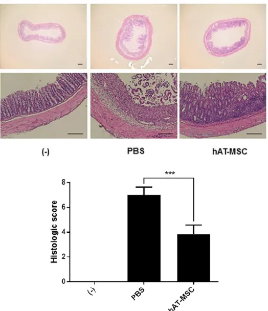
3). On day 10, the disease activity index (DAI) of mice treated with hAT- MSCs was significantly decreased, in comparison with that in the mice treated with PBS (Fig. 4). To evaluate length and histology of the colon, mice were sacrificed on day 10. Compared with that in the PBS-treated group, colon length was significantly improved in hAT-MSC-treated group (Fig. 5).
Histological examination of the DSS-induced colitis mouse colons showed severe submucosal or transmural thickening, destruction of entire epithelium, and severe inflammatory cell infiltration. In contrast, the extent of bowel wall thickening, crypt damage, and the infiltration of inflammatory cells were reduced in colon sections obtained from mice injected with hAT-MSCs, compared with those in the PBS-treated mice (Fig. 6). The analysis for haematoxylin and eosin (H&E)-stained colon sections showed a significant decrease in the histologic scores of hAT-MSC-treated group, in comparison with those in the PBS-treated group (Fig. 6).
30
hAT-MSCs reduce the inflammatory response by increasing the percentage of M2 macrophages in colon
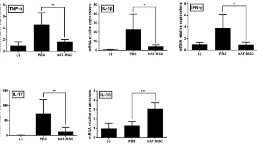
I explored whether intraperitoneally injected hAT-MSCs modulate inflammatory cytokines related to development of DSS-induced colitis. In colon tissues of the PBS-treated colitis mice, TNF-α, IL-1β, IFN-γ, and IL-17 mRNA expression levels were considerably increased, whereas that of IL-10 was increased slightly (Fig. 7). However, the administration of hAT-MSCs not only significantly decreased the expression levels of these genes but also significantly increased that of IL-10, in comparison with those in the PBS- treated animals (Fig. 7). Furthermore, protein levels of TNF-α and IL-10 in the colon samples also exhibited similar tendencies in the colitis mice (Fig.
8).
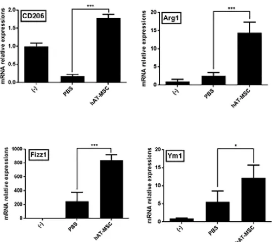
Considering that hAT-MSCs may affect the activity of inflammatory cytokines, I examined the presence of M2 macrophages in the colon tissues.
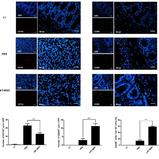
The expression levels of CD206, Arg1, Fizz1, and Ym1 (well-known M2 macrophage markers) were decreased or slightly increased in the colons of colitis model mice treated with PBS (Fig. 9). However, the injection of hAT- MSCs led to a significant increase in the expression of M2 macrophage markers, compared with that in the PBS-treated mice (Fig. 9). Furthermore, infiltrated total macrophages and M2 macrophages were detected in colon tissue sections, using the antibodies specific for CD11b and CD206,
31
respectively (Fig. 10). And I calculated the percentage of CD206-positive cells to CD11b-positive cells using colon sections from the same mice. The percentage of M2 macrophages in the PBS-treated group was 14.50% ± 2.65%, whereas it was 59.25% ± 1.71% in the hAT-MSC-treated group (Fig.
10).
Intraperitoneally injected hAT-MSCs do not migrate into the colon
I next tracked and quantified the hAT-MSCs administered intraperitoneally (2 × 10⁶ cells) by constructing standard curves using quantitative real time (qRT)-PCR (Figs. 11, 12, and Table 1). After 1 day of hAT-MSC administration, approximately 0.002%, 0.12%, 0.02%, 0.06% and 0.09% of the cells were detected in the heart, lung, liver, spleen, and kidney of the colitis mice, respectively (Fig. 13). After 3 days of hAT-MSC injection, percentages of the cells detected in the five organs were decreased compared with day 1 (Fig. 13). However, intraperitoneally infused hAT-MSCs were hardly detected in the brain and the inflamed colon tissues of the colitis mice at day 1 as well as day 3 (Figs. 11, 13).
hAT-MSC-produced TSG-6 induces the phenotypic switch to M2 macrophages in vitro
32
I further investigated whether TSG-6, one of well-known immunomodulatory factors secreted by hAT-MSCs, can alter macrophage phenotype to M2. To test the hypothesis, TNF-α-stimulated hAT-MSCs were used. TSG-6 gene expression was considerably induced in these cells, in comparison with that in the naïve hAT-MSCs (Fig. 14). Additionally, TSG-6 concentration determined in the supernatant collected from the culture of TNF-α-stimulated hAT-MSCs was significantly higher than that measured using the naïve hAT-MSCs (Fig. 14). Afterwards, lipopolysaccharide (LPS)- stimulated Raw 264.7 cells were co-cultured with the naïve or TNF-α- stimulated hAT-MSCs in a transwell system for 48 h, and M2 macrophage marker gene expression levels were shown to be significantly increased in the LPS-stimulated Raw 264.7 cells co-cultured with naïve hAT-MSCs, compared with those determined using the LPS-stimulated Raw 264.7 cells alone (Fig. 15). Interestingly, the expression levels of the M2 macrophage markers were shown to be further increased in the LPS-stimulated Raw 264.7 cells co-cultured with TNF-α-stimulated hAT-MSCs, in comparison with those in the cells co-cultured with naïve hAT-MSCs (Fig. 15). CD206 and Arg1 levels were determined as well, and LPS-stimulated Raw 264.7 cells co- cultured with naïve hAT-MSCs was shown to express CD206 and Arg1 (Fig.
16). Furthermore, the expression of both proteins was significantly increased in LPS-stimulated Raw 264.7 cells co-cultured with TNF-α-stimulated hAT-
33
MSCs compared with that in the cells co-cultured with naïve hAT-MSCs (Fig.
16).
Furthermore, TSG-6 expression in hAT-MSCs was knocked down by transient transfection with siRNAs. hAT-MSCs transfected with siRNAs maintained their fibroblast-like shape, proliferative ability, and differentiation potential in vitro (Fig. 2). TSG-6 expression was shown to be suppressed in hAT-MSCs transfected with TSG-6 siRNA (siTSG6-hAT-MSCs), whereas there was no significant change in TSG-6 expression levels in hAT-MSCs transfected with the control siRNA (siCTL-hAT-MSCs), compared with those in the naïve hAT-MSCs (Fig. 17). Additionally, the concentration of TSG-6 protein secreted by siTSG6-hAT-MSCs was significantly decreased compared with that by the naïve or siCTL-hAT-MSCs (Fig. 17). Afterwards, I co- cultured LPS-stimulated Raw 264.7 cells with naive or siRNA transfected hAT-MSCs in a transwell system for 48 h, and showed that M2 macrophage marker gene expression levels were significantly reduced in LPS-stimulated Raw 264.7 cells co-cultured with siTSG6-hAT-MSCs compared with those in the naïve hAT-MSCs (Fig. 18). Furthermore, CD206 and Arg1 levels were significantly decreased as well in the LPS-stimulated Raw 264.7 cells co- cultured with siTSG6-hAT-MSCs (Fig. 19). In contrast to this, siCTL-hAT- MSCs, with normal TSG-6 secretion levels, did not affect the gene or protein
34
expression of M2 macrophage markers in LPS-stimulated Raw 264.7 cells (Figs. 18, 19).
Intraperitoneal administration of hAT-MSCs with TSG-6 knockdown cannot alleviate IBD
I further investigated whether siRNA-transfected hAT-MSCs can alleviate DSS-induced colitis in mice. siTSG6-hAT-MSCs had no effect on body weight loss and DAI. In contrast, siCTL-hAT-MSCs were shown to significantly reduce body weight loss and DAI (Figs. 20, 21). Additionally, no significant improvement in the length and histologic score of colons obtained from the mice that received siTSG6-hAT-MSCs was observed.
However, the administration of siCTL-hAT-MSCs led to a significant improvement in length and histologic score of these mice (Figs. 22, 23).
Next, I assessed the expression level of M2 macrophages in colons of colitis mice. Intraperitoneal administration of siTSG6-hAT-MSCs had no effect on the expression level of the M2 macrophage markers in the colon, in comparison with those measured in the PBS-treated mice (Fig. 24). In contrast, siCTL-hAT-MSCs injection led to a significant increase in M2 macrophage marker expression in the colon, in comparison with that in the PBS-treated mice (Fig. 24). Furthermore, the percentage of CD206-positive M2 macrophages to CD11b-positive total macrophages was significantly
35
increased in the colon tissue sections of mice injected with siCTL-hAT-MSCs, whereas there was no significant difference in the percentage of M2 macrophages in the colons of mice injected with siTSG6-hAT-MSCs, compared with that in the PBS-treated mice (Fig. 25).
36
Discussion
Recently, hAT-MSCs, which can be obtained relatively easily in large quantities, have shown promising anti-inflammatory effects in studies that used different inflammatory disease models (Choi et al., 2016; González et al., 2009; Lopez‐Santalla et al., 2015; Shin et al., 2016; van den Broek et al., 2013). For chronic diseases, such as IBD, hAT-MSC administration may represent an important treatment option. Here, I showed that the application of hAT-MSCs to DSS-induced colitis mouse model may alleviate the symptoms of this disease, and that the weight loss and DAI were improved.
Furthermore, by measuring the length of colon and assigning the scores based on the microscopic observation of the colon tissue obtained during the mouse autopsy, I showed that the intraperitoneal administration of hAT-MSCs has therapeutic effects against DSS-induced colitis in mice. In addition, I analysed the mechanisms underlying this process, in order to help the development of efficient cell therapies and their clinical application.
For these experiments, I injected human MSCs into immunocompetent mice model of IBD. I used this strategy to focus on immune cells of mice after human MSC application. Also, human MSCs are immunoprivileged partly due to low expression of major histocompatibility complex class II (Le Blanc et al., 2003). Furthermore, similar strategy applying human MSCs to immunocompetent animal models has been
37
successfully performed by several groups, and obvious cross-species-induced immunological responses have not been reported (Kim et al., 2013; Kim et al., 2015; Qi et al., 2014; Roddy et al., 2011).
Macrophages observed in the inflamed tissue may appear as either M1 or M2 macrophages, with distinct roles in the regulation of inflammatory response(Jang et al., 2014; Nishikawa et al., 2014; Wang et al., 2007; Wynn et al., 2013; Zhu et al., 2014). Macrophages have very important roles in innate and acquired immune response, and consequently, macrophage type changes have a major impact on the inflammatory conditions in the inflamed tissue. It is well known that M2 macrophages can be induced by IL-4 and IL- 13, which induce the expression of CD206, Arg1, Fizz1, and Ym1 (Murray and Wynn, 2011; Sica et al., 2015). Moreover, recent studies demonstrated that MSCs might have ability to elicit the phenotypic switch from M1 to M2 macrophages in animal models for acute kidney injury, spinal cord injury and skin wound as well as in vitro (Cho et al., 2014; Geng et al., 2014; Nakajima et al., 2012; Vasandan et al., 2016; Ylöstalo et al., 2012; Zhang et al., 2010).
Consistent with these reports, I found here that colon tissues obtained from the hAT-MSC-injected group showed a considerably higher expression of M2 macrophage markers than the colon tissues obtained from the PBS-treated group. Moreover, the percentage of M2 macrophages to total macrophages identified using IF staining the in colon tissue sections was increased in the
38
hAT-MSC-treated group compared with the PBS-treated group. Gene expression levels of pro-inflammatory cytokines TNF-α, IL-1β, IFN-γ, and IL-17 were significantly reduced in the colons of the hAT-MSC-treated group, while the expression of anti-inflammatory cytokine IL-10 was significantly induced. These findings indicated that the intraperitoneally administration of hAT-MSCs modulates the expression of the inflammatory cytokines by inducing the cell differentiation into M2 macrophages in the colon, alleviation the inflammatory responses. In addition, my results might complement previous findings that MSCs infused into colitis mice reduced inflammation by altering macrophages(Anderson et al., 2012; Parekkadan et al., 2011).
According to previous studies that have revealed therapeutic effects of MSCs in the DSS-induced colitis mice model, I determined the optimal procedure, that is, 2 × 10⁶ hAT-MSCs were administered intraperitoneally (Kim et al., 2013; Wang et al., 2016). Also, I examined the distribution of hAT-MSCs intraperitoneally injected using qRT-PCR assays. At 1 day and 3 days after hAT-MSC administration, less than 0.5% of injected cells were detected in total for heart, lung, liver, spleen, kidney, and brain tissues. In addition, less than 0.001% of injected cells were detected in colon tissues despite inflammatory response. In addition, similar results obtained after siCTL- and siTSG6-hAT-MSC administration in colitis mice. These findings are related to several previous studies (Bazhanov et al., 2016; Sala et al.,
39
2015). Sala et al. reported that less than 1% of mouse bone marrow- and adipose-derived MSCs injected intraperitoneally to DSS-induced colitis mice model were detected in inflamed colon, whereas high frequency of injected cells formed aggregates with immune cells in the peritoneal cavity within 3 days (Sala et al., 2015). Bazhanov et al. also showed that the majority of human bone marrow-derived MSCs infused intraperitoneally into immunocompetent mice aggregated quickly and the aggregates were attached in the peritoneal cavity (Bazhanov et al., 2016). Based on these previous studies, my findings indicated that most of hAT-MSCs administered intraperitoneally might form aggregates in the peritoneal cavity and reduce colitis by inducing M2 macrophages distant from the inflamed colon through secretory factors.
Consistent with the findings, recent investigations have reported that MSCs regulate the inflammatory processes through the activity of various soluble factors, such as indoleamine 2,3-dioxygenase, TGF-β, prostaglandin E2 (PGE2), and TSG-6(Gonzalo-Gil et al., 2016; Kim et al., 2015; Liu et al., 2016; Liu et al., 2015). Among these factors, several studies have revealed that TSG-6 is pivotal in anti-inflammatory effects of MSCs against corneal inflammation, severe burn injury, acute lung injury, acute peritonitis, pancreatitis, and IBD(Broekman et al., 2016; Danchuk et al., 2011; Liu et al., 2016; Roddy et al., 2011; Sala et al., 2015; Wang et al., 2012). In addition,
40
Mittal et al. recently showed that TSG-6 prevented lung injury by inducing macrophage phenotype switch, although TSG-6 used in the study was not a protein released by stem cells (Mittal et al., 2016). Based on these previous findings and my results, it is tempting to speculate that TSG-6 secreted from hAT-MSCs plays a key role in inducing M2 macrophage polarization in the inflamed colon.
Thus, I conducted in vitro experiments to examine whether TSG-6 secreted by hAT-MSCs has ability to induce macrophage phenotype switch.
LPS-stimulated Raw 264.7 cells exhibit a conventional M1 macrophage pattern, and these cells were co-cultured with hAT-MSCs with TNF-α- induced or inhibited TSG-6 expression, or with the naive hAT-MSCs. The co- culturing of TSG-6 overexpressing hAT-MSCs with M1 macrophages led to a significant increase in the expression of M2 macrophage markers. However, because it is possible that other factors secreted by hAT-MSCs may affect this process as well, I performed additional experiments using TSG-6 targeting siRNAs. hAT-MSCs with TSG-6 knockdown were cultured with M1 macrophages, and the expression of M2 macrophage markers was shown to be decreased in these cells. Taken together, these findings suggest that TSG- 6 secreted by hAT-MSCs plays a crucial role in the regulation of M1 to M2 phenotypic switch in vitro.
41
In order to analyse effects of TSG-6 released by hAT-MSCs in vivo as well, I administered siRNA-treated hAT-MSCs to the experimental animals, and showed that, in comparison with those in the control group, siTSG6-hAT- MSC administration did not lead to significant changes in M2 macrophage marker expression levels in the colon. However, the expression of these markers was shown to be induced in the siCTL-hAT-MSC-treated group and naive hAT-MSC-treated group, in comparison with that in the PBS-treated group. Taken together, I confirmed that TSG-6 secreted by hAT-MSCs plays an important role in the alteration of macrophages to M2 phenotype in the inflamed mouse colons. Moreover, the percentage change of M2 macrophages to total macrophages in colon affected the severity of DSS- induced colitis symptoms in mice. In the siTSG6-hAT-MSC-treated group, weight loss rate, DAI, colon length, and histologic scores were not improved, in comparison with those in the PBS-treated group, but the animals treated with siCTL-hAT-MSCs showed considerable improvements in these parameters, which were similar to those observed in the naive hAT-MSC- treated group.
Even so, my results do not exclude the possibility that one or more other secretory factors released from hAT-MSCs increased ability to induce M2 macrophage polarization in colitis mice. Interestingly, recent investigations showed that human MSCs altered macrophage phenotype by
42
producing PGE2 in vitro (Vasandan et al., 2016; Ylöstalo et al., 2012).
Additional experiments related to other MSC-secreted factors such as PGE2 are required to determine their effects on macrophages in IBD models.
However, my findings suggest that TSG-6 plays a key role in M2 macrophage polarization in vitro and DSS-induced colitis mice.
In conclusion, I present here a possible mechanism underlying TSG- 6-induced effects in various inflammatory disease models, including IBD, which were previously described. I demonstrated that hAT-MSC-secreted TSG-6 induces the phenotypic switch of macrophages that infiltrated into colon to M2 type, which leads to the regulation of inflammatory cyt