저작자표시-비영리-변경금지 2.0 대한민국 이용자는 아래의 조건을 따르는 경우에 한하여 자유롭게
l 이 저작물을 복제, 배포, 전송, 전시, 공연 및 방송할 수 있습니다. 다음과 같은 조건을 따라야 합니다:
l 귀하는, 이 저작물의 재이용이나 배포의 경우, 이 저작물에 적용된 이용허락조건 을 명확하게 나타내어야 합니다.
l 저작권자로부터 별도의 허가를 받으면 이러한 조건들은 적용되지 않습니다.
저작권법에 따른 이용자의 권리는 위의 내용에 의하여 영향을 받지 않습니다. 이것은 이용허락규약(Legal Code)을 이해하기 쉽게 요약한 것입니다.
Disclaimer
저작자표시. 귀하는 원저작자를 표시하여야 합니다.
비영리. 귀하는 이 저작물을 영리 목적으로 이용할 수 없습니다.
변경금지. 귀하는 이 저작물을 개작, 변형 또는 가공할 수 없습니다.
이학박사 학위논문
Novel functions of lysyl-tRNA synthetase in invasive dissemination of colon cancer cells in 3D microenvironment
3차원적 콜라젠 환경에서 대장암세포의 침윤적 이탈을 위한 Lysyl-tRNA 합성효소의 기능
2017년 8월
서울대학교 대학원
자연과학대학 협동과정 유전공학전공
남 서 희
Novel functions of lysyl-tRNA synthetase in invasive dissemination of colon cancer cells in 3D
microenvironment
A THESIS
Submitted to the Graduate School of Seoul National University in Partial
Fulfillment of the Requirements for the Degree of
Doctor of Philosophy
By Seo Hee Nam
Interdisciplinary Graduate Program in Genetic Engineering College of Natural Science
Seoul National University
August, 2017
TABLE OF CONTENTS
CHAPTER 1
Noncanonical roles of membranous lysyl-tRNA synthetase in transducing cell-substrate signaling for invasive dissemination of
colon cancer spheroids in 3D collagen I gels. ... 1
Ⅰ. Abstract ... 2
Ⅱ. Introduction ... 3
Ⅲ. Experimental procedures ... 5
1. Cells ... 5
2. Spheroid formation and embedding into 3D collagen I gels ... 5
3. Extract preparation and Western blots ... 6
4. Normal 2D culture or replating on 2D ECM layer ... 7
5. Time-lapse FRET analysis for ERK activity ... 7
6. Time-lapse imaging of cells in 3D ECM gels ... 8
7. Coimmunoprecipitations ... 8
8. Indirect immunofluorescence ... 9
9. Chromatin immunoprecipitation (ChIP) ... 9
10. Generation of KRS heterozygote mouse ... 9
11. Measurement of metastasis in the MMTV-PyVT transgenic mice ... 10
12. Analyses of murine or human tissues ... 10
13. Immunohistochemistry ... 11
14. RT-PCR ... 11
15. Statistical methods ... 12
Ⅳ. Results ... 13
Incomplete EMT phenotype upon KRS suppression ... 13
KRS-dependent cell-laminin adhesion ... 13
KRS-dependent dissemination of HCT116 spheroids in 3D collagen I gels ... 15
KRS/p67LR/integrin a6β1 linkage correlates for ERKs activation ... 17
KRS-dependent cell dissemination require ERKs and paxillin phosphorylations ... 19
KRS-suppression-inhibited dissemination is recovered by ERK1 and/or paxillin expression ... 21
KRS appear to play pro-metastatic roles at the invasive margins of KRS-/+ mouse breast tumor and human colon tumor tissues ... 22
Ⅴ. Discussion ... 45
CHAPTER 2 Lysyl-tRNA synthetase (KRS) promotes dissemination from colon cancer spheroids via recruitment and reprogramming of macrophages in 3D collagen I gels.
...51
Ⅰ. Abstract ... 52
Ⅱ.Introduction ... 54
Ⅲ. Experimental Procedures ... 56
1. Cells. ... 56
2. Animals ... 57
3. Spheroid formation and embedding into 3D collagen I gels ... 57
4. Extract preparation and Western blots. ... 58
5. Time-lapse imaging of cells in 3D ECM gels. ... 59
6. Coimmunoprecipitations. ... 59
7. Indirect immunofluorescence. ... 59
8. Chromatin immunoprecipitation (ChIP). ... 60
9. Co-culture in 3D collagen I gels. ... 60
10. Cytokine antibody array. ... 61
11. Analysis of Subcellular Proteome Extraction. ... 61
12. Immunohistochemistry. ... 61
13. Real-Time PCR. ... 62
Ⅳ. Results ... 63
FGF2/GROa/M-CSF cause the dissemination from colon cancer spheroids embedded in 3D collagen I gels. ... 63
GAS6 from KRS-positive tumor cells causes the polarization of macrophages from M1 to M2 populations. ... 66
KRS translocated into nucleus promotes GAS6 mRNA level through transcription factor MiTF. ... 68
KRS-positive HCT116 tumor cells prefer to communicate with M2 macrophages rather than with M1 macrophages. ... 70
A positive relationship between KRS-positive colon cancer cells and M2 macrophages is important in favoring tumor microenvironment for KRS-positive metastatic phenotypes. ... 72
Ⅴ. Discussion ... 93
REFERENCES ... 98
ABSTRACT IN KOREAN ... 102
LIST OF FIGURES
Figure 1.1
KRS-dependent incomplete EMT phenotypes in normal 2D conditions.
Figure 1.2
KRS suppression impaired cell-ECM adhesion and signaling activities.
Figure 1.3
Impaired adhesion-related signaling in KRS-suppressed cells correlated to a lack of dissemination from spheroids in 3D collagen I gels.
Figure 1.4
KRS-mediated ERK activation correlated with the formation of a p67LR/integrin 61 complex.
Figure 1.5
KRS-dependent cell dissemination from spheroids in 3-D collagen I gels was blocked by anti-MEK/ERK U0126 or anti-KRS YH16899 treatment.
Figure 1.6
Blockade of dissemination by KRS-suppression was relieved by ERK and/or paxillin expression.
Figure 1.7
Positive relevance in tumor metastasis of KRS in KRS-/+ PyVT mouse and colon cancer
patient tissues.
Figure 2.1
Certain cytokines caused the dissemination phenotypes from colon cancer cell spheroids embedded in 3D collagen I gels.
Figure 2.2
Certain cytokines from KRS-positive tumor cells caused the polarization of macrophages from M1 to M2 populations.
Figure 2.3
KRS translocated into nucleus promotes GAS6 mRNA level and concomitantly KRS translocated on plasma membrane supports dissemination of colon cancer cells, respectively.
Figure 2.4
KRS-positive HCT116 tumor cells preferred to communicate with M2 macrophages rather than with M1 macrophages.
Figure 2.5
A positive relationship between KRS-positive colon cancer cells and M2 macrophages is important in favoring tumor microenvironment for KRS-positive metastatic phenotypes.
LIST OF ABBREBIATIONS
BSA Bovine Serum Albumin
CM Conditioned medium
COL Collagen
FN Fibronectin
IHC Immunohistochemistry
KRS Lysyl-tRNA synthetase
LN Laminin
Mono Monocyte
M1 M1 macrophages
M2 M2 macrophages
MSC Multi-tRNA synthetase complex
PXN Paxillin
qRT-PCR Quantitative Reverse Transcription PCR WCL Whole cell lysate
WT Wild-Type
3D 3-Dimensional
67LR 67kDa laminin receptor
37LRP 37kDa laminin receptor precursor
1
CHAPTER 1.
Noncanonical roles of membranous lysyl-tRNA synthetase in transducing cell-substrate signaling for invasive dissemination of colon cancer spheroids
in 3D collagen I gels.
2
Abstract
The cell-adhesion properties of cancer cells can be targeted to block cancer metastasis.
Although cytosolic lysyl-tRNA synthetase (KRS or KARS) functions in protein synthesis, KRS on the plasma membrane is involved in cancer metastasis. It may be hypothesized that KRS is involved in adhesion-related intracellular signaling for cancer cell migration and invasion. To test this hypothesis, colon cancer cells with modulated KRS protein levels were adhered on 2 dimension (2D) or embedded in 3D collagen I gel systems and analyzed for cell signaling and behaviors, in comparison to heterozygous KRS-/+ mice and clinical colon cancer tissues.
Although KRS suppression decreased expression of cell-cell adhesion molecules, cells still formed colonies without being scattered, supporting an incomplete EMT. Meanwhile, KRS- suppressed cells still exhibited focal adhesions on laminin, with Tyr397-phopshorylated FAK, but they lacked laminin-adhesion-mediated ERK and paxillin activation. Time-lapse imaging revealed KRS-dependent cell dissemination (e.g., outbound movement) from the spheroids, whereas KRS-suppressed spheroids remained static due to the absence of efficient cell-ECM adhesion-mediated outbound crawling movements. KRS, p67LR, and integrin 61 were found to interact, presumably to activate ERK for paxillin expression and activation even without involvement of FAK, so that specific inhibition of ERK or KRS in parental HCT116 spheroids blocked dissemination. Furthermore, KRS-suppressed spheroids recovered the capacity for dissemination upon the re-introduction of ERK or paxillin. The KRS/ERK/paxillin signaling axis was also observed in marginal areas of colon cancer or KRS-/+ mouse tissues.
Collectively, these observations indicate that KRS can cause cancer cell dissemination via the regulation of cell-cell and cell-ECM adhesion during KRS-mediated metastasis.
3
Introduction
Cancer metastasis is a complex process involving multiple steps, such as dissemination from the primary tumor mass via EMT, migration, and invasion, that occur even before intravasation processes [1]. Dissemination from the primary tumor mass can lead to a journey of metastatic cancer cells towards distal settlements, and the disseminated cancer cells migrate and invade through the stroma to reach vessels or lymph nodes along the way [2]. In addition to strategies aimed at the regression of the primary tumor mass through chemoprevention and/or radiation, anti-metastatic strategies that block dissemination process, which is an early step of metastasis, would be clinically important for achieving a higher cure rate for cancer.
Numerous signaling or adaptor molecules have been shown to play critical roles in facilitating cancer cells to metastasize. Of these molecules, we are interested in revealing the mechanisms underlying the roles of aminoacyl-tRNA synthetases in tumorigenesis and metastasis [3].
Cytosolic aminoacyl-tRNA synthetases are traditionally and importantly involved in protein translation. However, lysyl-tRNA synthetase (KRS) was recently shown to function in immune responses [4, 5] as well as in tumor metastasis [6, 7], clearly suggesting additional biological functions beyond protein translation. KRS is phosphorylated at the Thr52 residue by p38MAPK, which causes it to dissociate from the cytosolic multi-tRNA synthetase (MTS) complex and translocate to the plasma membrane, where it associates with and stabilizes a 67 kDa laminin receptor (p67LR) that is involved in migration and metastasis [6]. p67LR is known to associate with integrin 61 [8] or 64 [9], which have roles in cell adhesion to the ECM and tumor cell invasion [10]. The association of KRS with p67LR is sufficiently targeted to allow the inhibition of KRS-dependent metastasis in a subcutaneously cell-injected animal model, suggesting that KRS is a promising target for anti-cancer metastatic strategies [7].
4
Therefore, as part of our efforts to develop anti-metastatic therapeutics by targeting KRS- mediated cellular behaviors, we found that KRS might cause cell dissemination from spheroids embedded in an ECM-surrounded 3-dimensional (3D) environment. This system would be beneficial if the dissemination process observed in 3D gels could be mechanistically related to KRS-dependent dissemination in in vivo-like circumstances and could also generally be useful as a template screening system for anti-metastasis reagents. This study revealed that KRS appears to play regulatory roles in cell adhesion by activating ERK and paxillin during cancer cell dissemination from primary tumor masses.
5
Experimental procedures
1. Cells
HCT116 and SW620 colon cancer (American Type Culture Collection, Manassas, VA, USA) and other colon cancer (Korean Cell Bank, Seoul National University) cells were stably transfected with shRNA against KRS (transcript 1, NM_001130089, lysyl-tRNA synthetase MISSION® shRNA Plasmid DNA, Sigma) using Truefect (United Biosystems, Herndon, VA) before antibiotic selections. The target sequences of shKRS and the names of the KRS- suppressed cell clones are shown in table 1. HCT116 cells overexpressing KRS were established via the stable transfection of a myc-tagged KRS plasmid [6]. ERK1 and ERK2 (ERK clones) and paxillin (PXN clones) were also stably transfected, either separately or together (PEE clones for paxillin, ERK1, and ERK2), into KRS-suppressed cell clones. Stable myc-tagged KRS and different stable shKRS-transfected clones were established by using 250
g/ml G418 or 0.50 g/ml puromycin (AG Scientific, Inc., San Diego, CA). The cells were maintained in RPMI-1640 (WelGene, Daegu, Korea) containing 10% FBS and antibiotics (Invitrogen, Grand Island, NY, USA).
2. Spheroid formation and embedding into 3D collagen I gels
HCT116 or SW620 cells with modulated KRS expression levels, or other colon cancer cells were processed for spheroid formation using Perfecta3D® 96-well hanging-drop plates (3D Biomatrix, Ann Arbor, MI). Spheroids were selected to be less than 70 m in size using a cell strainer sieve (SPL Life Science Co., Pocheon-si, Korea) prior to being embedded into 3D collagen I gels (BD Biosciences, San Jose, CA, USA). For embedding, polydimethylsiloxane (PDMS) chambers with an 8-mm diameter, or 10 well-slips for 22 x 60
6
mm coverslips (model No.: WL-2260-10, Live Cell Instrument, Seoul, Korea), were filled with 30 l collagen I (2.0 mg/ml, BD Biosciences) to coat the bottom surfaces of the chamber.
PDMS (Sylgard 184, Dow Corning, Midland, MI) prepolymer was prepared using a 10:1 (w/w) mixture of PDMS base and curing agent, cast against the master and thermally cured to obtain a negative replica-molded piece. Spheroids were then mixed with ice-cold collagen I solutions (2.0 mg/ml), which were prepared by diluting a concentrated collagen I (BD Biosciences) stock with 10 × reconstitution buffer (260 mM sodium bicarbonate and 200 mM HEPES), and 10 × RPMI-1640 with phenol red [11]. The pH was adjusted in the range of 7.2 to 7.4 using ice-cold 2 N NaOH. The mixture was transferred to a 37ºC CO2 incubator to allow the collagen I to polymerize and form a fibrillar meshwork. The 3D culture was then refreshed each day with standard culture media containing 10 % FBS.
3. Extract preparation and Western blots
Colon cancer cells within collagen I gels were washed with ice-cold PBS and then homogenized with truncated pipette tips (3 times × 20 min on ice) in modified RIPA buffer (50 mM Tris-HCl, 150 mM NaCl, 1% NP-40, and 0.25% sodium deoxycholate) with a protease inhibitor cocktail (GenDepot, Barker, TX). The extracts were centrifuged for 30 min at 15,000 g and 4°C, and then the lysates were collected from the supernatant. The protein amounts were normalized with the band intensities for -tubulin or -actin via standard Western blots, before further immunoblotting with different antibodies. The primary antibodies used in the study were as follows: -tubulin, -SMA, talin, pY822vinculin, and vimentin (Sigma, St Louis, MO); pY416c-Src, ERKs, phospho-ERKs, FLAG, pS473Akt, pS425talin, caspase 3, Tensin 2, and Akt (Cell Signaling Technol. Danvers, MA); paxillin, N-cadherin, and FAK (BD Biosciences);
pY397FAK, p67LR, laminin, vinculin, and Twist1 (Abcam, Cambridge, UK); c-Src, pY118paxillin, pY577FAK, pY861FAK, pY925FAK, Snail1, E-cadherin, -catenin, and Slug
7
(Santa Cruz Biotech., Santa Cruz, CA); ZO1 (Zymed Laboratories, Camarillo); pY483Tensin 2 (Biorbyt, San Francisco, CA); vinculin, integrin 6, 1, and 4 (Millipore, Billerica, MA);
fibronectin (DAKO, Carpinteria, CA); and KRS (Atlas Antibodies, Stockholm, Sweden).
4. Normal 2D culture or replating on 2D ECM layer
Cells were kept in suspension or replated on ECM (10 g/ml, laminin or fibronectin, BD Biosciences)-precoated dishes or coverglasses in the absence (in the case of fibronectin) or presence of low serum (2%, in the case of laminin) for 1 h before being analyzed for cell-ECM adhesion signaling by standard Western blotting or for the formation of focal adhesions by indirect immunofluorescence, as described previously [12]. Function blocking antibodies against human integrin 6 (Millipore, 20 g/ml) were preincubated with cells by rocking at 37ºC for 1 h prior to replating. Pharmacological inhibitors were added to the culture media for 24 h or to replating media at the reseeding time. Cells were reseeded on coverglasses precoated with standard media containing 10% FBS, before imaging to monitor cell growth patterns or immunofluorescence to detect the formation of cell-cell contacts. Confluent cells were wounded with a pipette tip and washed twice with PBS. After treatment with DMSO, U0126 (10 M, LC Labs., Woburn, MA), or YH16899 (10 M) and incubation in a CO2 incubator for 24 h, the marginal edges of the wounds were imaged (CKX41, Olympus, Japan).
5. Time-lapse FRET analysis for ERK activity
HCT116 cells were cultured on a laminin-coated (5 g/ml) 35-mm glass-base dish and were transfected with FRET-based indicators [13] using the lipofectamine 2000 transfection reagent (Life Technologies, Grand Island, NY), according to the manufacturer’s instructions.
FRET images were obtained on a Nikon Ti-E inverted microscope (Nikon, Tokyo, Japan) equipped with PFS, a CoolSNAP HQ camera (Roper Scientific, Trenton, NJ), and excitation
8
and emission filter wheels. Images were acquired by using the 44 binning mode and 200-ms exposure time. All systems were controlled by MetaMorph software (Molecular Devices, Sunnyvale, CA). For dual-emission ratio imaging of the intramolecular FRET probe, we obtained images for CFP and FRET. After background subtraction, pseudo-colored images of FRET/CFP were created using eight colors from red to blue to represent the FRET/CFP ratio via the intensity modulated display (IMD) mode using MetaMorph software. Filters used for the dual-emission ratio imaging were purchased from Semrock (Rochester, NY): an FF01- 438/24-25 excitation filter, an FF458-Di02-2536 dichroic mirror, and two emission filters, FF01-483/32-25 for CFP and FF01-542/27-25 for FRET.
6. Time-lapse imaging of cells in 3D ECM gels
Parental or stable cells were transfected with mCherry-MT1-MMP for 24 h before spheroid formation. Three hours after spheroids were embedded in 3D collagen I gels, time- lapse images were collected for the indicated periods using IX81-ZDC microscope (Olympus, Tokyo, Japan). The microscope was equipped with a 10-well Chamlide Incubator system (Live Cell Instrument, Seoul, Korea), and an environmental chamber mounted on the microscope maintained constant conditions of 37°C, 5% CO2, and 95% humidity. Pharmacological inhibitors such as U0126 (50 M) or YH16899 [50 M, [7]] were mixed during the process of embedding the cells into 3D collagen I gels.
7. Coimmunoprecipitations
Whole-cell extracts prepared from cells in standard media containing 10% FBS or from cells kept in suspension or reseeded onto laminin (10 g/ml) in media containing 2% FBS for 2 h were immunoprecipitated overnight using anti-myc tag antibody-coated agarose beads (Sigma). The immunoprecipitated proteins were boiled in 2 × SDS-PAGE sample buffer before
9
standard Western blot analysis.
8. Indirect immunofluorescence
Cells reseeded on 2D normal culture media- or ECM-precoated glass coverslips or embedded in 3D collagen I gels within PDMS were immunostained using antibodies against E-cadherin, snail1, pERK, K14, or paxillin in addition to DAPI staining for the nucleus.
Immunofluorescence images were acquired on a fluorescence microscope (BX51TR, Olympus, Tokyo, Japan) or a confocal laser scanning microscope with a Nikon Plan- Apochromat 60×/1.4 N.A. oil objective (Nikon eclipse Ti microscope, Nikon, Tokyo, Japan), and then analyzed using the IMARIS imaging software (Bitplane AG, Zurich, Switzerland).
9. Chromatin immunoprecipitation (ChIP)
ChIP assays were performed using the ChIP-IT Express kit (Active Motif, Carlsbad, CA), following the manufacturer’s protocols. Briefly, cells embedded in collagen I gels for 24 h were fixed for 30 min with 2% formaldehyde, and the chromatin was then prepared by sonication shearing to aid nuclear release and enzymatic shearing. The sheared chromatin was immunoprecipitated using c-Jun (Cell Signaling) or Elk-1 (Santa Cruz Biotech.,) antibodies and captured with protein G magnetic beads. The chromatin was eluted from the purified immunoprecipitates, reversed cross-linked, and digested with proteinase K. The reaction was then stopped using proteinase K stop solution. The isolated chromatin was analyzed by 36 cycles of PCR. Ten microliters of each PCR product was separated via 2% agarose electrophoresis and visualized by ethidium bromide staining.
10. Generation of KRS heterozygote mouse
For the generation of KRS heterozygote animals, KRS genetrap ES cells (AJ0130) were purchased from the MMRRC and used to generate a chimeric mouse at the UCSD
10
transgenic mouse core facility. The disruption of the gene via the insertion of the genetrap cassette into intron 13 was confirmed by genomic PCR using primers flanking the breakpoint (forward: 5'- TGG GTT TCA TCC TGA GGT CT - 3'; reverse: 5'- GCT TTC TTT CCC AGG TCC TC - 3'; genetrap reverse: 5'-TGT CCT CCA GTC TCC TCC AC - 3'). No viable homozygous pups were born, nor were any viable embryos observed (up to embryonic day 9.5).
We confirmed the reduction of KRS expression levels in the heterozygote in multiple organs (Fig. S9). No gross phenotypic differences, including size, weight and movement behavior, were observed between wild-type and KRS heterozygote mice (data not shown). MMTV-PyVT transgenic mice were purchased from The Jackson Laboratory.
11. Measurement of metastasis in the MMTV-PyVT transgenic mice
MMTV-PyVT mice were crossed to KRS heterozygotes to generate the MMTV-PyVT;
KRS+/+ and MMTV-PyVT; KARS+/GT mice. Female mice were sacrificed at 16 and 18 weeks.
After the lungs were dissected out and fixed, each lobule was carefully examined under the microscope to count the total number of metastatic nodules. A white round bump of over 100
m on the lung surface was counted as a nodule.
12. Analyses of murine or human tissues
All procedures for animal and human tissues were performed in accordance with the procedures of the Seoul National University Laboratory Animal Maintenance Manual and Institutional Review Board (IRB) agreement approved by the Institute of Laboratory Animal Resources Seoul National University (ILARSNU) and SNUIRB, respectively. Human colon tissues were obtained with informed content from patients who received surgery at Samsung Medical Center (Sungkyunkwan University School of Medicine, Seoul, Korea) or National Biobank of Korea-Pusan National University Hospital (PNUH) in accordance with IRB agreements. Whole-tissue extracts were prepared with modified RIPA buffer [12] using a
11
Bullet Blender BBX24 surrounded by dry ice and containing 1.0 mm zirconium silicate beads (Next Advance, Inc. Averill Park, NY), prior to standard Western blots for the indicated molecules.
13. Immunohistochemistry
Dissected breast tumors were fixed and stored in 10% NBF. The tissues were processed, paraffin embedded, and sectioned at 6-μm thickness. After deparaffinization, antigen retrieval was performed by boiling the sections for 5 min in sodium citrate solution (0.01 M, pH 6). The sections were incubated he at 4οC with the indicated antibodies in PBST (0.3% Triton X-100 in PBS) containing 5% goat serum or 3% BSA. After washing, the sections were incubated with biotin-conjugated anti-rabbit, mouse or goat IgG (1:100, Sigma) antibody and ExtrAvidin-peroxidase (1:50, Sigma) at RT for 1 h each for 3,3′-diaminobenzidine detection. The sections were counterstained with hematoxylin and imaged under the microscope. Immunohistochemistry of serial sections of paired normal or tumor colon tissues was performed using normal rabbit IgG, normal mouse IgG, phospho-ERKs (Cell Signaling), KRS (Atlas Antibodies), and paxillin (BD Biosciences).
14. RT-PCR
Total RNA was extracted from cells in 2D normal 10% FBS-containing condition or ECM-precoated culture dishes with low (2%) serum-containing conditions, or in 3D collagen I gels, using TRIzol (Invitrogen) according to the manufacturer’s protocol. One microgram of total RNA was reverse transcribed using the amfiRivert Platinum cDNA Synthesis master mix (GenDepot). Primers were designed using Primer3 software as follows: human E-cadherin (CDH1) mRNA, forward 5’-TGCCCAGAAAATGAAAAAGG-3’ and reverse 5’- GTGTATGTGGCAATGCGTTC-3’; human N-cadherin (CDH2) mRNA, forward 5’- ACAGTGGCCACCTACAAAGG-3’ and reverse 5’-CCGAGATGGGGTTGATAATG-3’;
12
human Vimentin (VIM) mRNA, forward 5’-GAGAACTTTGCCGTTGAAGC-3’ and reverse 5’-GCTTCCTGTAGGTGGCAATC-3’; human Twist (TWIST) mRNA, forward 5’- GGAGTCCGCAGTCTTACGAG-3’ and reverse 5’-TCTGGAGGACCTGGTAGAGG-3’;
human paxillin (PXN) mRNA, forward 5’-GAAATCAGCTGAGCCTTCAC-3’ and reverse 5’- TTAGGCTTCTCTTTCGTCAGG-3’, Snail1, forward 5’-GGTTCTTCTGCGCTACTGCT-3’
and reverse 5’-TAGGGCTGCTGGAAGGTAAA-3’; slug, forward 5’- GGGGAGAAGCCTTTTTCTTG-3’ and reverse 5’-TCCTCATGTTTGTGCAGGAG-3;
human KRS (KARS) mRNA, forward 5’- CAATGCCCATGCCCCAGCCA-3’ and reverse 5’- ACCCCACCCTTCCGGCGAAT-3’, and human -actin (ACTB) mRNA, forward 5’-
TGACGGGGTCACCCACACTGTGCCCATCTA-3’ and reverse 5’-
CTAGAAGCATTTGCGGTGGACGACGGAGGG-3’.
15. Statistical methods
Student’s t-test was performed for statistical comparisons of mean values to determine significance. A p value less than 0.05 was considered statistically significant.
13
Results
Incomplete EMT phenotype upon KRS suppression
To explore the roles of KRS in metastatic dissemination, HCT116 colon cancer cells with endogenous, suppressed, or overexpressed KRS levels were analyzed for EMT phenotypes. Using an RT-PCR approach, we found that KRS suppression decreased E-cadherin mRNA (an epithelial marker) but increased the mRNAs of mesenchymal markers (Fig. 1.1A).
Furthermore, these KRS suppression-dependent changes in EMT markers also carried over to protein levels; epithelial markers including E-cadherin were expressed at higher levels in KRS- expressing parental and KRS-overexpressing HCT116 cells, whereas KRS-suppressed cell clones decreased epithelial marker expression and concomitantly enhanced the expression of mesenchymal markers (Fig. 1.1B). The increased levels of mesenchymal marker proteins after the suppression of KRS to certain levels, without caspase 3 activation, supported additional roles for KRS other than protein translation. Meanwhile, KRS-suppressed cell clones did not show altered expression of laminin and p67-laminin receptor (p67LR), which are important for KRS-mediated tumorigenic roles [3], or integrin chains 6, 1, or 4 compared with KRS- expressing cells (Fig. 1.1B). E-cadherin and -catenin were not clearly observed in cell-cell junctions when KRS was suppressed, as compared with KRS-expressing cells (Fig. 1.1C).
Therefore, the inverse relationship between KRS and epithelial marker expression may suggest the induction of EMT phenotypes. However, cell-cell contacts were not disrupted after KRS suppression (Figs. 1.1C and D), suggesting that KRS suppression caused an incomplete EMT.
KRS-dependent cell-laminin adhesion
We then examined the relationship between KRS expression and cell-ECM adhesion
14
properties. First, cells with various KRS expression levels were adhered under normal 10%
FBS-containing culture conditions and examined for adhesion-related signaling activities.
Under normal 10% serum-containing 2D culture conditions, KRS suppression abolished the signaling activities of FAK, ERK, c-Src, and paxillin (Fig. 1.2A) compared with KRS- expressing (or overexpressing) cells. Cell adhesion-related signaling activities were then analyzed after the cells were kept in suspension or reseeded onto ECM-precoated culture dishes. Because KRS has been shown to translocate to the plasma membrane in a laminin- dependent manner [6] and because KRS suppression did not change laminin expression (Fig.
1B), we explored the biological significance of KRS expression after reseeding onto laminin under reduced (2%) serum-containing conditions. The adhesion-dependent phosphorylation of FAK was not abolished by KRS suppression, nor was it affected by treatment with a KRS inhibitor [YH16899, which blocks the interaction between KRS and p67LR [7]], when compared with KRS-expressing cells (Fig. 2B, lanes 2, 5, 7, and 9). However, the phosphorylation of ERK, paxillin, and c-Src in KRS-expressing cells increased upon adhesion to laminin and was abolished by additional YH16899 treatment, whereas no effect was observed in KRS-suppressed cells (Fig. 1.2B). KRS-dependent FAK activity in 2D 10% serum- containing normal condition appeared not to be the case when HCT116 cells were manipulated to be newly adhered on laminin under a lower serum condition. When the cells were treated with the addition of soluble laminin to the culture media, paxillin expression and ERK phosphorylation were lower in KRS-suppressed cells than in KRS-expressing cells. We next examined the subcellular localization of phospho-ERK or phospho-Tyr118 paxillin in cells with various KRS expression levels. When the cells were reseeded onto laminin-precoated coverglasses in 2% serum-containing media, KRS-positive (but not KRS-suppressed) cells showed obvious phospho-ERK staining in the nucleus (Fig. 1.2C). Phospho-Tyr118 paxillin was also clearly localized to focal adhesions in a KRS-dependent manner (Fig. 1.2D).
15
Interestingly, replating cells on laminin for a shorter (2 h) or a longer (24 h) period showed phospho-Tyr397 FAK independent of KRS expression, although ERKs phosphorylation and paxillin expression and phosphorylation depended on KRS expression (Figs. 1.2E), suggesting that FAK activity upon laminin stimulation in 2D condition might not be relevant for KRS expression. Given that KRS suppression induced fibronectin (Fig. 1.1B), we also examined how the cells would respond to fibronectin simulation. Reseeding KRS-suppressed cells onto fibronectin-precoated dishes did not result in any significant difference in Tyr397- phosphorylated FAK, compared with KRS-expressing cells, although ERK phosphorylation depended on KRS expression. Meanwhile, wounds were made in confluent cells in normal 10% FBS-containing 2D conditions, and KRS-expressing parental cells exhibited clear outbound movement at the wound boundary, whereas KRS-suppressed cells did not (Fig. 1.2F).
These KRS-dependent differential movements toward free spaces might be intrinsically correlated with non-effective cell-ECM adhesion for crawling out.
KRS-dependent dissemination of HCT116 spheroids in 3D collagen I gels
Because KRS-expressing (but not KRS-suppressed) cells efficiently moved toward free spaces during wound healing, we wondered whether KRS could be important for cell dissemination from the tumor masses. In particular, we explored the significance of KRS expression in cell dissemination by time-lapse monitoring of HCT116 spheroids (with various KRS expression levels) embedded into 3D collagen I gels. Hanging drops of HCT116 cell clones with various KRS expression levels efficiently formed spheroids. When spheroids with diameters between 70 and 100 m were sieved and embedded into collagen I (2.0 mg/ml) gels, the KRS-expressing cells exhibited outbound movement or dissemination of single or as well as small groups of cells (Fig. 1.3A, top and bottom panels). However, KRS-suppressed cell clones did not show any such dissemination even when monitored for 28 h (Fig. 1.3A, middle
16
panels). Because imaging commenced 3 h after embedding, signaling activities were also examined at 3 or 24 h after separate embedding into collagen I gels. Consistent with the laminin-adherent 2D condition, c-Src, ERK, and paxillin (but not FAK) activity were similarly dependent on KRS expression, decreasing with time after embedding, although only phospho- Tyr118 paxillin increased as time passed after the embedding (Fig. 1.3B). Decreases in ERK and paxillin signaling activities upon KRS suppression were also observed in another colon cancer cell line, SW620, in 3D collagen gels (Fig. 1.3C). SW620 cells also showed dissemination from spheroids in a KRS expression-dependent manner. Furthermore, KRS- suppressed cells showed increased mesenchymal markers and decreased epithelial markers 24 h after embedding (Fig. 1.3D), although they did not disseminate (Fig. 1.3A). The lack of dissemination from spheroids by KRS-suppressed cells in 3D collagen I gels might be related to an incomplete EMT. Consistent with the absence of outbound movements and lack of scattering in 2D conditions (Fig. 1.1), no dissemination from or cell scattering of spheroids in 3D collagen I gels was observed, although the cells showed reduced E-cadherin at cell-cell contacts (Fig. 1.3E). When one epithelial marker (cytokeratin 14, K14) was immunostained in the spheroids in 3D collagen I gels, KRS-expressing parental or KRS-overexpressing cells were positive for K14 staining in cells at the dissemination margins, whereas KRS-suppressed or U0126- or YH16899-treated parental cells were negative for K14 (Fig. 1.3F), suggesting a role for K14 in dissemination. Furthermore, ERK phosphorylation and paxillin expression in KRS- expressing parental HCT116 spheroids in 3D collagen I gels were also decreased by KRS- suppression (Figs. 1.3G and H). Meanwhile, KRS suppression did not alter other focal adhesion molecules such as vinculin, talin, and tensin 2 expression and talin phosphorylation levels but decreased vinculin and tensin 2 phosphorylations (Fig. 1.3I), suggesting specific effects on paxillin expression and FA molecule phosphorylations via KRS suppression. These observations indicate that KRS-mediated dissemination appears to involve cell-cell adhesion
17
properties, as well as cell-ECM adhesion signaling for ERK activity and paxillin expression/activity-mediated phosphorylations of FA molecules.
KRS/p67LR/integrin a6β1 linkage correlates for ERKs activation
Because KRS-suppression decreased ERK phosphorylation, we attempted to measure the in situ ERK activity using an ERK biosensor. On laminin-precoated coverglasses in 2%
serum-containing media, KRS-expressing parental or KRS WT-overexpressing cells showed greater FRET signals, indicating highly active ERK with activity oscillations, compared to KRS-suppressed cells, which showed gradual declines (Figs. 1.4A). This KRS-dependent ERK activation was consistent with the observation that ERK phosphorylation was increased by KRS overexpression (Fig. 1.2A). Comparising of the mean FRET signal intensities showed that ERK activity clearly depended on KRS expression (Fig. 1.4B). Interestingly, transient transfection of ERK1 or 2 into KRS-suppressed cells resulted in somewhat increased paxillin expression and Tyr118 phosphorylation, but not FAK Tyr397 phosphorylation, in addition to dramatically increased phospho-ERK levels (Fig. 1.4C). This observation suggests the presence of a link between pERK and paxillin expression/phosphorylation in KRS-expressing cells. Given that KRS expression led to ERK phosphorylation, we examined how ERK could be activated through KRS. KRS can translocate to the plasma membrane after Thr52- phosphorylation-dependent disassociation from cytosolic MSC and associate and collaborate with p67LR in the presence of laminin [6]. Different HCT116 cell clones with various KRS expression levels did not show altered laminin, p67LR, or integrin 6, 1, and 4 expression levels (Fig. 1.1B). Integrin 61 and 64 are responsive to laminin and are known to bind p67LR [8, 9]. Furthermore, integrins are well-known to activate ERK for different cell functions in many cell and tissue systems [14]. Therefore, to determine whether the interaction between KRS, p67LR, and integrin 61 could be correlated to ERK activation, we examined
18
possible physical interactions between these proteins via coimmunoprecipitation. Myc-KRS prepared from normal 10% FBS-containing culture conditions co-precipitated with integrin 6 and p67LR, and this interaction was reduced by anti-KRS inhibitor (YH16899) treatment (Fig.
1.4D). To further analyze the significance of laminin stimulation for this association, myc-KRS immunoprecipitates were prepared from cells kept in suspension or reseeded onto laminin- precoated dishes in culture media containing 2% FBS. Whereas p67LR was shown to bind to KRS in lysates from suspended cells, KRS binding to integrin 6 and 1 was observed to be adhesion-dependent, and was disrupted by additional treatment with YH16899 (Fig. 1.4E).
Those molecules including KRS, laminin, p67LR, and integrin 6 were positively correlated around the invasive marginal area of breast tumor tissues from the PyVT mouse model (Fig.1.
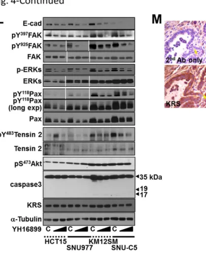
4F). We then examined how ERK activity could be related to paxillin levels. First, we found that c-Jun expression and Ser63 phosphorylation, but not Elk-1, p38, or JNKs expression and phosphorylation, were correlated with KRS expression (Fig. 14G). In addition, the suppression of Elk-1, an ERK-mediated transcription factor, did not down-regulate paxillin expression and Tyr118 phosphorylation (Fig. 1.4H). Thus, we speculated that c-Jun might be involved in ERKs activity-mediated paxillin expression in a KRS-dependent manner. We therefore examined whether ERK activity could be linked to paxillin transcription via c-Jun by chromatin immunoprecipitation approaches. Promoter regions that bind c-Jun (Region 1 with two putative binding sites at -380 or -460 base pairs upstream of the starting point +1, and nonbinding control Region 2) or Elk-1 (putative binding sites at CG-enriched -10 base pair and -498 base pair for Region 3 and nonbinding control Region 4) following ERK activation were identified (Fig. 1.4I). Chromatin immunoprecipitated with anti-c-Jun, but not normal IgG, showed a PCR product for the PXN promoter region, which was abolished in KRS-suppressed or YH16899- treated HCT116 cells (Fig. 1.4J). Meanwhile, Chromatin immunoprecipitated with anti-Elk-1 did not show any amplified PCR product (Fig. 1.4K), indicating that ERK activated in KRS-
19
expressing cells may transcriptionally induce paxillin via c-Jun rather than Elk-1. In addition to HCT116 cells, other colon cancer cells showed decreases in ERK and paxillin activities and paxillin expression upon YH16899 treatment (Fig. 1.4L), suggesting that this signaling axis depends on the compound to regulate the interaction between KRS, p67LR, and integrin 61.
Furthermore, in the PyVT breast tumor mouse model, staining of the invasive marginal area showed a positive relationship for these signaling molecules (Fig. 4M). Therefore, the interaction between KRS and integrin 61 via p67LR may be correlated with KRS expression-mediated ERK activity and paxillin expression/activity during dissemination from cell masses.
KRS-dependent cell dissemination require ERKs and paxillin phosphorylations
We then explored whether KRS-dependent ERK activation was important for dissemination. KRS-expressing parental HCT116 spheroids exhibited the dissemination of single cells or cell groups, and this was not observed when the spheroids were embedded in the presence of a MEK inhibitor (U0126, leading to ERK inhibition) or YH16899 [to block the interaction between KRS and p67LR, as shown in Figs. 4D and 4E and in a previous study [7], Supplementary movie 14], although outbound processes or spikes still formed (Figs. 1.5A).
Treating SW620 cells with U1026 or YH16899 also blocked dissemination. Furthermore, the L142A KRS mutant, which does not interact with p67LR [6], caused decreased ERK activity and paxillin expression and Tyr118 phosphorylation and did not cause dissemination, much like KRS suppression. In addition, whereas EGF treatment of KRS-expressing parental or KRS-overexpressing cells showed more dramatic dissemination, KRS-suppression-inhibited dissemination was not recovered by exogenous EGF treatment, indicating that growth factor- mediated ERKs activation might not be sufficient for dissemination under KRS-suppressed conditions. Being consistent, EGF treatment-mediated ERK and Elk-1 (but not c-Jun) activities
20
could not be linked to paxillin induction in KRS-suppressed cells. Therefore, these observations suggested that the complex formed by KRS, p67LR, and integrin 61 upon cell- laminin adhesion might also be important for ERKs activation for dissemination. Treating KRS-expressing cells with either U0126 or YH16899 decreased both paxillin and E-cadherin mRNA levels (Fig. 1.5B). Each inhibitor decreased ERK phosphorylation, and paxillin expression and phosphorylation without activating caspase 3 and resulted in the loss of epithelial markers (Figs. 1.5C and D). These inhibitor-mediated effects were very similar to the effects of KRS suppression (Figs. 1.1B, 1.3C, and 1.3D). Meanwhile, YH16899 treatment of KRS-expressing cells did not result in changes in focal adhesions containing Tyr397- phosphorylated FAK or in cellular morphology (Fig. 1.5E) but decreased phospho-ERK (Fig.
1.5F) and phospho-Tyr118 paxillin under 2D laminin-precoated conditions (Fig. 1.5G) or phospho-ERK in 3D collagen I gels (Fig. 1.5H). YH16899 or U0126 treatment decreased paxillin protein levels in KRS-suppressed cells in 3D collagen I gels (Fig. 1.5I). Interestingly, U1026 or YH16899 treatment also decreased the K14 epithelial protein that was localized along the outbound dissemination (Fig. 1.5J). Furthermore, it was tested whether the lack of dissemination in KRS-suppressed cells might be attributed to no protease activity in these cells.
MT1-MMP-transfection in KRS-suppressed cells did not cause dissemination, although MT1- MMP transfected cells were disseminated from parental HCT116 spheroids (Fig. 1.5K, upper panels). However, the treatment of U0126 and YH16899 blocked the dissemination of parental cells even after MT1-MMP transfection (Fig. 1.5K, lower panels). Thus, the lack of dissemination in KRS-suppressed cells might be irrelevant to MMP-based proteolytic activity.
These observations therefore suggest that the KRS-dependent dissemination of HCT116 spheroids in 3D collagen I gels may be regulated by ERK and paxillin expression and activity.
21
KRS-suppression-inhibited dissemination is recovered by ERK1 and/or paxillin expression
We explored whether ERK activity in KRS-expressing cells depended on FAK.
Because KRS-dependent ERK activity appeared to occur via the formation of a signaling complex between KRS, p67LR, and integrin 61, we examined whether and how functionally blocking of integrin 6 affected the phosphorylation of FAK and ERK. When HCT116 parental cells were preincubated with neutralizing anti-human integrin 6 antibodies, ERK activity that was dependent on cell adhesion to laminin for 24 h decreased, as did paxillin expression and Tyr118 phosphorylation, but not FAK phosphorylation (Fig. 1.6A). To examine whether FAK was important for dissemination, we examined the correlation between FAK activity, ERK activity, and paxillin expression and activity downstream of KRS. First, the expression of kinase-dead (R454) FAK in KRS-expressing cells decreased FAK phosphorylation, but not ERK activity, paxillin expression or Tyr118 phosphorylation (Fig. 1.6B, lanes 1 and 2). In addition, the expression of active FAK [N-terminal deleted FAK [15]] or wild-type FAK in KRS-suppressed cells increased FAK phosphorylation, but did not alter ERK activity or paxillin expression and Tyr118 phosphorylation (Fig. 1.6B, lanes 3 to 8). The lack of a link between FAK and ERK/paxillin activity and expression downstream of KRS was also observed for the dissemination of cells from spheroids in 3D collagen I gels; although R454 FAK expression in KRS-expressing cells did not significantly block dissemination, expressing active FAK in KRS-suppressed cells also did not cause dissemination (Fig. 1.6C). In addition, L37A mutant FAK, which is shown to be active [15], was stably transfected into KRS-suppressed cells. The expression of L37A FAK did not cause ERK activation, paxillin expression or Tyr118 phosphorylation or dissemination (Fig. 1.6C). These experiments suggested that KRS- dependent dissemination might involve FAK-independent ERK activation. Because KRS- dependent cell dissemination was inhibited by ERK inhibition and because pERK might induce
22
paxillin expression, we examined whether the inefficient dissemination of KRS-suppressed cells in 3D collagen I gels could be recovered by the overexpression of ERK1 alone, paxillin alone, or both together. Stable transfection of ERK1 resulted in ERK activation, and a slight induction of paxillin expression and Tyr118 phosphorylation (Fig. 1.6D), as shown also in Fig.
4C. In addition, stable transfection of paxillin alone or together with ERK1 resulted in moderately increased Tyr118 phosphorylation in paxillin (Fig. 1.6D). The stable transfection of ERK1 alone, paxillin alone, or both together caused the KRS-suppressed cell clones to undergo dissemination from the spheroids (Fig. 1.6E). Therefore, the blockade of dissemination in KRS-suppressed cells may be due to decreased ERK and paxillin activity in these cells.
KRS appear to play pro-metastatic roles at the invasive margins of KRS-/+ mouse breast tumor and human colon tumor tissues
Next we explored the roles of KRS in lung metastasis of breast tumors using KRS- heterozygous MMTV-PyVT mice. To generate the KRS genetrap mouse, a genetrap cassette was inserted into intron 13 to disrupt KRS gene expression. A comparison of KRS immunoblots in various organs from wild-type and heterozygous mice demonstrated the overall reduction of KRS. When breast tumor sections in PyVT mice were stained with hematoxylin-eosin or immunostained for KRS, the compact core region of the tumor section showed very weak KRS- staining, whereas marginal regions with highly complex lumen structures showed strong KRS- staining (n = 4, Fig. 1.7A). KRS-heterozygous PyVT mice showed a lower incidence of metastasis than KRS wild-type mice, as determined by counting metastatic lung nodules in the PyVT mice (TG) with the indicated krs genotype at 16 or 18 weeks of age (Fig. 1.7B).
Therefore, this KRS-/+ mouse model suggested that KRS might play important roles at the invasive margins of tumor masses. We then examined the correlation between KRS, pERK,
23
and paxillin in clinical colon cancer tissues. Compared with normal colon tissues from colon cancer patients, tumor tissues showed higher KRS and paxillin expression (7/7, Fig. 1.7C), suggesting a positive correlation between KRS and paxillin, which support the KRS-mediated metastatic dissemination phenotypes as well as the positive staining of invasive margins in KRS-/+ mouse tumor models. Interestingly, pERK was not easily detected to access the correlation with KRS or paxillin expression in either normal or tumor colon tissue extracts (Fig.
1.7C), presumably because the extracts were from mixed populations of different cell types around the tumor lesions. Furthermore, an online database (i.e., Oncomine, www.oncomine.org) showed a positive correlation between the significant overexpression of KRS and paxillin in colon carcinoma samples (Fig. 1.7D). Therefore, we examined the molecular correlation in serial sections of normal and tumor colon tissues. Tumor tissues identified as grade II or III clearly showed marginal and local staining for pERK, whereas KRS and paxillin were observed not only in local areas but also in massive core areas (Fig. 1.7E).
These observations indicated that KRS might mediate ERK and paxillin activation around invasive margins during an early metastatic step.
24
25
Fig. 1.1. KRS-dependent incomplete EMT phenotypes in normal 2D conditions.
(A and B) RT-PCR (A) or standard Western blots (B) for the indicated epithelial or mesenchymal markers from KRS-expressing parental HCT116 (P), stably myc-KRS wildtype- overexpressing HCT116 (KRS WT), and stably KRS-suppressed HCT116 cell clones (2-1, 2- 2, 2-3, and 5-4 for shKRS) normally cultured 1n 2D 10% FBS-containing condition. (C) Cells were cultured on coverglasses precoated with 10% FBS-containing culture media until confluence and then immunostained for E-cadherin or -catenin along with DAPI costaining.
(D) Subconfluent cells from the normal 2D condition were imaged using a phase contrast microscope. Data shown represent at least three independent experiments.
26
27
Fig. 1.2. KRS suppression impair cell-ECM adhesion and signaling activities.
(A) HCT116 cells with various KRS expression levels [parental P, KRS-suppressed 2-1 and 5- 4 clones, and myc-KRS WT overexpressed (WT) cells] were analyzed for the indicated molecules via standard Western blots. KRS-suppressed cells in normal 2D conditions showed decreased FAK, ERKs (A), c-Src, and paxillin phosphorylation, compared with KRS- expressing cells. (B to D) Cells were kept in suspension (S) or reseeded onto laminin (Ln) - precoated dishes or coverglasses with DMSO (C) or YH16899 (YH) treatment for 2 h, prior to harvesting for standard Western blots for the indicated molecules (B) or immunostaining for pERK (C) or Tyr118-phsophorylated paxillin (D) together with phalloidin staining for F-actin.
(E) Cells were replated onto laminin-precoated culture dishes containing reduced (2%) FBS (to make cells healthy while minimizing effects from serum components) for 2 or 24 h, prior to immunostaining using anti-Tyr397-phosphorylated FAK (green), phalloidin (red), and DAPI (blue) staining. (F) Confluent cells grown under normal 2D conditions were wounded, washed, and treated with either DMSO or the indicated inhibitor. Fifteen hours later, cells near the wound edges were imaged. Data shown represent three independent experiments.
28
29
30
Fig. 1.3. Impaired adhesion-related signaling in KRS-suppressed cells correlates to a lack of dissemination from spheroids in 3D collagen I gels.
(A) Spheroids of HCT116 colon cancer cells (parental, stable shKRS-transfectant clones of 2- 1, 2-2, 2-3, and 5-4, and stably myc-KRS WT overexpressing cells) embedded in 3D collagen I gels were imaged by time-lapse microscopy for 28 h. Note that KRS-expressing parental and myc-KRS WT cells showed dissemination of single cell or small groups of cells from the spheroids. (B to D) HCT116 (B and D) or SW620 (C) cells embedded in 3D collagen I gels for 3 (B) or 24 h (B, C, and D) were harvested for whole-cell extracts prior to performing standard Western blots for the indicated molecules. (E) Spheroids of HCT116 parental cells (left) or HCT116 shKRS cells (2-1 clone, middle), and HCT116-KRSWT (right) cells embedded in 3D collagen I gels for 24 h were double-stained with DAPI (blue) and anti-E-cadherin (green) antibody. Note that spheroids in the 3D environment retained their shapes without dispersion.
(F) Cells were embedded in 3D collagen I gels for 24 h before fixation, permeabilization, and immunostaining for K14 (green) in parallel with phalloidin (red) and DAPI (blue) staining.
Arrow heads depict cells presumably disseminating with K14 expression. (G and H) Cell spheroids embedded in 3D collagen I gels for 24 h were immunostained for pERK (G, red) or paxillin (H, green), with DAPI (blue) costaining. (I) Cells were embedded in 3D collagen I gels for 24 h were harvested and processed for standard Western blots for the molecules. Data represent three independent experiments.
31
32
33
Fig. 1.4. KRS-mediated ERK activation correlates with the formation of a p67LR/integrin
61 complex.
(A) Cells reseeded onto laminin-precoated cover glasses for 24 h were processed for ERK activity analysis using an ERK1 FRET biosensor. (B) The mean fluorescence intensities of each cell from separate cell images (n= separately imaged cell numbers) were measured using the MetaMorph software and presented as a graph of the mean ± standard deviation (SD) of the FRET value/CFP ratio. (C) In addition to KRS-expressing parental (P) and KRS WT- overexpressing cells, KRS-suppressed cells were transiently transfected with mock (M),
34
paxillin (Px), or ERK1/2 (Ek) expression vectors for 24 h, and then their spheroids were embedded into 3D collagen I gels for another 24 h, before harvesting and standard Western blotting for the indicated molecules. (D and E) Myc-KRS WT cells grown in normal 2D conditions (D), kept in suspension (S), or reseeded onto laminin (Ln)-precoated dishes in the presence of DMSO (C) or YH16899 (YH) treatment were harvested for whole cell lysates (WCL) prior to immunoprecipitation using normal IgG (IgG) or anti-myc antibody and standard Western blotting for the indicated molecules. (F) Serially-sectioned breast tumor tissues from PyVT mice were stained for the indicated molecules and visualized at ×200 magnification. Black-dotted boxes depict the marginal area invasive to mesenchyme. (G and H) Subconfluent cells (G) or cells transiently transfected with siRNA against a control sequence (siControl) or Elk-1 (siElk-1) (H) were harvested for whole-cell lysates and normalized and processed for standard Western blots for the indicated molecules. (I) Schematic representation of the promoter regions of the human paxillin gene with putative c-Jun or Elk-1 binding sites and the PCR amplification region of the chromatin immunoprecipitates. (J and K) Chromatin immunoprecipitated from cells using normal IgG or anti-c-Jun (H) or anti-Elk- 1 (I) antibodies without or with U0126 or YH16899 treatment were processed for PCR using primers for the PXN promoter regions (5 putative c-Jun binding sites within Region 1, a putative binding site in Region 3 for Elk-1) or control regions without binding sites (Region 2 for c-Jun, Region 4 for Elk-1). Bp depicts the DNA ladders. (L) Subconfluent colon cancer cells were treated with DMSO (C) or YH16899 (increasing doses at 50 and 100 M) for 24 h, prior to harvesting of whole-cell lysates and immunoblotting for the indicated molecules. (M) Immunohistochemistry of PyVT breast tumor tissues with invasive marginal area (boxed) for the indicated molecules. Data represent three independent experiments.
35
36
37
38
Fig. 1.5. KRS-dependent cell dissemination from spheroids in 3-D collagen I gels is blocked by anti-MEK/ERK U0126 or anti-KRS YH16899 treatment.
(A) Spheroids of HCT116 parental cells embedded in 3D collagen I gels in the presence of the ERK inhibitor U0126 (50 µM) or the KRS inhibitor YH16899 (50 µM) were imaged by time- lapse microscopy after embedding for 32 h. (B to D) HCT116 parental spheroids in 3D collagen I gels were treated with U0126 (50 or 100 µM) or YH16899 (50 or 100 µM) for 24 h, and the cells were then processed for RT-PCR for the indicated molecules (B) or harvested for standard Western blots, using antibodies against the indicated molecules (C and D). (E and G) Cells were reseeded onto laminin-precoated coverglasses for 2 h in the presence of YH16899 (50
M) treatment before immunostaining for Tyr397 FAK-phosphorylated FAK (E, green), pERK (F, green), or Tyr118-phosphorylated paxillin (G, green) with phalloidin for F-actin (red) and DAPI for DNA (blue) costaining. (H) HCT116 parental spheroids in 3D collagen I gels were immunostained for pERK (red) with DAPI (blue) costaining. (I and J) Spheroids embedded in 3D collagen I gels for 24 h in the presence of DMSO (vehicle control), U0126, or YH16899 were immunostained for paxillin together with DAPI staining (I) or the epithelial marker K14 (green, J) together with F-actin (red) and DAPI staining (I). Right panels are enlarged from the red boxes in the corresponding left panels (I). Arrow heads depict cells presumably disseminating with K14 expression (J). (K) Cells transiently transfected with mCherry- conjugated MT1-MMP (red) plasmid were embedded into 3D collagen I for 3 h and then live- imaged for dissemination in the absence or presence of U0126 or YH16899 treatment. Data represent three independent experiments.
39
40
41
Fig. 1.6. Blockade of dissemination by KRS-suppression is relieved by ERK and/or paxillin expression.
(A) HCT116 parental cells were suspended in serum-free BSA containing media, preincubated with normal IgG or functional anti-human integrin 6 blocking antibody (20 g/ml) for 30 min, and then kept in suspension or reseeded onto laminin-precoated culture dishes with 2%
serum-containing media for 2 or 24 h. Whole-cell lysates were immunoblotted for the indicated molecules. (B and C) Cells were transduced with adenovirus (Ad-HA) for R454 (kinase dead) FAK, ΔN(1-100) FAK, or FAK WT for 12 h, or stably transfected with the L37A FAK mutant.
The cells were embedded in 3D collagen I gels, and 3 h later, cell lysates were prepared for immunoblotting (B) or time-lapse imaging was performed for a further 28 h ( 1:04:00, C). (D and E) KRS-expressing cells and KRS-suppressed HCT116 cell spheroids (2-1, 2-3 and 5-4 clones) with stable transfection of ERK1 (ERK), paxillin (PXN), or ERK1/paxillin (PEE) were embedded in 3D collagen I gels for 24 h, prior to harvesting whole-cell lysates for immunoblotting for the indicated molecules (A). Alternatively, the spheroids were embedded for 3 h and then time-lapse imaged for another 24 h (B). Note that KRS-suppressed cells did not disseminate, whereas spheroids with ERK1, paxillin, or ERK1/paxillin overexpression showed dissemination. Data represent three independent experiments.
42
43
44
Fig. 1.7. Positive relevance in tumor metastasis of KRS in KRS-/+ PyVT mouse and colon cancer patient tissues.
(A) Breast tumor sections of the compact core or marginal region in PyVT mice were stained with hematoxylin-eosin (HE, upper panels) or immunostained for KRS (KRS IHC, lower panels). (B) Metastatic lung nodules were counted in the PyVT mice (TG) with the indicated Kars genotypes (WT or KRS-/+ heterozygosity, Het) at 16 or 18 weeks of age. (C) Serial sections (6 μm) from normal (N) or tumor (T) tissues from grade II or grade III colon cancer patients were processed for immunohistochemistry using anti-pERK, KRS, or paxillin antibodies in addition to controls using normal rabbit (Rb) or mouse (MS) IgG. (D) Data from Oncomine online cancer genomics data analysis tools showed the significant overexpression of KRS and paxillin in a positively-correlated manner in colon carcinoma, compared with normal colon tissues. (E) Normal (N) or tumor (T) colon tissues from different clinical colon cancer patients were harvested for whole-tissue extracts at 4ºC, prior to standard Western blotting for the indicated molecules. Data shown represent three independent experiments. (F) Scheme to represent KRS-mediated cell dissemination from spheroids in 3D collagen I gels via regulations of cell-cell and cell-ECM adhesion. Laminin expressed in an autocrine manner binds to p67LR, which forms a complex with KRS and integrin 61, leading to ERK activation that in turn causes paxillin expression, via c-Jun-dependent transcriptional activation, and Tyr118 phosphorylation. Activated paxillin can mediate cell-ECM adhesion- related signaling and/or force generation to facilitate dissemination or crawling out.
45
Discussion
The observations in this study, made using diverse systems, such as in vitro cells adherent on 2D layers or embedded in 3D collagen I gels, KRS-/+ heterozygous mice, and clinical colon tissues, support the notion that KRS functions together with p67LR/integrin
61 receptors to mediate cell dissemination from (tumor) cell masses via the laminin adhesion-dependent regulation of ERK and paxillin expression and activity. During the regulation of dissemination, KRS was also involved in regulating cell-cell contacts and cell- ECM adhesion. The outbound movement or dissemination from the boundaries of KRS- expressing cell spheroids appeared to be supported by KRS-dependent signaling to facilitate outbound crawling upon the adhesion of cells to laminin. ERK activity and paxillin expression/activity leading to efficient FA formation and signaling, following the formation of a complex between KRS, p67LR and integrin 61, were important for cell dissemination from colon cancer spheroids embedded in 3D collagen I gels (Fig. 7F). Inactivating ERK or disrupting complex formation blocked dissemination and the reintroduction of ERK or paxillin in KRS-suppressed cells restored dissemination. Although FAK-mediated ERK activity in cell adhesion is well known [16] and KRS-expressing A549 cells activate FAK upon being reseeded on laminin [6], here FAK appears not to paly importantly in the 3D dissemination of HCT116 cells. KRS-suppressed HCT116 cells showed still detectable FAK activity, whereas KRS- overexpressed HCT116 cells showed more FAK activity than parental HCT116 cells, indicating a correlation between KRS and FAK might be valid only when KRS is highly expressed.
Furthermore, FAK activity was not relevant for KRS-dependent ERK activation and dissemination. This KRS/ERK/paxillin signaling axis was implicated in marginal invasiveness and metastasis in mouse animal and in clinical colon tissue models. This study provides further
46
evidence of a platform for KRS-dependent cell dissemination from tumor masses in 3D collagen I gels, which may be useful to screen anti-metastatic reagents against KRS-dependent colon cancers.
This study revealed that KRS was involved in the regulation of cell-cell contacts as part of its pro-metastatic functions. However, such a role for KRS has never been explored.
Here, we observed that whereas KRS suppression (to certain levels) did not cause any apoptotic caspase 3 activation, KRS suppression modulated the epithelial-mesenchymal properties of cells; KRS suppression decreased the expression of epithelial markers (i.e., E-cadherin, - catenin, ZO1, etc.) and concomitantly increased mesenchymal markers (i.e., Vimentin, N- cadherin, Twist, Snail1, etc.), although it is not currently known how KRS causes these alterations. However, KRS suppression did not cause cell scattering, supporting an incomplete EMT phenotype in KRS-suppressed cells, and indicating that KRS-suppressed cells retained certain characteristics of epithelial cells that prevent them from scattering. Furthermore, the lack of proteolytic activity of MMPs such as MT1-MMP appeared not to be the cause of no dissemination in KRS-suppressed cells. KRS-suppressed cells could not move outbound or disseminate from the boundaries of cell masses, presumably because losses of epithelial markers would not be all enough for the outward movement or dissemination, with further cell- ECM adhesion capacity also being required for these processes. Consistent with this hypothesis, cells disseminating from spheroids in a KRS-dependent manner were positive for the epithelial marker K14, although its role in dissemination as an epithelial marker protein remains to be explained.
We observed that KRS-dependent dissemination might not depend solely on either epithelial or mesenchymal characteristics; KRS-suppressed cells could not efficiently disseminate from cell masses in either 2D or 3D conditions, although they obviously lost E-
47
cadherin and other epithelial markers. It is well established that circulating tumor cells (CTCs) originating from primary breast tumors consist of cells with epithelial, mesenchymal, or both characteristics [1, 17]. The EMT is a key process that results in morphological changes and altered adhesion characteristics and is crucial to the metastasis of CTCs [18]. Several previous studies have examined biomarkers from colorectal cancers (CRCs)-derived CTCs, which include hepatocyte growth factor receptor c-Met, melanoma antigen gene-A3 family (MAGEA3), 1-4-N-acetylgalactoaminyltransferase (GalNAc-T), and cytokeratin-20 (CK20) [19], or human telomerase reverse transcriptase (hTERT), cytokeratin-19 (CK19)/cytokeratin- 20 (CK20), and carcinoembryonic antigen (CEA) [20]. In these reports, CTCs derived from colorectal cancer cells could commonly be marked by CK20, which is a typical epithelial marker, suggesting that epithelial characteristics may also be at least somewhat important for metastasis, presumably by playing roles during colonization and proliferation at secondary metastatic sites. Therefore, it may be likely that KRS-expressing cells with certain epithelial characteristics resulted in dissemination for eventual metastasis.
In addition to regulating cell-cell adhesion, KRS was involved in the regulation of cell- ECM adhesion as part of its pro-metastatic functions. KRS translocates from the cytosolic MSC to the plasma membrane after p38 MAPK-dependent Thr52 phosphorylation upon extracellular laminin stimulation, where it protects p67LR from ubiquitination-mediated degradation [6].
Furthermore, the interaction between KRS and p67LR is critically involved in the lung metastasis of subcutaneously-injected mouse breast carcinoma 4T1 cells with KRS overexpression [7]. We observed here that the KRS/p67LR complex also included integrin 6, allowing KRS-expressing cells to transduce intracellular signals under extracellular laminin- stimulated conditions. Although the 3D culture in this study was performed using collagen I gels supplied with 10% FBS-containing culture media, cells efficiently expressed laminin