저작자표시-비영리-변경금지 2.0 대한민국 이용자는 아래의 조건을 따르는 경우에 한하여 자유롭게
l 이 저작물을 복제, 배포, 전송, 전시, 공연 및 방송할 수 있습니다. 다음과 같은 조건을 따라야 합니다:
l 귀하는, 이 저작물의 재이용이나 배포의 경우, 이 저작물에 적용된 이용허락조건 을 명확하게 나타내어야 합니다.
l 저작권자로부터 별도의 허가를 받으면 이러한 조건들은 적용되지 않습니다.
저작권법에 따른 이용자의 권리는 위의 내용에 의하여 영향을 받지 않습니다. 이것은 이용허락규약(Legal Code)을 이해하기 쉽게 요약한 것입니다.
Disclaimer
저작자표시. 귀하는 원저작자를 표시하여야 합니다.
비영리. 귀하는 이 저작물을 영리 목적으로 이용할 수 없습니다.
변경금지. 귀하는 이 저작물을 개작, 변형 또는 가공할 수 없습니다.
의학석사 학위논문
The effect of tension on the phy sical characteristics and migratio
n of monocytes
장력이 단핵구의 이동성 및 물리적 특성에 미치는 영향
2020 년 8 월
서울대학교 대학원 의과학과 면역학 전공
신 하 은
Master’s Thesis of Medical Science
The effect of tension on the phy sical characteristics and migratio
n of monocytes
장력이 단핵구의 이동성 및 물리적 특성에 미치는 영향
August 2020
Department of Biomedical Sciences Seoul National University
Major in Immunology
Ha-Eun Shin
i
Abstract
The effect of tension on the physical characteristics and migration of monocytes
Ha-Eun Shin
Department of Biomedical Sciences
The Graduate School
Seoul National University
Monocyte plays an important role in the early stages of the innate
immune response. Monocytes are generated in hematopoietic organs
and actively migrate to inflamed site. Surface adhesion molecules on
the monocyte are known to participate migration and extravasation
through its interactions with various ligands. Recently, studies
showed that the tension caused by receptor-ligand interaction
ii
functionally regulates immune cells. I hypothesized that inflammation
caused by infection alters the physical characteristics of monocyte
and consequently affects early innate immune responses. Using a
tension gauge tether platform, I confirmed altered tension in
monocytes isolated from Streptococcus pneumoniae (SP) infected
mice compare to one that from mock infected mice. To determined
global gene profile change of monocyte from infected mice,
expression of transcription factors, cell adhesion molecules,
cytoskeleton and signaling related proteins were analyzed. Finally,
migration chip assay was conducted to show the actual change in cell
mobility after SP infection. Increased cell mobility will contribute to
accelerated monocyte emigration from the bone marrow ensuring
sufficient cell supply to infected tissue.
Keyword: Monocytes, Cellular adhesion molecule, Migration, Immune
iii
response, Tension, Receptor-ligand interaction, Physical properties
Student number: 2018-26037
iv
Table of Contents
Introduction ··· 1
Material and Method ··· 5
Results ··· 14
Discussion ··· 34
Reference··· 40
Abstract in Korean ··· 45
v
List of figures
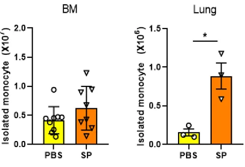
Fig. 1. The comparison of cell number in PBS and SP infected mice
··· 15
Fig. 2. Schematics of TGT tension sensor
··· 17
Fig. 3. Adhesion and migration of monocyte on TGT surfaces
18
Fig. 4. The comparison of ruptures pattern in PBS, SP, Flu and Sal infected mice after 1 hr incubation
··· 20
Fig. 5. The comparison of ruptures pattern in PBS and SP infected mice after 30 min, 1 hr and 2 hr incubation
··· 22
Fig. 6. Image of integrin mediated adhesion of monocytes derived from BM and lung in PBS and SP infected mice on TGT surfaces after 1hr
incubation
··· 23
Fig. 7. The comparison of ruptures pattern from BM and lung in PBS and SP infected mice after 1 hr incubation
··· 24
Fig. 8. Image of integrin mediated adhesion of monocytes derived from BM in PBS and SP infected mice on TGT surfaces after 30 min, 1 hr
vi
and 2 hr incubation
··· 27
Fig. 9 Strategy of gating for FACS sorting
··· 28
Fig. 10 Heatmap for comparison of transcription factors in PBS and SP infected group
··· 29
Fig. 11 Chip to show migration of monocytes
··· 32
1
Introduction
Inflammation is caused by stressed and damaged condition, such as
infection [1]. Innate immune cells, including monocytes, play a key
role in initiating inflammation and pathogen clearance by recognizing
pathogen-associated molecular patterns [2]. Monocytes also move
to the site of infection and differentiates into macrophages during
inflammation [3, 4]. Monocytes and monocyte-derived macrophages
play an important role in both innate and adaptive immune response,
including inflammation, tissue remodeling, vascular homeostasis and
pathogen clearance [5, 6].
Monocytes are originated from the bone marrow (BM) where most
of immune cells develop. Stromal cell, endothelial cells and red blood
2
cells are also present in BM cavity [7]. Infection can induce alteration
of physical characteristics including adhesion molecules, surface
proteins in many cell populations. This changes are related to
mobilization of monocytes [3, 4, 8-10]. Integrins expressed on most
of BM cells are seminal in cell migration after infection. Integrin is
transmembrane receptor composed of alpha and beta subunit that link
immune cells with ECM (extracellular matrix) or mediate cell-cell
interactions [11, 12]. Integrins also transduce mechanical
information that generated from the ECM into attached cells [13].
Integrins are subdivided into various classes [14] and monocytes
express adhesion molecules, such as αLβ2, αMβ2, and αVβ3
[8].
3
Mechanical tension caused by integrin-mediated cell-cell
interaction functionally regulates immune responses by various
immune cells, including macrophages and T cells [15-18]. Whereas,
mechanobiological property of monocyte which potentially
contributes to cell migration and functional regulation has not been
studied.
Tension gauge tether (TGT) platform is fine mechanical sensor
which is composed of two complementary DNA strands. TGT allows
to measure the single molecule forces generated by receptor-ligand
binding in cellular level [13]. Typically, one strand has a ligand which
binds a target receptor on the cell surface and the other strand is
biotinylated for immobilization on the substrate [19]. I applied TGT
4
platform to confirm mechanical property of monocyte depends on
different strength of mechanical tension. As the infection triggers the
production of regulatory factors in BM, monocytes generated during
the infection are functionally different compare to one that from
healthy condition. I hypothesized that infection shift mechanical
tension between monocyte integrin and their receptor, which can
affect immune response. I confirmed that monocyte from infected
animal has weakened integrin-ligand interaction.
5
Material and Method
Mice
Female C57BL/6 wild-type (WT) mice aged 6-8 weeks were
purchased from Orient (Seongnam, South Korea). These mice were
maintained under specific pathogen free conditions at the Asan
Institute for life science. This study was approved by the Institutional
Animal Care and Use Committee (2018-12-302). All animal
experiments were conducted in accordance with the approved
guidelines.
Infection
Streptococcus pneumonia (SP) and Salmonella Typhimurium (Sal)
were provided by Dr. Gabriel Nunez (University of Michigan).
Influenza (Flu) was provided by Dr. Baik Lin Seong (Yonsei
University). Mice were anaesthetized by injecting 100 μl of
6
ketamine (25 mg/ml) and xylazine (2 mg/ml) mixture and intranasally
inoculated with 1×10⁷ CFU of SP, 5×10⁷ CFU of Sal and 1×10⁵ PFU
of Flu, respectively. Control mice were treated same volume of
phosphate-buffered saline (PBS; Welgene, LB001-02) instead of
pathogen.
BM monocyte extraction
Three days after infection, femur and tibias were removed and cut
both edges of the bones to expose the diaphysis. The BM was
obtained by flushing the bone with PBS using a 26G needle and 10 ml
syringe. To isolate monocytes, I used mouse monocyte isolation kit
(EasySepTM, Stem Cell, 19861), following the manufacturer’s
procedures. Cells were labeled with antibody cocktail and magnetic
particles and applied for negative selection of monocytes using the
magnet (BioLegend).
7 Monocyte extraction from lung tissue
Lung of PBS and infected mouse was taken off and chopped with
surgical scissor. Then, diverse cell types were extracted from the
lung through incubation with lung isolation buffer (RPMI 1640
medium (Hyclone, SH30027.01) containing 3% heat-inactivated
fetal bovine serum (FBS) (Hyclone, SH30919.03), collagenase Ⅳ
(Sigma, C5138-1g) and DNase-Ⅰ (Roche, 11284932001). 75%
percoll (GE Healthcare, 17-0891-01) was placed at the bottom of a
15 ml conical tube and mixture of 40% percoll and cell suspension
was stacked above the 75% percoll solution. Then, the tube was
centrifuged and pure monocyte layer was appeared at the middle of
the mixed solution. Only the monocyte layer was isolated to 15 ml
conical tube with transfer pipette and centrifuged again with full of
lung isolation buffer. After the centrifugation, removed the
supernatant and add isolation buffer. then cells were isolated with
8 Monocytes Isolation Kit.
Flow cytometry
Flow cytometry was performed using an FACS canto (BD
Biosciences). Cells were blocked with BD Fc Block Receptor Binding
Inhibitor (BD Bioscience) for 15 min on ice, followed by staining with
fluorochrome-conjugated antibodies for 15 min on ice in the dark.
Monocytes (CD11b+Siglec-F-Ly6C+Ly6G-) were separated using
monoclonal antibodies conjugated with fluorochrome: CD11b (M1/70,
FITC), Siglec-F (S17007L, PE), Ly6C (AL21, APC), Ly6G(1A8,
PE-Cy7), all purchased from BioLegend.
Flow cytometry sorting
Flow cytometry sorting was performed using an FACS Aria Sorter
(BD Biosciences). Single cell solutions were blocked with BD Fc
9
Block Receptor Binding Inhibitor (BD Bioscience) for 15 min on ice,
followed by staining with fluorochrome-conjugated antibodies for 15
min on ice in the dark. For the sorting of monocytes, the following
antibodies were used Table 1.
10 Table 1.
Antibodies used for FACS sorting.
Antibody Specificity Clone Fluorochrome Company / Cat No.
CD11b Monocytes/
Neutrophils
M1/70 FITC BioLegend
/ 101205 Siglec-
F
Eosinophils/
Neutrophils
E50- 2440
PE BD Biosciences / 552126 Ly6C Monocytes/
Neutrophils
AL-21 APC BD Biosciences
/ 560595 Ly6G Neutrophils/
Granulocytes
1A8 PE-Cy7 BioLegend
/ 127618
11 RNA sequencing
RNA of sorted monocytes were extracted with the RNeasy mini Kit
(Qiagen, Cat No. 74106), according to the manufacturer’s protocol.
RNA quality and quantity was confirmed using a Nano-drop. RNA
sequencing was performed at Macrogen Inc. (www.macrogen.com)
TGT test
TGT platform was designed by co-worker from Incheon University.
Primary monocytes were seeded on each well of the 8 well chamber
and incubated at 200 ul density on TGT surfaces at 37℃ and 5% for
30 min, 1hr and 2 hr. After the incubation, cells were fixed with 4%
paraformaldehyde for 10 min and washed 3 times by PBS. Bright field
and fluorescent images were acquired using 60x objective of Nikon
Ti2.
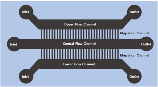
12 Immune cell migration assay
After ECM coating, M-CSF containing media and serum-free media
were introduced with constant flow rate (40 ul/hr) into the upper and
the lower flow channel through the tubing connected to the inlet of
the upper and the lower flow channel, respectively. monocytes were
injected into the central flow channel. M-CSF gradient was
generated throughout from the upper to the lower channels by
diffusion of the attractant.
Microscopy and Imaging
Time-lapse images were acquired on a Ti2 (Nikon) inverted
microscope equipped with an on-stage incubation chamber which
maintained the temperature at 37℃. And the mixed gas was injected
to the incubation chamber that maintaining the concentration at 5%.
To maintain humidity inside of the chamber, deionized water
13
container was placed in the chamber. Time-lapse phase-contrast
images were acquired by a camera (Andor, iXon Life) using NIS
elements software.
14
Results
To determine the changes of monocytes number in BM and lung
following SP infection, C57BL/6J mice were treated with PBS or
infected with Streptococcus pneumoniae (SP). Mice were then
sacrificed 3 days after infection and monocytes were isolated using
magnetic-based negative isolation method. The number of isolated
monocytes per mice was measured. Although mean bone marrow
(BM) monocytes number of SP-infected mice were higher than that
of PBS group, it was not statistically significant. Whereas, the lung
had significantly increased monocyte after infection (Fig. 1).
Based on previous studies that suggesting the role of cellular
tension on adhesion, migration, and proliferation of immune cells [11,
13, 16, 19], I hypothesized that the tension caused by receptor-
ligand interaction would affect the immunological character of
monocytes. To test this hypothesis, I used tension gauge tether
15
Fig. 1. The comparison of cells in PBS and SP infected mice.
C57BL/6J mice were treated for 3days with PBS and Streptococcus
pneumoniae (SP). Monocytes were isolated by negative selection kit
and measured. The number of isolated monocytes in BM and lung
from PBS and SP-infected group. Monocytes from SP group has
more than PBS group in lung.
16
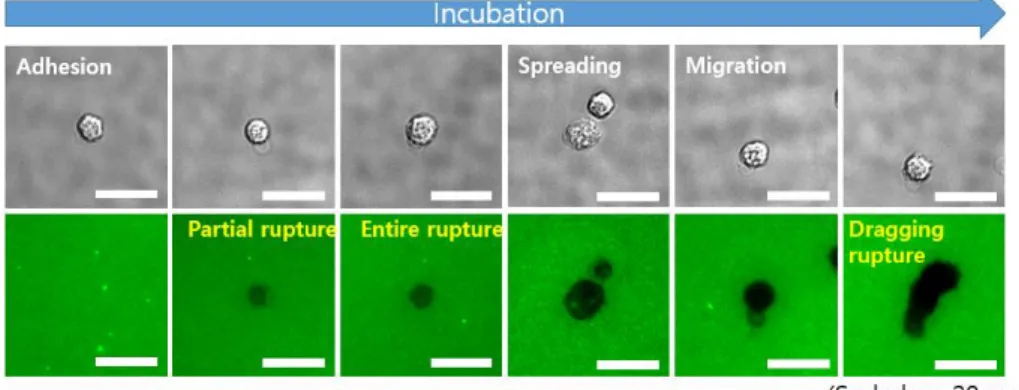
(TGT) platform, which can measure tension and mobility of cells (Fig.
2). To confirm the migration of monocytes, BM and lung monocytes
from naive and SP-infected mice were seed on the TGT. Digital
image correlation (DIC) and fluorescence images were obtained and
used to classify morphology of monocytes in terms of adhesion,
spreading, and migration states (Fig. 3). When tension of integrin-
ligand bond is larger than tension tolerance, cells form several types
of rupture on TGT [11, 20, 21]. I classified the rupture pattern into
partial rupture, entire rupture and dragging rupture and counted the
number of rupture depending on each criterion.
Initially, I expected to see increase adhesion and the number of
ruptures with monocyte isolated from SP-infected mice because
monocytes undergo an extravasation process to migrate inflamed
tissue through a receptor-ligand interaction with high affinity during
infection [3-6]. However, the number of ruptures was reduced with
17 Fig. 2. Schematics of TGT tension sensor
The schematic show that a ligand for integrin receptor is immobilized
on the TGT surface. Ligand-TGT constructs were immobilized by
Biotin. Ruptures occur when the tension applied by the cell through
the receptor is larger than its Ttol.
18
Fig. 3. Adhesion and migration of monocyte on TGT surfaces
Analysis of TGT rupture pattern. Initially, single cell was adhered on
TGT surface and spread over time. Adhesive cell and spreading cell
could be confirmed by partial rupture and entire rupture on TGT. If
cell spread on TGT, cell can migrate on TGT surface. Migration could
be confirmed by rupture as referred to dragging rupture on TGT.
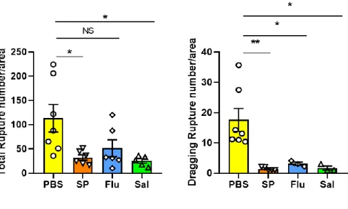
19
monocyte isolated from SP-infected mice as well as Salmonella
Typhimurium (Sal)-infected mice (Fig. 4). Monocytes isolated from
influenza (Flu) virus infected mice showed comparable total rupture
with that of PBS-infected mice but exhibited significantly reduced
dragging rupture formation (Fig. 4). The ability to form dragging
rupture reflecting the mobility of cells was reduced to significant
level in monocytes isolated from infected mice. My results suggested
that induced tension by receptor-ligand interaction affects cell
mobility during infection. In later experiment, I used SP infection
model as it showed the most significant difference among Sal, Flu,
and SP models.
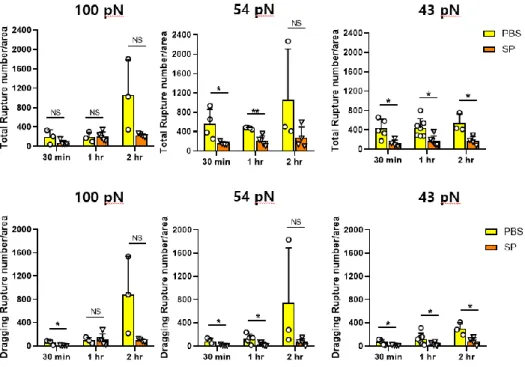
Isolated monocytes from PBS and SP-infected mice were applied
on the TGTs with Ttol = 43 pN, 54 pN and 100 pN for 30 min, 1 hr
and 2 hr. Integrins expressed on monocytes was bound to the top
DNA strand of TGT. Monocytes can pull ligands through cellular
20
Fig. 4. The comparison of ruptures pattern in PBS, SP, Flu and Sal
infected mice after 1 hr incubation at 54 pN
Comparison of the number of total rupture and dragging rupture for
PBS, Streptococcus pneumonia (SP), Influenza (Flu), Salmonella
Typhimurium (Sal)-treated group. ruptures analyzed based on two
variables: total rupture and dragging rupture. The number of rupture
after 1 hr incubation was counted using Image J.
21
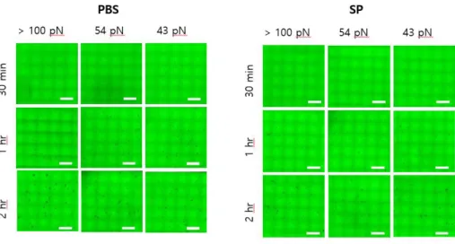
forces transmitted by integrins, which lead to rupture on TGT surface.
Although no significant difference could be identified at 100 pN (Fig.
5). Total and dragging ruptures on 54 pN had significant differences
at 30 min, 1 hr, excluding 2 hr, while at 43 pN, significant differences
were observed at 30 min, 1 hr, and 2 hr (Fig. 5). Hereafter, I used
1hr for further experiment.
When TGT test was performed at different tolerance for 1 hr,
less rupture was produced on all TGT condition in SP group than PBS
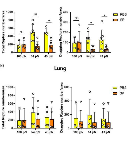
group (Fig. 6). In monocytes isolated from BM, total rupture and
dragging rupture did not show significant difference on TGT having
100 pN tolerance, but that of 54 pN and 43 pN showed significant
decrease in the SP group (Fig. 7A). In the lung, it also was showed
that the number of ruptures in the SP group decreased, having similar
tendency with BM (Fig. 7B). Since there were slight differences on
each pN, the migration ability of monocytes separated from BM in
22
Fig. 5. The comparison of ruptures pattern in PBS and SP infected
mice after 30 min, 1 hr and 2 hr incubation
Comparison of the number of total rupture and dragging rupture in
BM from PBS and SP infected group. The number of rupture after 30
min, 1 hr, 2 hr incubation was counted. Ruptures analyzed based on
two variables: total rupture and dragging rupture. The number of
rupture was counted using Image J on 54 pN. The most significant
result was obtained when incubated at 54 pN for 1 hr incubation.
23
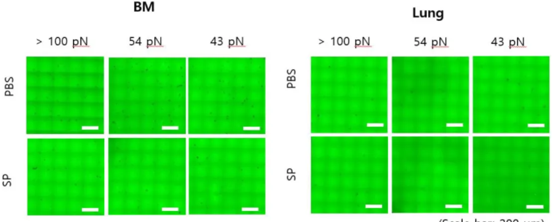
Fig. 6. Image of integrin mediated adhesion of monocytes derived from
BM and lung in PBS and SP infected mice on TGT surfaces after 1 hr
incubation
The image show rupture on TGT surfaces (incubation time, t = 1 hr).
Cells adhere on 100, 54 and 43 pN TGT that we made. On all
tolerance, the number of ruptures of naïve group is more than SP
group in BM and lung. The TGT assay in each condition was repeated
at least three times.
24
Fig. 7. The comparison of ruptures pattern from BM and lung in PBS
and SP infected mice after 1 hr incubation
Comparison of the number of total rupture and dragging rupture in
BM (A) and lung (B) from PBS and SP-infected group. The number
25
of rupture after 2 hr incubation was counted. Ruptures analyzed
based on two variables: total rupture and dragging rupture. The
number of rupture after 1 hr incubation was counted using Image J.
26
PBS and SP group was confirmed via TGT according to pN and time
(Fig. 8).
To link activation of monocyte by infection with their altered
migratory phenotype, I analyzed gene expression profile RNA
sequencing. To obtain maximum purity, monocytes were isolated by
FACS sorting. CD11b+Siglec-F-Ly6C+Ly6G- cells were gated to
sort monocytes (Fig. 9). Forces exerted by integrin receptor-ligand
are transmitted to cell membrane and alters expression of surface
protein [22].
Heatmap was prepared for three different criteria: (a)
transcription factors that related to cell adhesion molecules (CAMs),
focal adhesion, (b) cytoskeletal regulation, (c) signaling molecules
(Fig. 10). Transcription changes were considered significant when
expression number were different more or less than doubled fold
compared to the control group. RNA sequencing data revealed that
27
Fig. 8. Image of integrin mediated adhesion of monocytes derived from
BM in PBS and SP infected mice on TGT surfaces after 30 min, 1 hr
and 2 hr incubation.
The image show rupture on TGT surfaces in BM from PBS and SP
treated group (incubation time, t = 30 min, 1 hr, 2 hr). Cells adhere
on 100, 54 and 43 pN TGT that we made. The number of ruptures
increased over time. The number of rupture in naïve group is more
than SP group.
28 Fig. 9. Strategy of gating for FACS sorting
FACS sorting strategy for purification of monocytes population from
BM. Monocytes were sorted on the makers as known monocytes
expressed. To monocyte isolation, CD11b+, Siglec-F-, Ly6C+,
Ly6G- gating used. The sorted populations were used for RNA
sequencing.
29
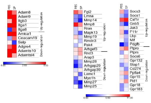
Fig. 10. Heatmap for comparison of transcription factors in PBS and
SP infected group
Heatmap of the differentially expressed transcription factors from
RNA sequencing data in PBS and SP treated group. Data represents
P-value (log10). Up-regulated genes and down-regulated genes
are shown in color.
30
disintegrin and metalloproteinase (ADAM) subtype, a factor that
interferes with the function of integrin, was up-regulated. Meanwhile,
Selectin and Ceacams related to cell adhesion were down-regulated.
Among the factors related to adhesion molecules, several types of
Rho GTPase were downregulated in the SP group. Recent studies
have focused on how the tension between these integrins and
receptors affects multiple immune cells. I hypothesized that many
genes expressed in cells change during infection, and as a result,
tension caused by changing receptor ligand interactions will affect
the immune response. Although TGT test and cDNA microarray
analysis were performed to determine the changes in the gene
expression pattern after infection. Because TGT test use fixed cell
at specific time points, it could not be compared exactly. Instead, a
migration chip that can see kinetics of a single cell was additionally
executed. The M-CSF containing media and serum-free media were
31
added with constant flow rate (40 ul/hr) into the upper and the lower
flow channel, respectively (Fig. 11). Isolated monocytes were added
into the central flow channel. It can be compare cell motility between
PBS and SP group.
32 (A)
Fig. 11. Designed microfluidic chip for single-cell migration. (A)
Schematic of the chip.
The schematic show that chip for cell migration. The Migration chip
is useful to confirm the cell’s kinetics. It can also be confirmed that
the result according to TGT data. In order to inducing gradient inside
the migration chip, Upper flow channel was added RPMI medium
containing chemoattractant M-CSF and lower flow channel was
added pure RPMI medium. Monocytes were added to central flow
33 channel.
34
Discussion
Despite a long history of immunological studies, it was not that
long ago when researches start to investigate the mechanobiological
involvement in functional characterization of immune cells.
Mechanical force that occurs during receptor-ligand interaction has
critical roles in cellular biological process including differentiation,
proliferation, survival, and activation of various cell types [23-30].
Integrins are especially critical to cope with bacterial and viral
infection in terms of microbial pathogenesis and functional maturation
[23, 24, 31-34]. M. tuberculosis infection increases expression of
integrin αVβ3, which promotes monocyte adhesion to ECM [8].
RNA-sequencing data from my research also showed that integrin
αVβ3 was up-regulated in SP-infected monocyte. However,
overall receptor-ligand interaction between ligand and SP-infected
monocyte integrin was weaker when it compared to interaction with
35
control monocyte isolated from PBS group. The reason for weaker
cellular adhesion by SP-infected monocyte, even with higher
expression of αVβ3, may be ascribed to less efficient integrin
clustering. Indeed, previous study showed that expression level of
integrins are similar between immune cells isolated from infected and
non-infected animals [34]. But infected monocyte showed disrupted
integrin clustering which affects integrin activation, strength of
binding, and phosphorylation [34]. I also confirmed that expression
level of αLβ2 and αMβ2 on monocyte are not changed after SP
infection using flow cytometry. Further researches are needed to
elucidate factors related to control of focal adhesion after infection.
T. gondii-infected monocyte has defects in cell spreading,
clustering of integrin, and focal adhesion kinase. These defects
allows monocyte migrate at a higher speeds than uninfected
monocytes [35]. However, lipopolysaccharide or Escherichia coli
36
infection did not induce impairment of integrin clustering, indicating
mechanobiological property of monocyte may vary depending of the
pathogen they encounter [35]. Furthermore, N. gonorrhoeae can bind
to Ceacams, molecule that involved in bacteria-cell adhesion [32,
36]. In contrast, nonpathogenic Neisseria cinerea can lead to
adhesion of Ceacams-expressing cells. In my setting, Ceacams were
down-regulated suggesting receptor-ligand interaction is weakened
in SP-infected mice. The result also suggests that infection-
triggered alteration of signaling and gene expression can modulate
cell adhesion and migration following infection. It seems that SP
infection makes focal adhesion weaker or disrupts integrin clustering
formation.
Integrin is critical mediators of viral and bacterial infections, and
integrin-mediated adhesion as well as pathogenic processes [32].
Other study confirmed that monocytes and endothelial cells are
37
activated by infection, which lead to enhancing expression of
adhesion molecules, including E-selectin, fibronectin-1 [9]. To
know exactly how the monocyte react upon infection, it is important
to understand which integrin subtypes are changed in their
expression levels and whether these changes affect cellular
processes during infection. Other study suggested that the T. gondii-
induced disruption in integrin-mediated adhesion is linked to a
dysregulation of components of the focal adhesion complex. My
results also indicate that SP infection weakens receptor-ligand
interaction. Based on previous studies and my results, I speculated
that adhesion of SP-infected monocyte is weaker maybe since focal
adhesion could not made well.
Several studies have shown that physical pressure on the cell
stimulate cell to activate pro-inflammatory responses. The PIEZO
ion channel can induce a pro-inflammatory response by sensing
38
cyclical pressure in myeloid cells [37]. It is similar to the content
that a proper cellular reaction occurs by recognizing stimuli such as
pressure and tension in surrounding environment of the cell. Although
a pro-inflammatory response occurs through pressure, I thought that
low tension is important for the migration of cells from bone marrow
to peripheral blood based on the results of my experiment.
Previous studies mainly used TGT test to analyze cells in a
stationary state after incubation [13, 19]. Although TGT test was
efficient to measure tension in single cell level, the study did not
show whether the change in tension alters migration profile of live
monocytes. As the migration pattern and speed are closely related to
immune response, I used system to confirm the mobility of live cells
utilizing migration chip. This technique was efficient to monitor the
live monocyte migration and kinetics using microscopy time lapse. In
addition, I coated the chip with cyclic RGD which were used for TGT
39
test to assess the cellular tension is involved in mobile characteristics
of infected monocyte. By combining the fields of immunology and
mechanobiology, the inter-relation between altered mechanical
characteristic and cell migration under host’s infection can be
studied extensively. As I used live imaging with migration chip to
show leukocyte behavior, such as crawling and swarming, further
efforts on introducing new mechanobiological techniques will allow
researchers in immunology field to understand cellular
communication and in vivo characteristics better in the future.
40
References
1. Medzhitov, R., Origin and physiological roles of inflammation.
Nature, 2008. 454(7203): p. 428-35.
2. Bekkering, S., et al., In Vitro Experimental Model of Trained Innate Immunity in Human Primary Monocytes. Clin Vaccine Immunol, 2016. 23(12): p. 926-933.
3. Imhof, B.A. and M. Aurrand-Lions, Adhesion mechanisms regulating the migration of monocytes. Nat Rev Immunol, 2004.
4(6): p. 432-44.
4. Ley, K., et al., Getting to the site of inflammation: the leukocyte adhesion cascade updated. Nat Rev Immunol, 2007. 7(9): p. 678- 89.
5. Narasimhan, P.B., et al., Nonclassical Monocytes in Health and Disease. Annu Rev Immunol, 2019. 37: p. 439-456.
6. Kratofil, R.M., P. Kubes, and J.F. Deniset, Monocyte Conversion During Inflammation and Injury. Arterioscler Thromb Vasc Biol, 2017. 37(1): p. 35-42.
7. Vandoorne, K., et al., Imaging the Vascular Bone Marrow Niche During Inflammatory Stress. Circ Res, 2018. 123(4): p. 415-427.
8. Brilha, S., et al., Monocyte Adhesion, Migration, and Extracellular Matrix Breakdown Are Regulated by Integrin alphaVbeta3 in Mycobacterium tuberculosis Infection. J Immunol, 2017. 199(3): p.
982-991.
9. Thomas-Ecker, S., et al., Alteration in the gene expression pattern
41
of primary monocytes after adhesion to endothelial cells. Proc Natl Acad Sci U S A, 2007. 104(13): p. 5539-44.
10. Meisel, S.R., et al., Increased expression of neutrophil and
monocyte adhesion molecules LFA-1 and Mac-1 and their ligand ICAM-1 and VLA-4 throughout the acute phase of myocardial infarction: possible implications for leukocyte aggregation and microvascular plugging. J Am Coll Cardiol, 1998. 31(1): p. 120-5.
11. Wang, X.F., et al., Integrin Molecular Tension within Motile Focal Adhesions. Biophysical Journal, 2015. 109(11): p. 2259-2267.
12. Calderwood, D.A., S.J. Shattil, and M.H. Ginsberg, Integrins and actin filaments: reciprocal regulation of cell adhesion and signaling.
J Biol Chem, 2000. 275(30): p. 22607-10.
13. Wang, X.F. and T. Ha, Defining Single Molecular Forces Required to Activate Integrin and Notch Signaling. Science, 2013. 340(6135):
p. 991-994.
14. Chen, K. and X.Y. Chen, Integrin Targeted Delivery of Chemotherapeutics. Theranostics, 2011. 1: p. 189-200.
15. Kang, H., et al., Immunoregulation of macrophages by dynamic ligand presentation via ligand-cation coordination. Nat Commun, 2019. 10(1): p. 1696.
16. Rossy, J., J.M. Laufer, and D.F. Legler, Role of
Mechanotransduction and Tension in T Cell Function. Front Immunol, 2018. 9: p. 2638.
17. Pageon, S.V., et al., Mechanoimmunology: molecular-scale forces govern immune cell functions. Mol Biol Cell, 2018. 29(16): p.
42 1919-1926.
18. McWhorter, F.Y., C.T. Davis, and W.F. Liu, Physical and mechanical regulation of macrophage phenotype and function. Cell Mol Life Sci, 2015. 72(7): p. 1303-16.
19. Jo, M.H., W.T. Cottle, and T. Ha, Real-Time Measurement of Molecular Tension during Cell Adhesion and Migration Using Multiplexed Differential Analysis of Tension Gauge Tethers. Acs Biomaterials Science & Engineering, 2019. 5(8): p. 3856-3863.
20. Wang, Y. and X. Wang, Integrins outside focal adhesions transmit tensions during stable cell adhesion. Sci Rep, 2016. 6: p. 36959.
21. Roein-Peikar, M., et al., Ultrasensitivity of cell adhesion to the presence of mechanically strong ligands. Physical Review X, 2016.
6(1): p. 011001.
22. Zhu, C., et al., Mechanosensing through immunoreceptors. Nature Immunology, 2019. 20(10): p. 1269-1278.
23. Basu, R., et al., Cytotoxic T Cells Use Mechanical Force to Potentiate Target Cell Killing. Cell, 2016. 165(1): p. 100-110.
24. Giannone, G., Super-resolution links vinculin localization to function in focal adhesions. Nature Cell Biology, 2015. 17(7): p.
845-847.
25. Bashour, K.T., et al., CD28 and CD3 have complementary roles in T-cell traction forces. Proceedings of the National Academy of Sciences of the United States of America, 2014. 111(6): p. 2241- 2246.
26. Wehner, S., et al., Mechanical strain and TLR4 synergistically
43
induce cell-specific inflammatory gene expression in intestinal smooth muscle cells and peritoneal macrophages. Am J Physiol Gastrointest Liver Physiol, 2010. 299(5): p. G1187-97.
27. Irwin, E.F., et al., Modulus-dependent macrophage adhesion and behavior. J Biomater Sci Polym Ed, 2008. 19(10): p. 1363-82.
28. Refai, A.K., et al., Effect of titanium surface topography on
macrophage activation and secretion of proinflammatory cytokines and chemokines. J Biomed Mater Res A, 2004. 70(2): p. 194-205.
29. Pugin, J., et al., Activation of human macrophages by mechanical ventilation in vitro. Am J Physiol, 1998. 275(6): p. L1040-50.
30. Rich, A. and A.K. Harris, Anomalous preferences of cultured macrophages for hydrophobic and roughened substrata. J Cell Sci, 1981. 50: p. 1-7.
31. Moreno-Layseca, P., et al., Integrin trafficking in cells and tissues.
Nat Cell Biol, 2019. 21(2): p. 122-132.
32. Bachmann, M., et al., Cell Adhesion by Integrins. Physiol Rev, 2019. 99(4): p. 1655-1699.
33. Zhang, Y. and H. Wang, Integrin signalling and function in immune cells. Immunology, 2012. 135(4): p. 268-75.
34. Hogg, N., et al., T-cell integrins: more than just sticking points.
Journal of Cell Science, 2003. 116(23): p. 4695-4705.
35. Cook, J.H., N. Ueno, and M.B. Lodoen, Toxoplasma gondii disrupts beta 1 integrin signaling and focal adhesion formation during monocyte hypermotility. Journal of Biological Chemistry, 2018.
293(9): p. 3374-3385.
44
36. Muenzner, P., et al., CEACAM engagement by human pathogens enhances cell adhesion and counteracts bacteria-induced
detachment of epithelial cells. J Cell Biol, 2005. 170(5): p. 825-36.
37. Solis, A.G., et al., Mechanosensation of cyclical force by PIEZO1 is essential for innate immunity. Nature, 2019. 573(7772): p. 69-74.
45
국문 초록
장력이 단핵구의 이동성 및 물리적 특성에 미치는 영향
단핵구들은 일반적으로 혈류 내에서 순환하지만, 병원체가 침입하거나
염증이 발생하면 표적 부위로 이동하여 대식세포로 분화하여 선천 면역
반응의 초기에 중요한 역할을 한다. 단핵구들이 표적 부위로 이동할 때
혈관 내벽 세포와 단핵구가 발현하고 있는 세포부착 분자들을 이용한다
고 알려져있다. 수용체-리간드의 상호작용에서 발생하는 장력, 리간드
밀도 등이 대식세포의 기능 조절에 미치는 영향은 비교적 잘 연구되어
있지만, 단핵구에서는 분화에 미치는 영향 외에는 연구가 잘 되어있지
않다. 또한, 세포 주변 환경으로부터의 물리적 자극이 대식세포와 T세포
를 비롯한 일부 면역 세포의 기능 조절에 미치는 영향이 잘 연구되어 있
기때문에, 단핵구에서 또한 주변 환경이 면역기능에 영향을 미칠 것이라
가설을 세웠다. TGT라는 플랫폼을 이용하여 리간드-수용체 상호작용시
46
발생하는 장력을 조절하고 세포의 이동성을 확인하였다. 마우스에
Streptococcus pneumoniae (SP) 박테리아를 감염시켰을 때, 세포부착
분자들이 증가하고 그에 의해 세포의 이동성이 확연하게 증가할 것이라
고 생각했었다. 하지만 예상한 바와 달리 감염군에서 대조군에 비해 세
포의 이동성이 감소하였다. SP 뿐만 아니라, 살모넬라와 인플루엔자 감
염에서도 유사한 경향성을 보였다. 이와 같은 결과를 토대로 감염시 발
생하는 리간드-수용체 상호작용 장력이 세포의 이동성에 영향을 준다는
것을 확인하였다. 감염시 세포에 일어나는 물리적 변화를 확인하고, 리
간드-수용체 상호작용에 의해 세포에 전달되는 장력이 단핵구의 혈관외
유출 및 세포의 기능에 어떠한 영향을 주는지 밝히기 위해 RNA
sequencing을 통해 대조군과 감염군에서 발현하고 있는 세포부착 분자
간의 차이를 확인하였고 이후 migration chip 연구를 통해 세포 이동성
의 변화를 확인하고자 한다.
주요어 : 단핵구, 세포부착분자, 리간드-수용체 상호작용, 세포의 이동성,
47 면역반응, 장력, 물리적 특성
학 번 : 2018-26037