저작자표시-비영리-변경금지 2.0 대한민국 이용자는 아래의 조건을 따르는 경우에 한하여 자유롭게
l 이 저작물을 복제, 배포, 전송, 전시, 공연 및 방송할 수 있습니다. 다음과 같은 조건을 따라야 합니다:
l 귀하는, 이 저작물의 재이용이나 배포의 경우, 이 저작물에 적용된 이용허락조건 을 명확하게 나타내어야 합니다.
l 저작권자로부터 별도의 허가를 받으면 이러한 조건들은 적용되지 않습니다.
저작권법에 따른 이용자의 권리는 위의 내용에 의하여 영향을 받지 않습니다. 이것은 이용허락규약(Legal Code)을 이해하기 쉽게 요약한 것입니다.
Disclaimer
저작자표시. 귀하는 원저작자를 표시하여야 합니다.
비영리. 귀하는 이 저작물을 영리 목적으로 이용할 수 없습니다.
변경금지. 귀하는 이 저작물을 개작, 변형 또는 가공할 수 없습니다.
i
의학석사 학위논문
Pretreatment with epidermal growth factor promotes primary hair recovery in the
chemotherapy-induced alopecia
화학요법으로 유발된 탈모에서 표피성장 인자 전처치에 의한 일차모발회복
2013년 2월
서울대학교 대학원
임상의과학과 전공
백 승 환
Abstract
Pretreatment with epidermal growth factor promotes primary hair recovery in the chemotherapy-induced alopecia
Seung Hwan Paik, M.D.
(Director: Professor, Oh Sang Kwon, M.D., Ph.D.)
Clinical Medical Science The Graduate School Seoul National University
Epidermal growth factor (EGF) is not only a potent cell growth stimulant but also has a catagen-inducing effect on hair follicles. Because chemotherapeutic agents primarily damage anagen hair follicles, it would be important to investigate whether catagen inducers have beneficial effects in the prevention of chemotherapy-induced alopecia (CIA). I pretreated hair follicles with a vehicle or EGF (50 mg/ml or 100 mg/ml) before treatment with a
chemotherapeutic agent, and I observed the severity of hair follicle damage after chemotherapy and examined the recovery patterns after CIA by using the C57BL/6 mouse model of cyclophosphamide-induced alopecia. To validate the catagen-inducing property and damage response pathway after CIA, I evaluated the hair cycle score in histological sections and the number of TdT-mediated dUTP nick end labeling (TUNEL)-positive cells in the hair bulbs of control and EGF-treated mice. To follow gross changes as well as to assess the progression of CIA and the recovery pattern after CIA, photographs of the back skin of mice were captured and analyzed. I confirmed that topical EGF application induced a catagen-like stage in anagen hair follicles after depilation; I found that catagen-like hairs induced by EGF were protected from chemotherapy-mediated damage. Moreover, my results showed that EGF treatment favored primary hair recovery via the dystrophic anagen pathway after CIA. Given that hair follicles subjected to less severe chemotherapeutic insult enter the dystrophic anagen pathway followed by primary recovery, rather than entering the dystrophic catagen pathway followed by secondary recovery, the results of this study suggest that catagen inducers could be useful as a new alopecia-protection strategy, especially in the context of CIA.
Keywords: Epidermal growth factor, chemotherapy-induced alopecia, primary hair recovery, dystrophic anagen pathway
Student Number: 2011-21991
ii
Contents
Chapter 1 Introduction --- 1
Chapter 2 Materials and Methods--- 4
2.1 Materials--- 4
2.2 Animals--- 4
2.3 Pretreatment with rhEGF and CYP injection ---5
2.4 Photographic and histomorphometric analyses---6
2.5 Immunofluorescent staining---7
2.6 Statistical analysis---8
Chapter 3 Results---9
3.1 Topical application of EGF induces catagen-like effects in the C57BL/6 mouse model of CIA---9
3.2 Pretreatment with EGF decreased the chemotherapeutic damage to hair follicles and preferentially activated the dystrophic anagen pathway ---10
3.3 Pretreatment with EGF favored primary hair recovery after CIA---11
Chapter 4 Discussion---17
References--- 21
Abstract--- 23
iii
List of Figures
Figure 1. Experimental scheme
Figure 2. EGF induced catagen-like effect in hair follicles of depilated C57BL/6 mice
Figure 3. Pretreatment with EGF protected against chemotherapy-induced hair follicle damage and progression toward dystrophic catagen
Figure 4. EGF retards the progression of CIA and promotes primary recovery after CIA
1
Chapter 1. Introduction
Chemotherapy-induced alopecia (CIA) is one of the unsolved problems in clinical oncology.
Because chemotherapeutic drugs can directly damage rapidly dividing cells, the toxicities of these drugs can be broadly divided into 3 classes: bone marrow suppression, gastrointestinal disturbance, and alopecia (1). Substantial efforts have been invested into the use of drugs for managing bone marrow suppression and gastrointestinal disturbance; however, several effective treatment options for preventing or overcoming CIA remain to be explored.
To develop methods for the management of CIA, it is necessary to understand the mechanism of hair loss in CIA. CIA is categorized as acute diffuse shedding caused by anagen effluvium, which refers to the pathologic loss of anagen hairs (2). Normal human hair follicles undergo 3 phases of growth: anagen, catagen, and telogen, corresponding to the growing phase, degenerative phase, and resting phase, respectively. Anagen hair follicles have high mitotic activity in their bulb region, thus are the most vulnerable to chemotherapeutic agents (3).
Therefore, anagen hairs are damaged and shed after systemic chemotherapy.
It is also important to know the patterns of recovery following CIA. Previous studies have revealed 2 specific pathways depending on the severity of chemotherapeutic damage: the
2
dystrophic anagen pathway and the dystrophic catagen pathway (4, 5). The less severe chemotherapeutic damage, the higher is the number of hair follicles that go through the dystrophic anagen pathway following primary hair recovery. In contrast, more severe chemotherapeutic damage induces the dystrophic catagen pathway following secondary hair recovery (4, 5).
In the dystrophic anagen pathway, hair follicles damaged due to chemotherapy enter the dystrophic anagen cycle directly, thus leading to primary hair recovery, which is characterized by incomplete regeneration of the pigmentary system histologically manifested as a slightly disrupted banding pattern and grossly as white-to-gray colored hair. These do not enter the telogen period. In contrast, in the dystrophic catagen pathway hair follicles enter the dystrophic catagen cycle followed by a shortened dystrophic telogen period, and then normal-looking hair is regenerated, in a process called “secondary recovery” (4, 5).
Previous studies showed that several substances promote the dystrophic anagen pathway or the dystrophic catagen pathway. Cyclosporine, FK506, and PTH/PTHrp 7-34 promoted the dystrophic anagen pathway, whereas calcitriol, glucocorticoids, and PTH 1-34 promoted the dystrophic catagen pathway (4, 6-8).
Given that CIA is anagen effluvium, it is clinically meaningful to investigate the effect of hair cycle modifiers in alleviating the chemotherapeutic damage to hair follicles. Epidermal growth
3
factor (EGF) is a protein composed of 53 amino acids (molecular weight, 6054 Da) and is known to induce catagen-like effects on hair follicles (9). In addition, it has been reported that all EGF receptor inhibitors currently in use induce trichomegaly (10-12), which might be a consequence of the reversal effect of EGF.
To the best of my knowledge, there has been no study investigating the effect of the catagen transition induced by EGF on patterns of hair recovery after CIA. Herein, I pretreated hair follicles with EGF to induce a catagen-like effect, and monitored the severity of the damage to hair follicles after chemotherapy and the recovery patterns after CIA in an effort to devise strategies to prevent CIA.
4
Chapter 2. Materials and Methods
2.1 Materials
Liposomal solutions loaded with vehicle or recombinant human epidermal growth factor (rhEGF) were obtained from the Daewoong Pharmaceutical Co. Ltd. (Yongin, South Korea). The rhEGF-loaded liposomes were formulated as previously described (13). Briefly, the water phase (sodium ascorbyl phosphate, EDTA, and rhEGF) and the oil phase (triglycerides, hydrogenated phospholipid, diethylamine cetyl phosphate, and butylated hydroxyl toluene dissolved in ethanol) were mixed and passed through a high-pressure homogenizer (Emulsiflex-C3; AVESTIN, Canada). Two different concentrations of rhEGF (50 and 100 mg/ml) were used in this study, and the vehicle alone was used as the control treatment.
To induce CIA, I used cyclophosphamide (CYP; Endoxan, Baxter Oncology, Germany) and injected each test mouse with 120 mg/kg body weight of CYP.
2.2 Animals
Seven-week-old, female C57BL/6 mice (18–20 g body weight) were purchased from KOATECH (Pyeongtaek, Korea). Mice were housed in groups of 3 or 4 animals, under conventional standardized conditions. Animals were fed tap water and commercial mouse chow ad libitum.
The hair follicles of the mice were in the telogen stage, represented by pink skin on the back,
5
when the experiments were performed. After a 1-week adjustment period, 24 8-week-old mice were subdivided into 3 groups for treatment with the vehicle control, or rhEGF at 2 different concentrations. All animal procedures were approved by the Institutional Animal Care and Use Commitee of Seoul National University Hospital Biomedical Research Institute.
To induce the anagen phase, the back skins of the mice in the telogen stage were depilated as previously described (4). Briefly, a wax/rosin mixture was melted and applied to the back skin.
After hardening, it was peeled off, thus plucking all the telogen-stage hair shafts, after which anagen VI hair follicles were induced within 9 days after depilation. Whereas spontaneous anagen hair develops in a wave-like pattern, depilation-induced anagen is fully synchronized over the entire depilated area; therefore, it can be used as a mouse model of CIA making vigorous anagen effluvium over that area. On day 17 after depilation, the catagen transition first appeared at the neck region and subsequently reached the tail region about 2 days later, i.e., on day 19 (Figure 1A) (14).
2.3 Pretreatment with rhEGF and CYP injection
Starting on day 9 after depilation, when the hair follicles entered anagen VI, 0.5 ml of EGF (50 mg/ml, or 100 mg/ml) or the vehicle was administered twice daily with a cotton applicator to the back skin of the mice for 4 days (Figure 1B). After application of the topical agents, the back skins were immediately covered with an occlusive dressing (Tegaderm; 3M, London, UK) to
6
increase the percutaneous absorption of drugs. A previous report showed that within 4 days, rhEGF could induce anagen- to catagen-like morphological changes (9); therefore, I pretreated mice with EGF for 4 days before injecting the chemotherapeutic agent.
On day 13 after depilation, a single intraperitoneal injection of CYP was administered (120 mg/kg body weight, freshly dissolved in distilled water) to all 24 mice. Topical treatments (EGF or vehicle control) were applied for 2 more days after administration of the CYP injection for maintaining the catagen-like state during the washout period of CYP (Figure 1B).
2.4 Photographic and histomorphometric analyses
The back skins of all animals were photographed using a digital camera (PowerShot S51S;
Canon, Japan) up to 30 days after depilation. The photographs were documented, and the percentage of primary recovery in the depilated back area was assessed using an image analysis program (Image J; NIH, USA).
The mouse skin was harvested on days 11, 13, and 17 after depilation, which corresponds to days 2 and 4 after the application of topical agents and day 4 after injection of the chemotherapeutic drug, respectively (Figure 1B). Sections were prepared from the harvested skin and stained with hematoxylin and eosin. Histological morphometry was performed on the stained sections to evaluate the catagen-like changes in the tissues induced by the application of topical agents on days 11 and 13 after depilation and to assess the protection from CIA after the injection
7
of CYP on day 17 after depilation. To evaluate the catagen-inducing property and the damage response pathway after CIA following EGF treatment, the hair cycle score (HCS) was assessed as previously described (6). Briefly, randomly selected hair follicles were scored according to their hair cycle stages (0: anagen VI, 1: catagen I-III, 2: catagen IV-V, 3: catagen VI-VIII). The percentage of hair follicles in each of the stages was calculated. The HCS of an individual mouse was calculated by multiplying the percentage of follicles at each stage with the respective score for each stage and then summing the resulting values. Hair cycle stages of a minimum of 50 hair follicles per mouse were assessed and assigned to the corresponding scores.
2.5 Immunofluorescent staining
As previously described (15), I performed a TdT-mediated dUTP nick end labeling (TUNEL) assay (In Situ cell death detection kit with fluorescein; Roche Diagnostics, Mannheim, Germany) to measure the proportion of apoptotic cells. An anti-digoxigenin fluorescein isothiocyanate (FITC)-conjugated antibody was used to visualize TUNEL-positive cells and a 4′,6-diamidino-2- phenylindole (DAPI) mounting media kit (Vector Laboratories, Burlingame, CA, USA) was used for nuclear counterstaining. Cells labeled using the TUNEL method were then observed under an Olympus BX61 fluorescent microscope (Olympus, Tokyo, Japan) using the FITC filter and photographed with a Leica Application Suite (LAS) v. 3.8 (Leica Microsystems, Barcelona, Spain).
8
For quantitative analyses, I counted the number of TUNEL-positive cells in the bulb region of hair follicles in the control and EGF-treated groups. A minimum of 30 hair follicles per mouse were analyzed.
2.6 Statistical analysis
Data from different groups of mice were compared, and the statistical significance of the differences was determined using ANOVA and Scheffe’s tests. All statistical analyses were performed with SPSS version 18 (IBM Corporation, Somers, NY, USA), and p-values lower than 0.05 were considered statistically significant.
9
Chapter 3. Results
3.1 Topical application of EGF induces catagen-like effects in the C57BL/6 mouse model of CIA
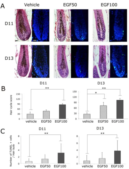
Progression to catagen-like stages was induced by treatment of a topical EGF liposomal solution. Figure 2A shows the images of hematoxylin and eosin-stained and TUNEL-stained sections of mouse skin tissue on days 11 and 13 after depilation, which correspond to days 2 and 4 after the application of EGF or the vehicle. To access the hair cycle stages of the test groups, I used the criteria for hair cycle classification set forth by Muller-Rover S et al. (14). Compared with the vehicle-treated hair, which was in the anagen phase on both days 2 and 4 after treatment, a representative hair follicle of the group treated with EFG (50 mg/ml) was in the catagen I phase, as characterized by 2 more apoptotic cells in the bulb with TUNEL staining. Hair follicles of mice treated with 100 mg/ml EGF were in the catagen II phase, with a smaller bulb and a narrower dermal papilla, compared with anagen VI/catagen I-stage hair, when observed under a light microscope, and with up to 4 apoptotic cells as observed by TUNEL staining.
When I assessed the HCS values, on both days 11 and 13, the HCS increased in an EGF concentration-dependent manner and almost every hair follicle in the group treated with EGF (100 mg/ml) had reached the early catagen stage (HCS >85). On both days 11 and day 13, the
10
HCS was significantly higher in the group treated with EGF (100 mg/ml) than in the control group (p < 0.01, Figure 2B).
EGF substantially induced apoptotic cells in the bulb in a concentration-dependent manner. On days 11 and 13 after depilation, the mean numbers of apoptotic cells per hair follicle increased with increasing concentrations of EGF. On both days, the group treated with 100 mg/ml EGF showed a statistically significant difference in the mean number of apoptotic cells per hair follicle compared to that in the control group (p < 0.01, Figure 2C).
3.2 Pretreatment with EGF decreased the chemotherapeutic damage to hair follicles and preferentially activated the dystrophic anagen pathway
Topical EGF protected mice from chemotherapy-induced follicular dystrophy. On day 17 after depilation (day 4 after chemotherapy), a representative hair follicle of the control group showed advanced dystrophic changes of intrafollicular and perifollicular ectopic melanin clumps, swollen dermal papilla, and follicular distortion, whereas that of the group treated with EGF (100 mg/ml) showed mild dystrophy, with only intrafollicular ectopic melanin clumps (Figure 3A). To evaluate keratinocyte apoptosis induced by the chemotherapeutic insult, I performed TUNEL staining of hair follicles. Hair follicles of the control group showed more apoptotic cells than that of the group treated with EGF (100 mg/ml) group (Figure 3B). When the number of TUNEL-
11
positive cells in the bulb were counted, apoptotic cells decreased in the EGF-treated groups, and a statistically significant difference was observed between the control and EGF group (treated with 100 mg/ml EGF) (p < 0.05, Figure 3C). Furthermore, EGF treatment resulted in retardation of follicle progression to dystrophic catagen. Classification criteria for two damage response pathways of hair follicles have been reported, the dystrophic anagen pathway and the dystrophic catagen pathway (5). Representative hair follicles of the control and EGF-treated group (treated with 100 mg/ml EGF) showed dystrophic catagen and dystrophic anagen hair, respectively (Figure 3A). When the HCS values were assessed, almost every hair follicle in the control group was in the late dystrophic catagen stage (HCS >250). The HCS of the group treated with EGF (100 mg/ml) group was significantly lower than that of the control group (p < 0.05, Figure 3D).
3.3 Pretreatment with EGF favored primary hair recovery after CIA
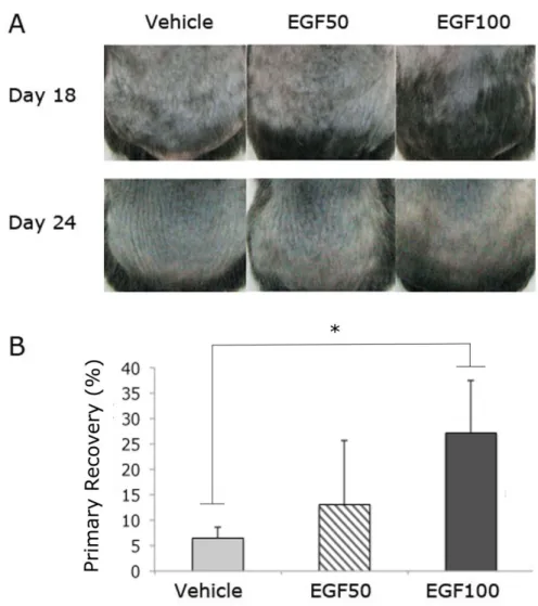
Pretreatment with EGF caused delayed progression of CIA. On day 18 after depilation, a substantial reduction of the alopecic area after CYP treatment was observed in the EGF-treated mice, and the alopecic areas reduced in a concentration-dependent manner (Figure 4A). The group treated with EGF (100 mg/ml) showed the maximal reduction of alopecia, to about half of the depilated back skin.
Pretreatment with EGF favored primary recovery after CIA. Although the CIA developed in all control and test mice after all, hairs in the corresponding areas protected from CIA grew
12
through a primary recovery pattern on day 18 after depilation (Figure 4A, representative mice shown). Primary recovery after induction of the dystrophic anagen pathway is characterized by depigmented hairs and is relatively faster than secondary recovery. On day 24 after depilation, the EGF-treated groups showed rapid recovery from alopecia, with gray-colored hair, whereas the control group had no recovered hair in the alopecic area.
Photographic analysis revealed a significant progression toward primary recovery in the EGF- treated test group. When the percentage of primary recovery area in the depilated back skin area of the mice was analyzed, the groups treated with EGF (100 mg/ml) showed significantly higher primary recovery than the control group (p < 0.05), (Figure 4B).
13
Figure 1. Experimental scheme A: Time-scale for the hair growth cycle in depilated C57BL/6 mice (14)
B: Schedules for treatment with topical agents, injection of cyclophosphamide, and skin harvesting. From days 9 to 15 after depilation, vehicle or EGF (50 mg/ml or 100 mg/ml) was topically applied to the back skin of the mice. On day 13 after the depilation, cyclophosphamide (120 mg/kg body weight) was intraperitoneally injected. For the histological analysis, skin was harvested on days 11, 13, and 17 after depilation.
EGF, epidermal growth factor; IP, intraperitoneal injection
14
Figure 2. EGF induced catagen-like effect in hair follicles of depilated C57BL/6 mice
A: Histological appearance of representative hair follicles of mice from the 3 groups (a control group and 2 groups treated with 50 mg/ml and 100 mg/ml EGF) on days 11 and 13 after depilation, which correspond to days 2 and 4 after the application of EGF or vehicle (hematoxylin and eosin staining image: magnification, x400; TUNEL:
magnification, x400; scale bars indicate 20 μm). On both days, representative hair follicles of mice from the control group, and the group treated with EGF (50 mg/ml or 100 mg/ml) were at the anagen VI, catagen I, and catagen II stages, respectively.
B: Hair cycle scores (HCSs) of mice from the 3 groups on days 11 and 13 after depilation. The HCS increased in an EGF concentration-dependent manner, and a statistically significant difference was observed between the control group and the group treated with EGF (100 mg/ml) on both days (p < 0.01).
C: The number of TUNEL-positive cells in the hair bulbs of mice from the 3 groups on days 11 and 13 after depilation.
TUNEL-positive cells increased in an EGF concentration-dependent manner, and a statistically significant difference was observed between the control group and the group treated with EGF (100 mg/ml) on both days (p < 0.01).
15 Figure 3. Pretreatment with EGF protected against chemotherapy-induced hair follicle damage and progression toward dystrophic catagen A: Representative hair follicles of the control and EGF- treated (100 mg/ml) groups on day 17 after depilation, which corresponds to day 4 after administration of the
cyclophosphamide injection. A representative hair follicle of a mouse from the control group showed advanced dystrophy (intrafollicular and perifollicular ectopic melanin clumps, swollen dermal papilla, follicular distortion), whereas that of a mouse form the group treated with EGF (100 mg/ml) showed mild dystrophy with only intrafollicular ectopic melanin clumps (hematoxylin and eosin-staining images:
magnification, x400; scale bars indicate 20 μm).
B: TUNEL-stained hair follicles of mice from the control and EGF-treated (100 mg/ml) groups on day 17 after depilation. A hair follicle of a mouse from the control group showed a higher number of apoptotic cells in the hair bulb than that of the mouse from the group treated with EGF (100 mg/ml). (TUNEL: magnification, x400; scale bars indicate 20 μm).
C: The number of the TUNEL-positive cells in hair bulb of mice of the 3 groups (a control group and 2 groups treated with 50 mg/ml and 100 mg/ml EGF) on day 17 after depilation. Apoptotic cells in the hair bulb induced by cyclophosphamide decreased in an EGF concentration- dependent manner, and a statistically significant difference was observed between the control group and the group treated with EGF (100 mg/ml) (p < 0.05).
D: Dystrophic hair cycle scores (HCSs) of mice of the 3 groups on day 17 after depilation. The progression toward the dystrophic catagen stage was retarded in an EGF
concentration-dependent manner, and a statistically significant difference was observed between the control and the group treated with EGF (100 mg/ml) (p < 0.05).
16
Figure 4. EGF retards the progression of CIA and promotes primary recovery after CIA
A: Representative mice of the 3 groups (a control group and 2 groups treated with 50 mg/ml and 100 mg/ml EGF) on days 18 and 24 after depilation. Note that mice in the control group show more areas of back skin affected by alopecia than those in the 2 groups treated with EGF (50 mg/ml and 100 mg/ml) on day 18 after depilation. These areas of relative protection from CIA were recovered through a primary hair recovery pattern on day 24 after depilation. Note that mice in the EGF-treated groups showed rapid recovery of alopecia, with gray-colored hair, whereas those in the control group showed no recovered hair in the alopecic area.
B: The percentage of primary recovery area in the depilated back skin area of mice of the 3 groups on day 24 after depilation. Progression toward primary recovery was observed in an EGF concentration-dependent manner, and a statistically significant difference was observed between the control group and the group treated with EGF (100 mg/ml) (p < 0.05).
17
Chapter 4. Discussion
There have been many attempts to prevent or minimize CIA (16), and physical and pharmacological means are the 2 main attempts. First, in the physical means, the scalp cooling method has been most widely studied (17). The rationale for this method is that the vasoconstriction of the scalp blood vessels decreases the proportion of the administered chemotherapeutic agent that reaches the hair follicles and the reduced biochemical activity of the hair follicle. Second, several pharmacological measures to prevent CIA have been evaluated, and some promising results were seen (18). The pharmacological agents include drug-specific antibodies, hair growth-cycle modifiers, cytokines and growth factors, antioxidants, inhibitors of apoptosis, and cell cycle and proliferation modifiers. However, there has been no attempt to induce the catagen cycle in hair to reduce the chemotherapy-induced hair follicle damage. The hair growth-cycle modifiers that are used for overcoming CIA are indeed anagen inducers that reduce the duration and severity of the CIA, but cannot prevent the development of CIA.
One of the most important factors determining the severity of CIA is the hair cycle stage of the affected hair follicles during treatment with chemotherapeutic agents. Whereas anagen hairs are damaged and shed, catagen- and telogen-staged hairs are much less affected. This “anagen effluvium” is reflected in the clinical characteristics of CIA. As 90% of the human scalp hair is in
18
the anagen cycle, the alopecic area of CIA usually covers almost the entire scalp. In addition, the eyebrows and eyelashes, in which the percentage of anagen hair is low, usually show much milder hair loss than that seen in the scalp hairs. Therefore, it is reasonable to assume that a hair growth-cycle modifier that induces the catagen or telogen phase decreases the damage to hair follicles in the context of CIA.
In my study, I demonstrated catagen-like transition in an in vivo mouse model by using topical application of EGF (Figure 2). My results are consistent with those of previous studies, which showed anagen-to-catagen-like transition in an in vivo sheep model (19) and in an in vitro human hair follicle culture model (9). The catagen-like hairs induced by EGF are characterized by “club hair” like morphology along with decreased proliferation and increased apoptosis in the hair matrix (9), which are normally seen in spontaneous catagen development. Because of decreased proliferative activity in the hair matrix, catagen-like hairs induced by EGF are likely protected from chemotherapy-induced damage, and my study provides convincing evidence on this front.
First, EGF decreased the chemotherapy-induced follicular dystrophies. Second, EGF retarded the progression of CIA. Third, EGF promoted primary recovery after the CIA via the dystrophic anagen pathway. Treatments that promote the dystrophic anagen pathway could be considered for decreasing chemotherapy-induced damage to hair follicles because a lower dose of chemotherapeutic agents is known to lead to primary recovery via the dystrophic anagen pathway,
19
rather than secondary recovery via the dystrophic catagen pathway (4).
Compared with spontaneous catagen development, catagen-like hairs induced by EGF had a major difference. During normal development, after the catagen phase, hairs enter the telogen phase, but catagen-like hairs induced by EGF re-enter the anagen phase (19). This difference makes them more appropriate for overcoming CIA because they will re-enter the anagen phase after discontinuation of EGF without passing through the telogen phase, which might delay the recovery of hairs after CIA. Therefore, I supposed that these hairs would evade chemotherapy- mediated damage in an induced catagen-like phase, resulting in the regeneration of new hairs with a less severely damaged form, without passage through a resting phase.
A previous study showed that EGF has a protective effect against 1-β-d- arabinofuranosylcytosine-induced alopecia in a rat model (20). However, the mechanism of the protective property has not yet been identified. I suggest that the catagen-inducing property of EGF reduces chemotherapy-induced damage to hair follicles, thereby demonstrating a protective effect against CIA.
Although further studies will be required to prove whether other catagen-inducing agents can also protect against CIA, the results of my study suggest that it is possible to alleviate CIA by pretreatment with a catagen inducer. In previous studies, several substances, including transforming growth factor-beta 2, dickkopf protein 1, and interferon-gamma, were shown to
20
induce the catagen stage in hair follicles (21-23); therefore, it would be interesting to explore whether these agents are also effective in CIA. In addition, to overcome CIA, it is also necessary to screen for substances that have ideal characteristics for expediting catagen progression during chemotherapy, as well as for inducing rapid re-entry into the anagen phase after chemotherapy.
In conclusion, I demonstrated that topical application of EGF not only induces the catagen-like transition in an in vivo mouse model of CIA but also decreases chemotherapy-induced hair follicle dystrophies and the progression toward the dystrophic catagen pathway. I also showed that EGF retards the progression of CIA and favors primary recovery after CIA, thereby supporting the potential of EGF as a new alopecia-protection strategy, especially in decreasing CIA.
21
References
1. Trueb RM. Chemotherapy-induced alopecia. Seminars in cutaneous medicine and surgery. 2009;28(1):11-4. Epub 2009/04/04.
2. Paus R, Cotsarelis G. The biology of hair follicles. The New England journal of medicine. 1999;341(7):491-7. Epub 1999/08/12.
3. Paus R. Principles of hair cycle control. The Journal of dermatology.
1998;25(12):793-802. Epub 1999/02/17.
4. Paus R, Handjiski B, Eichmuller S, Czarnetzki BM. Chemotherapy-induced alopecia in mice. Induction by cyclophosphamide, inhibition by cyclosporine A, and modulation by dexamethasone. The American journal of pathology. 1994;144(4):719-34.
Epub 1994/04/01.
5. Hendrix S, Handjiski B, Peters EM, Paus R. A guide to assessing damage
response pathways of the hair follicle: lessons from cyclophosphamide-induced alopecia in mice. The Journal of investigative dermatology. 2005;125(1):42-51. Epub 2005/06/29.
6. Maurer M, Handjiski B, Paus R. Hair growth modulation by topical immunophilin ligands: induction of anagen, inhibition of massive catagen development, and relative protection from chemotherapy-induced alopecia. The American journal of pathology.
1997;150(4):1433-41. Epub 1997/04/01.
7. Peters EM, Foitzik K, Paus R, Ray S, Holick MF. A new strategy for modulating chemotherapy-induced alopecia, using PTH/PTHrP receptor agonist and antagonist. The Journal of investigative dermatology. 2001;117(2):173-8. Epub 2001/08/21.
8. Paus R, Schilli MB, Handjiski B, Menrad A, Henz BM, Plonka P. Topical calcitriol enhances normal hair regrowth but does not prevent chemotherapy-induced alopecia in mice. Cancer research. 1996;56(19):4438-43. Epub 1996/10/01.
9. Philpott MP, Kealey T. Effects of EGF on the morphology and patterns of DNA synthesis in isolated human hair follicles. The Journal of investigative dermatology.
1994;102(2):186-91. Epub 1994/02/01.
10. Braiteh F, Kurzrock R, Johnson FM. Trichomegaly of the eyelashes after lung cancer treatment with the epidermal growth factor receptor inhibitor erlotinib. Journal of clinical oncology : official journal of the American Society of Clinical Oncology.
2008;26(20):3460-2. Epub 2008/07/10.
11. Vaccaro M, Pollicino A, Barbuzza O, Guarneri B. Trichomegaly of the eyelashes following treatment with cetuximab. Clinical and experimental dermatology.
2009;34(3):402-3. Epub 2009/01/06.
12. Pascual JC, Banuls J, Belinchon I, Blanes M, Massuti B. Trichomegaly following
22
treatment with gefitinib (ZD1839). The British journal of dermatology.
2004;151(5):1111-2. Epub 2004/11/16.
13. Jeon SO, Hwang HJ, Oh DH, Seo JE, Chun KH, Hong SM, et al. Enhanced percutaneous delivery of recombinant human epidermal growth factor employing nano- liposome system. Journal of microencapsulation. 2012;29(3):234-41. Epub 2012/01/05.
14. Muller-Rover S, Handjiski B, van der Veen C, Eichmuller S, Foitzik K, McKay IA, et al. A comprehensive guide for the accurate classification of murine hair follicles in distinct hair cycle stages. The Journal of investigative dermatology. 2001;117(1):3-15.
Epub 2001/07/10.
15. Yoon SY, Kim KT, Jo SJ, Cho AR, Jeon SI, Choi HD, et al. Induction of hair growth by insulin-like growth factor-1 in 1,763 MHz radiofrequency-irradiated hair follicle cells. PloS one. 2011;6(12):e28474. Epub 2011/12/14.
16. Chon SY, Champion RW, Geddes ER, Rashid RM. Chemotherapy-induced alopecia. Journal of the American Academy of Dermatology. 2012;67(1):e37-47. Epub 2011/12/20.
17. Grevelman EG, Breed WP. Prevention of chemotherapy-induced hair loss by scalp cooling. Annals of oncology : official journal of the European Society for Medical Oncology / ESMO. 2005;16(3):352-8. Epub 2005/01/12.
18. Wang J, Lu Z, Au JL. Protection against chemotherapy-induced alopecia.
Pharmaceutical research. 2006;23(11):2505-14. Epub 2006/09/15.
19. Hollis DE, Chapman RE, Panaretto BA, Moore GP. Morphological changes in the skin and wool fibres of Merino sheep infused with mouse epidermal growth factor.
Australian journal of biological sciences. 1983;36(4):419-34. Epub 1983/01/01.
20. Jimenez JJ, Yunis AA. Protection from 1-beta-D-arabinofuranosylcytosine- induced alopecia by epidermal growth factor and fibroblast growth factor in the rat model. Cancer research. 1992;52(2):413-5. Epub 1992/01/15.
21. Ito T, Ito N, Saathoff M, Bettermann A, Takigawa M, Paus R. Interferon-gamma is a potent inducer of catagen-like changes in cultured human anagen hair follicles. The British journal of dermatology. 2005;152(4):623-31. Epub 2005/04/21.
22. Soma T, Tsuji Y, Hibino T. Involvement of transforming growth factor-beta2 in catagen induction during the human hair cycle. The Journal of investigative dermatology.
2002;118(6):993-7. Epub 2002/06/13.
23. Kwack MH, Kim MK, Kim JC, Sung YK. Dickkopf 1 promotes regression of hair follicles. The Journal of investigative dermatology. 2012;132(6):1554-60. Epub
2012/02/24.
23
국문초록
화학요법으로 유발된 탈모에서 표피성장인자 전처치에 의한 일차 모발 회복
서울대학교 대학원 임상의과학과 임상의과학 전공
백 승 환 (지도: 권 오 상 교수)
표피성장인자는 강력한 세포 성장 자극제일 뿐만 아니라, 모낭에서 퇴행기 유도 효과를
가지는 물질이다. 항암제는 주로 생장기 모낭을 손상시키기 때문에, 퇴행기 유도제가
화학요법 유발 탈모의 예방에 있어 유익한 효과가 있는지 조사하는 것은 중요할 것이다.
이번 연구에서는 항암제의 하나인 사이클로포스파마이드로 유도된 탈모 C57BL/6 생쥐 동물 모델에서 항암제 처리 전에 위약 혹은 표피성장인자 (50 mg/ml 혹은 100 mg/ml) 를 전처치하고,
24
항암제에 의한 모낭 손상의 정도와 화학요법 유발 탈모후 모발 회복 양상을 관찰했다.
표피성장인자의 퇴행기 유도 특징과 화학요법 유발 탈모 후 모낭손상반응경로를 알아보기
위하여, 조직학적으로 모발주기점수와 털망울에서의 TUNEL 양성 세포 수를 평가하였다.
거시적인 변화와 화학요법 유발 탈모의 진행, 화학요법 유발 탈모후의 회복을 평가하기
위하여, 실험 동물의 등 피부를 사진 기록하고 평가하였다. 이번 연구는 표피성장인자의
도포가 발모(depilation)로 유도된 생장기 모발을 퇴행기 유사단계로 이행시킨다는 것을 밝힐
수 있었고, 표피성장인자로 유도된 퇴행기양 모발은 항암제에 의한 손상으로부터 보호된다는
것을 발견하였다. 더욱이, 표피성장인자의 도포가 화학요법 유발 탈모이후 이영양성 생장기
경로를 통하여 일차모발회복양상으로 모발회복을 유도한다는 증거를 보여주었다. 소량의
항암제 투여가 이영양성 퇴행기 경로 후 이차모발회복보다 이영양성 생장기 경로 후
일차모발회복을 유도한다는 점을 고려하면, 이번 연구 결과는 퇴행기 유도제가 특히
화학요법 유발 탈모 상황에서 새로운 탈모보호전략으로 유용할 수 있음을 시사해준다.
주요어: 표피성장인자, 화학요법 유발 탈모, 일차모발회복, 이영양성 생장기 경로 학번: 2011-21991