저작자표시-비영리-변경금지 2.0 대한민국 이용자는 아래의 조건을 따르는 경우에 한하여 자유롭게
l 이 저작물을 복제, 배포, 전송, 전시, 공연 및 방송할 수 있습니다. 다음과 같은 조건을 따라야 합니다:
l 귀하는, 이 저작물의 재이용이나 배포의 경우, 이 저작물에 적용된 이용허락조건 을 명확하게 나타내어야 합니다.
l 저작권자로부터 별도의 허가를 받으면 이러한 조건들은 적용되지 않습니다.
저작권법에 따른 이용자의 권리는 위의 내용에 의하여 영향을 받지 않습니다. 이것은 이용허락규약(Legal Code)을 이해하기 쉽게 요약한 것입니다.
Disclaimer
저작자표시. 귀하는 원저작자를 표시하여야 합니다.
비영리. 귀하는 이 저작물을 영리 목적으로 이용할 수 없습니다.
변경금지. 귀하는 이 저작물을 개작, 변형 또는 가공할 수 없습니다.
의학 석사 학위논문
Prognostic Value of Late Gadolinium Enhanced Magnetic Resonance Imaging in Patients with and without Left Ventricular
Dysfunction Who Underwent Coronary Artery Bypass Graft Surgery
관상동맥우회술을 시행 받은 관상동맥 질환 환자에서 LGE-MRI 의 예후 평가에 대한 고찰
2016
년2
월서울대학교 대학원 의학과 내과학 전공
이 승 아
ABSTRACT
Prognostic Value of Late Gadolinium Enhanced Magnetic Resonance Imaging in Patients with and without Left Ventricular
Dysfunction Who Underwent Coronary Artery Bypass Graft Surgery
Seung-Ah Lee College of Medicine, Internal Medicine The Graduate School Seoul National University
Purpose To evaluate the long-term prognostic value of late gadolinium
enhanced (LGE) magnetic resonance imaging (MRI), based on the presence or absence of LV dysfunction, in patients with coronary artery disease (CAD) who undergo coronary artery bypass graft surgery (CABG).
Materials and Methods The institutional review board approved the study and
waived the need for written informed consent. One hundred forty-six consecutive patients (age, 64 ± 9 years; male, 72%) underwent cine- and LGE-MRI before CABG. Adverse cardiac events were cardiac death, nonfatal myocardial infarction, heart failure, and unstable angina. The Cox proportional hazards model was used in event-free survival analysis.
Results During a median follow up of 9.4 years, 44 (30.1%) patients
experienced adverse cardiac events. In the overall study population, LGE
presence (adjusted hazard ratio [HR], 2.58; P = 0.027), score (per score;
adjusted HR, 1.06; P < 0.001) and extent (per percent; adjusted HR, 1.08; P
< 0.001) were independent predictors of adverse cardiac events. The LGE presence (adjusted HR, 4.48; P = 0.007), score (adjusted HR, 1.14; P <
0.001), and extent (adjusted HR, 1.18; P < 0.001) were independently associated with adverse cardiac events in patients with LVEF ≥50%. Only LGE extent was an independent predictor of adverse cardiac events (adjusted HR, 1.16; P = 0.038) in patients with LVEF <50%.
Conclusions The qualitative and quantitative analysis of myocardial scar using LGE-MRI provides long-term prognostic information after surgical revascularization. The LGE extent was a strong predictor of adverse cardiac events, independent of LV function.
Keywords: coronary artery disease, cardiac magnetic resonance, coronary artery bypass grafting, myocardial viability
Student Number: 2013-21687
LIST OF TABLES AND FIGURES
Figure 1. Composition of the study subjects ... 4
Figure 2. Cine- and LGE-MRI in a 58-year-old male with 3-vessel coronary artery disease. Cine-MRI obtained in a. diastole and b. systole demonstrates LV ejection fraction of 60%. c. and d. LGE-MRI demonstrates myocardial scar with LGE score of 14 and LGE extent of 10%. ... 5
Figure 3. Kaplan-Meier event free survival curves for patients with and without a. LV dysfunction, b. late gadolinium enhancement (LGE) and c.
viability ... 9
Figure 4. Kaplan-Meier event free survival curves, stratified according to the presence of late gadolinium enhancement (LGE) in patients with and without LV dysfunction ... 11 Table 1. Baseline characteristics ... 13 Table 2. Adverse cardiac events ... 14 Table 3. Univariate analysis of factors associated with adverse cardiac events ... 15
Table 4. Multivariate analysis of factors associated with adverse cardiac events ... 16
LIST OF ABBREVIATIONS
CABG = coronary artery bypass graft surgery CAD = coronary artery disease
CI =confidence intervals
DES = dobutamine stress echocardiography FOV = field of view
HR = hazard ratio
LGE = late gadolinium enhancement LV = left ventricle
LVEF = left ventricular ejection fraction MI = myocardial infarction
MRI = magnetic resonance imaging
SPECT = single photon emission computed tomography TR/TE = repetition time/echo time
WMA = wall motion abnormality
INTRODUCTION
Late gadolinium enhanced (LGE)-magnetic resonance imaging (MRI) can accurately delineate irreversible myocardial injury that is associated with adverse cardiac events such as death and recurrent myocardial infarction (MI) (1-4). The extraordinary spatial resolution of LGE-MRI enables discriminating between a subendocardial scar and transmural scar. This feature of LGE-MRI is valuable because the transmural extent of myocardial injury predicts functional recovery after coronary artery bypass graft surgery (CABG). (5, 6) However, to date, the impact of LGE-MRI on prognosis after CABG remains incompletely understood. A previous study (6) reported that the transmural extent of LGE predicts improvement in regional function after CABG, but that study did not evaluate the prognostic value of LGE-MRI after revascularization. In a recent study, Gerber et al. (3) reported that the detection of dysfunctional viable myocardium by LGE-MRI was an independent predictor of mortality only in medically treated patients, but not in revascularized patients. However, the study only included patients with severe left ventricular (LV) dysfunction (LV ejection fraction [LVEF] ≤35%) and did not provide a difference in the prognostic value of LGE-MRI, based on LV function. In addition, survival was only compared by the presence or absence of myocardial viability, which was defined as four or more dysfunctional segments with the transmurality of LGE ≤50% during midterm follow up. Thus, the predictive power of LGE-MRI was not comprehensively evaluated. In the present study, we primarily aimed to evaluate the long-term prognostic value of LGE-MRI, based on the presence or absence of LV dysfunction in patients with coronary artery disease (CAD) who underwent
CABG. Inaddition, we evaluated event-free survival according to LGE presence, score, and extent, and the presence of dysfunctional viable myocardium.
MATERIALS AND METHODS
Study Population
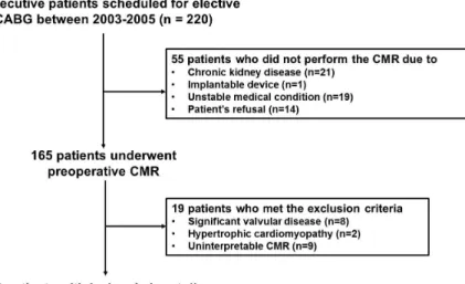
The medical records of 220 consecutive adult patients who underwent CABG between June 2003 and July 2005 at our institution were retrospectively reviewed. One hundred sixty-five patients underwent cardiac MRI to evaluate myocardial viability within 1 month before CABG. Fifty-five patients could not undergo MRI because of acute coronary syndrome with cardiogenic shock or acute pulmonary edema (19 patients), chronic kidney disease (21 patients), cardiovascular implanted electronic device (1 patient), and refusal (14 patients). We excluded patients with hypertrophic cardiomyopathy (two patients), significant valve disease (grade ≥2 stenosis or insufficiency [eight patients]), or uninterpretable MRI (nine patients). The remaining 146 patients formed the study cohort (figure 1). Before undergoing MRI, all patients gave written informed consent. The institutional review board committee approved this retrospective study and waived the need for additional written informed consent.
Figure 1. Composition of the study subjects
Image Acquisition
A 1.5-T MRI system (Intera CV release 10; Philips Healthcare) with five-channel cardiac coils was used. All images were acquired with electrocardiogram-gating and breath-holding. Steady-state free-precession cine-MRI included vertical long-axis images, four-chamber view images, and a set of short-axis images covering the entire LV (figure 2). The sequence parameters were field of view (FOV), 350–400 mm; repetition time/echo time (TR/TE), 3.0–3.6/1.5–1.8 ms; flip angle, 60°; and slice thickness, 8 mm. Fifteen minutes after administering intravenous gadodiamide (0.2 mmol/kg; Omniscan, GE Healthcare Waukesha, WI), an inversion recovery prepared, T1-weighted, gradient-echo sequence was used for LGE-MRI in the same planes as cine-MRI (figure 2). The LGE-MRI parameters were FOV, 350–400 mm; TR/TE, 4.5–4.6/1.3–1.5 ms; flip angle, 15°; inversion time,
200–300 msec; and slice thickness, 8 mm. The inversion time was adjusted to nullify the signal of the normal myocardium.
Figure 2. Cine- and LGE-MRI in a 58-year-old male with 3-vessel coronary artery disease. Cine-MRIs obtained in a. diastole and b. systole demonstrates LV ejection fraction 60%. c. and d. LGE-MRI demonstrate myocardial scar with LGE score of 14 and LGE extent of 10%.
Image Analysis
Imaging data were analyzed using a commercial postprocessing workstation (Mass; Medis, Leiden, the Netherlands). Endocardial and epicardial contours were prescribed manually on short-axis cine-MRI of the LV at end diastole and end systole to obtain the LV volumes, mass, and LVEF. A LVEF <50% indicated LV systolic dysfunction (7). Wall motion abnormality (WMA) on cine-MRI and LGE on contrast-enhanced MRI were assessed in each of 16 segments using the American Heart Association model (8) by the consensus of two observers blinded to patient history and clinical outcome. The WMA was graded visually on a three-level Likert scale (normal contraction = 0; hypokinesia = 1; akinesia = 2). The WMA score was the sum of the WMA grade of each segment. For the semiquantitative analysis of LGE, the maximal transmural extent of LGE was determined on a
five-point Likert scale (absent = 0; 1%–25% = 1; 26%–50% = 2; 51%–75%
= 3; 76%–100% = 4). The LGE score was the sum of the transmurality scores of the 16 segments. A viable myocardium contained four or more viable dysfunctional segments (the transmurality of LGE ≤50%) (3). We also measured the LGE mass by using the full-width at half-maximum technique (9). Summing the LGE mass of all slices yielded the total mass of LGE.
The extent of LGE was expressed as the percentage of the total LV mass.
For statistical analysis, the LGE score and extent were divided into three categories: 0, 1–10, and >10; and 0%, 1%–10%, and >10%, respectively.
Follow-Up
Clinical information was obtained from medical records and telephone interviews. Adverse cardiac events included (1) cardiac death, (2) progressive heart failure requiring hospitalization, (3) new acute MI, and (4) unstable angina requiring hospitalization. In patients experiencing more than one adverse cardiac event, the first event was used. All deaths were classified as cardiac or noncardiac.
Statistical Analysis
All statistical analyses were performed using SPSS version 22 software (IBM Corporation, Somers, NY, USA). Continuous variables were expressed as the mean ± standard deviation and categorical variables as the count and percentage. Student’s t test was used to test for differences in normally distributed continuous variables. The Wilcoxon rank-sum test was used to compare variables that were not normally distributed. Categorical
variables were compared using the chi-square test or Fisher’s exact test, as appropriate. A two-sided value of P < 0.05 was considered statistically significant. Kaplan–Meier curves were used to estimate the distribution of time to adverse cardiac events, based on a LVEF of 50%, the presence of LGE, and the presence of dysfunctional viable myocardium. Between-group differences were evaluated with log-rank statistics. Annualized event rates were calculated by dividing the 11-year Kaplan–Meier event rate by 11. The association of clinical and MRI variables with adverse cardiac events was investigated with the Cox proportional hazard model using univariate and multivariate procedures. The Cox model was used to estimate the risk of given variable as expressed by hazard ratios (HR) with corresponding 95%
confidence intervals (CIs).
RESULTS
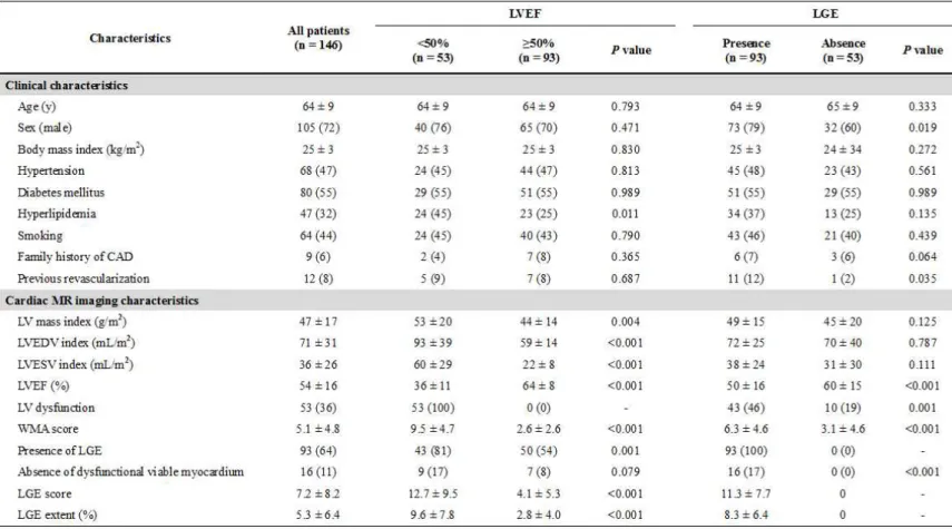
One hundred forty-six patients (105 men; 64 ± 9 years) were available for the final analysis (Table 1). The entire study cohort’s mean LVEF was 54% ± 16%. Fifty-three (36%) patients had LVEF <50% and 96 (64%) patients had LVEF ≥50%. Ninety-three (64%) patients had LGE in the LV myocardium. Patients with LVEF <50% were more likely to have WMA or LGE and a history of hyperlipidemia, and had significantly greater LV volume and mass indices. Patients with LGE were more likely to be male and have a history of previous revascularization, and had significantly more WMA and lower LVEF (Table 1).
At the end of follow-up (median, 9.4 years; interquartile range 8.1–
10.2 years), 44 of 146 patients had adverse cardiac events (Table 2). Eleven patients had noncardiac deaths: malignancy (four patients); pneumonia (three patients); septic shock (two patients); stroke (one patient); and suicide (one patient). These patients were censored at the time of death.
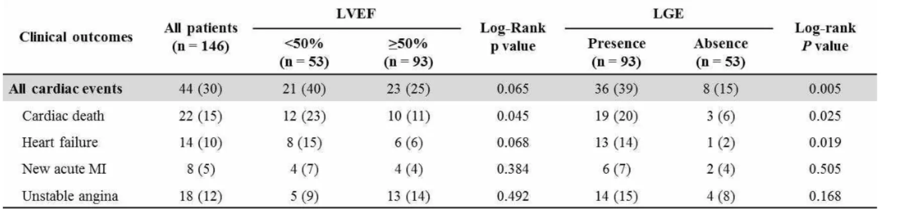
Patients with LVEF <50% had a higher event rate (21 of 53 patients; annualized event rate, 4.5%), compared to patients with LVEF ≥50%
(23 of 96 patients; annualized event rate, 2.5%). The difference was not statistically significant (P = 0.065). Event-free survival curves became parallel after the first 4 years (figure 3a). Patients with LGE had a significantly higher event rate (36 of 93 patients; annualized event rate, 3.9%) compared to patients without LGE (8 of 53 patients; annualized event rate, 1.5%; P = 0.005). The two event-free survival curves diverged over time (figure 3b).
Patients without viable myocardium had a significantly higher event rate (8 of 16 patients; annualized event rate, 5.0%), compared to patients with viable
myocardium (36 of 130, annualized event rate 2.8%). This difference was statistically insignificant (P = 0.072) (figure 3c).
Figure 3. Kaplan-Meier event free survival curves for patients with and without a. LV dysfunction, b. late gadolinium enhancement (LGE) and c.
viability
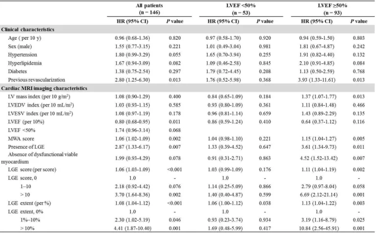
Table 3 summarizes the results of univariate analysis of clinical and MRI variables and adverse cardiac events. In the overall study population, previous revascularization was the only significant clinical predictor (unadjusted HR, 2.80; 95% CI, 1.25-6.30; P = 0.013). The LVEF, WMA score, and LGE presence, score, and extent were significant predictors of adverse cardiac events. A 10% increase in the LVEF was associated with 20% hazard decrease (per 10%; unadjusted HR, 0.80; 95% CI, 0.68-0.95; P
= 0.011); however, LV dysfunction (LVEF <50%) was not significantly associated with adverse cardiac events (unadjusted HR, 0.98; 95% CI, 0.96-3.02; P=0.092). The presence of LGE demonstrated an approximately three-fold hazard increase (unadjusted HR, 2.87; 95% CI, 1.33-6.17; P = 0.007). Further hazard increase occurred in patients with a LGE score >10 (unadjusted HR, 3.70; 95% CI, 1.64-8.36; P = 0.002) and in patients with
LGE extent >10% (unadjusted HR, 4.41; 95% CI, 1.87-10.40; P = 0.001), compared to patients without LGE. However, absence of dysfunctional viable myocardium was not a significant predictor of adverse cardiac events (unadjusted HR, 0.50; 95% CI, 0.23-1.08; P = 0.078).
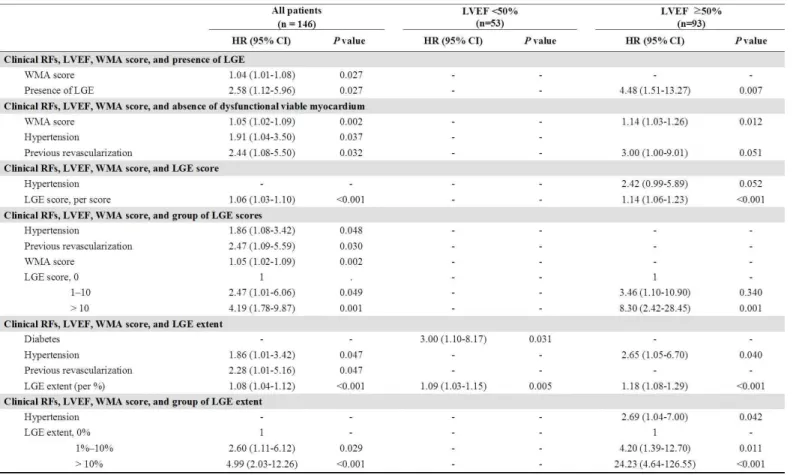
Multivariate analysis included clinical risk factors (age, sex, hypertension, diabetes, and previous history of revascularization), LVEF (per 10%), WMA score, and LGE (Table 4). The LGE presence (adjusted HR, 2.58; 95% CI, 1.12-5.96; P = 0.027), score (per score; adjusted HR 1.06;
95% CI, 1.03-1.10; P<0.001), and extent (per percent; adjusted HR 1.08;
95% CI, 1.04-1.12; P<0.001) were independent predictors of adverse cardiac events; however, the LVEF (per 10%) was not independently associated with adverse cardiac events. Stratifying the LGE score and extent by group showed stepwise increases in hazard for adverse outcome with increasing LGE score (0, 1–10, and >10) and with increasing LGE extent (0%, 1%–
10%, and >10%). However, the absence of dysfunctional viable myocardium was not an independent predictor of adverse cardiac events.
We performed subgroup analysis to evaluate the predictive value of LGE in the presence or absence of LV dysfunction (LVEF <50%). The LGE presence was a significant predictor of adverse cardiac events only in patients with LVEF ≥50% (unadjusted HR, 3.6; 95% CI, 1.34-9.73; P = 0.011), but not in patients with LVEF <50% (unadjusted HR, 1.33; 95% CI, 0.39-4.52;
P=0.647) (Table 3). In patients with LVEF ≥50%, the LGE score and extent
were significant predictors of adverse cardiac events (unadjusted HR, 1.11;
95% CI, 1.04-1.19; P = 0.002, and unadjusted HR, 1.13; 95% CI, 1.04-1.22;
P = 0.003, respectively). LGE score >10 and extent >10% were associated
with further hazard increase (unadjusted HR, 6.69; 95% CI, 2.12-21.14;
P=0.001; and unadjusted HR, 10.84; 95% CI, 2.56-45.91; P = 0.001, respectively). Absence of dysfunctional viable myocardium was associated with hazard increase (unadjusted HR, 4.52; 95% CI, 1.52-13.42; P = 0.007).
In patients with LVEF <50%, only LGE extent were significant predictor of adverse cardiac events (unadjusted HR, 1.06; 95% CI, 1.00-1.12; P = 0.038), whereas LGE presence and LGE score were not. Kaplan–Meier analysis revealed that LGE presence was associated with worse event-free survival in patients with LVEF ≥50% (P = 0.007) but not in patients with LVEF <50%
(P = 0.646). Patients with LVEF ≥50% and with LGE had unfavorable event-free survival curves comparable to those of patients with LV dysfunction (P = 0.731) (figure 4).
Multivariate analysis included clinical risk factors, LVEF, WMA score, and LGE in each group (Table 4). In patients with LVEF ≥50%, LGE Figure 4. Kaplan-Meier event free survival curves, stratified according to the presence of late gadolinium (LGE) in patients with and without LV dysfunction
presence maintained a greater than four-fold hazard increase (adjusted HR, 4.48; 95% CI, 1.51-13.27; P = 0.007), and LGE score and extent were significantly associated with adverse cardiac events (adjusted HR, 1.14; 95%
CI, 1.06-1.23; P < 0.001; and adjusted HR, 1.18; 95% CI, 1.08-1.29; P
<0.001, respectively). In addition, stepwise increases in hazard for adverse outcome occurred with increasing LGE score and extent by group. However, in patients with LVEF <50%, only LGE extent (per percent; adjusted HR, 1.16; 95% CI, 1.00-1.12; P = 0.038) remained as an independent predictor of adverse cardiac events. Absence of dysfunctional viable myocardium was not independently associated with adverse cardiac events in either group.
Table 1. Baseline characteristics
Table 2. Adverse cardiac events
Table 3. Univariate analysis of factors associated with adverse cardiac events
Table 4. Multivariate analysis of factors associated with adverse cardiac events
DISCUSSION
The present study demonstrated (1) LGE presence, score, and extent
—rather than the absence of dysfunctional viable myocardium—were significant independent predictors of adverse cardiac events in patients who undergo CABG; (2) the prognostic power of LGE was alleviated in patients with LV dysfunction; and (3) LGE extent was a strong independent predictor of adverse cardiac events, regardless of LV systolic function.
The LGE presence had a significant prognostic value over clinical risk factors for adverse cardiac events (10-12). In a meta-analysis (4), the presence and extent of a myocardial scar evaluated by LGE-MRI was an independent predictor of adverse cardiac events in patients with suspected or known CAD. However, sparse data exist on the impact of LGE-MRI on long-term prognosis after CABG. We found that LGE presence, score, and extent were significant predictors of adverse cardiac events after CABG—
independent of clinical risk factors, WMA score, and LVEF—during a median 9.4-year follow-up period.
In clinical practice, LGE-MRI is important for risk stratification and for treatment decisions because transmural scar extent detected by LGE-MRI allows the prediction of LV functional recovery after revascularization (5).
Viable myocardium may improve outcome and LV function after revascularization. Identifying viable myocardium by single-photon-emission computed tomography (SPECT), positron emission tomography, and low-dose dobutamine stress echocardiography (DSE) predicts improved survival after CABG (13). However, the multicenter Surgical Treatment for Ischemic Heart Failure (STICH) trial (14), which prospectively and randomly assigned
patients to receive medical therapy alone or medical therapy plus CABG, demonstrated that viable myocardium detected by SPECT or DSE was not independently associated with a greater likelihood of survival. No significant interaction existed between viability status and treatment assignment for mortality and adverse cardiovascular events. The investigators posed an important limitation of the study, which defined viability by combining SPECT and DSE because of the fundamental differences between the two modalities. Gerber et al. (3) evaluated the impact of myocardial viability assessment by LGE-MRI and revascularization therapy on survival in patients with ischemic heart failure. They found that revascularization therapy improved survival in patients with dysfunctional viable myocardium. However, in contrast to previous expectations, survival in the revascularized subgroup was similar, regardless of whether the myocardium was viable. Because both studies exclusively enrolled patients with severe LV systolic dysfunction (LVEF ≤35%), we hypothesized that the impact of LGE-MRI on prognosis after revascularization would be alleviated if LV systolic dysfunction were present. Thus, we performed a subgroup analysis based on the presence of LV systolic dysfunction (LVEF ≤50%), and found that the prognostic power of LGE was alleviated in patients with LV dysfunction. We could not divide our cohort by LVEF of 35% because of the small number of patients with severe LV systolic dysfunction; however, we demonstrated that the absence of dysfunctional viable myocardium and LGE presence and score were significantly associated with adverse cardiac events only in patients with LVEF ≥50% (Table 3). This result suggests that outcome was not worse when LGE was present in patients with LV dysfunction (LVEF <50%), who
already had increased risk in this study population. However, the finding does not indicate that LGE-MRI is useless for patients with LV dysfunction who undergo CABG because LGE extent is a strong independent predictor of adverse cardiac events in patients with and without LV dysfunction.
We also evaluated whether the presence or absence of dysfunctional viable myocardium evaluated by LGE-MRI was associated with the outcome after CABG (3). In concordance with previous research (3), the absence of dysfunctional viable myocardium was not independently associated with an adverse outcome after revascularization in patients with a low LVEF.
Although there was an association with adverse cardiac events in patients with LVEF ≥50%, but statistical significance was not maintained when adjusting for clinical risk factors, LVEF, and WMA score. The finding suggests that a parameter based on scar burden rather than transmural extent was a valuable predictor for adverse cardiac outcome. In our study population, a LGE score based on the transmural extent also failed to independently predict adverse outcome in patients with LVEF <50%, whereas LGE extent was an independent predictor in patients with LVEF <50% and LVEF ≥50%. Further studies are required to confirm our findings.
This study had several limitations. First, our single-center study was limited by the relatively small number of patients, which affects the precision and statistical power of our analyses. Our study population was small;
therefore, we could not evaluate the prognostic value of LGE-MRI in patients with severe LV systolic dysfunction (LVEF <35%), as in previous studies (3).
The results need to be confirmed in a larger patient population. To our knowledge, this is the first study to evaluate the long-term prognostic value
of various parameters from LGE-MRI exclusively in patients who underwent CABG. We demonstrated that the prognostic power of LGE was alleviated in patients with LV dysfunction with LVEF <50%. Second, this study had the limitations inherent to retrospective analysis. After the cardiac MRI results were available, subsequent treatments were at the discretion of the treating physician. In addition, selection bias may limit the generalizability of our results. With such limitations being inevitable in an observational study, the current results do not allow determining whether the physicians’ knowledge of cardiac MRI results may have influenced the prognostic association of LGE to adverse cardiac events.
In conclusion, LGE-MRI can provide long-term prognostic information in patients who undergo CABG. The prognostic power of LGE was alleviated in patients with LV dysfunction, although LGE extent remains a strong independent predictor of adverse cardiac events, regardless of LV function. Our findings suggest that LGE-MRI, which has superior spatial and contrast resolution over other currently available noninvasive imaging tests, may improve the risk stratification of CAD patients who undergo CABG.
REFERENCE
1. Kelle S, Roes SD, Klein C, et al. Prognostic value of myocardial infarct size and contractile reserve using magnetic resonance imaging. J Am Coll Cardiol. 2009;54(19):1770-7.
2. Kwon DH, Halley CM, Carrigan TP, et al. Extent of left ventricular scar predicts outcomes in ischemic cardiomyopathy patients with significantly reduced systolic function: a delayed hyperenhancement cardiac magnetic resonance study. JACC Cardiovasc Imaging. 2009;2(1):34-44.
3. Gerber BL, Rousseau MF, Ahn SA, et al. Prognostic value of myocardial viability by delayed-enhanced magnetic resonance in patients with coronary artery disease and low ejection fraction: impact of revascularization therapy. J Am Coll Cardiol. 2012;59(9):825-35.
4. El Aidi H, Adams A, Moons KG, et al. Cardiac magnetic resonance imaging findings and the risk of cardiovascular events in patients with recent myocardial infarction or suspected or known coronary artery disease: a systematic review of prognostic studies. J Am Coll Cardiol.
2014;63(11):1031-45.
5. Kim RJ, Wu E, Rafael A, et al. The use of contrast-enhanced magnetic resonance imaging to identify reversible myocardial dysfunction. N Engl J Med. 2000;343(20):1445-53.
6. Selvanayagam JB, Kardos A, Francis JM, et al. Value of delayed-enhancement cardiovascular magnetic resonance imaging in predicting myocardial viability after surgical revascularization. Circulation.
2004;110(12):1535-41.
7. Cheong BY, Muthupillai R, Wilson JM, et al. Prognostic significance of delayed-enhancement magnetic resonance imaging: survival of 857 patients with and without left ventricular dysfunction. Circulation.
2009;120(21):2069-76.
8. Cerqueira MD, Weissman NJ, Dilsizian V, et al. Standardized myocardial segmentation and nomenclature for tomographic imaging of the heart. A statement for healthcare professionals from the Cardiac Imaging Committee of the Council on Clinical Cardiology of the American Heart Association. Circulation. 2002;105(4):539-42.
9. Schulz-Menger J, Bluemke DA, Bremerich J, et al. Standardized image interpretation and post processing in cardiovascular magnetic resonance:
Society for Cardiovascular Magnetic Resonance (SCMR) board of trustees task force on standardized post processing. J Cardiovasc Magn Reson.
2013;15:35.
10. Kwong RY, Sattar H, Wu H, et al. Incidence and prognostic implication of unrecognized myocardial scar characterized by cardiac magnetic resonance in diabetic patients without clinical evidence of myocardial infarction. Circulation. 2008;118(10):1011-20.
11. Yoon YE, Kitagawa K, Kato S, et al. Prognostic significance of unrecognized myocardial infarction detected with MR imaging in patients with impaired fasting glucose compared with those with diabetes. Radiology.
2012;262(3):807-15.
12. Kwong RY, Chan AK, Brown KA, et al. Impact of unrecognized myocardial scar detected by cardiac magnetic resonance imaging on event-free survival in patients presenting with signs or symptoms of coronary artery disease. Circulation. 2006;113(23):2733-43.
13. Allman KC, Shaw LJ, Hachamovitch R, Udelson JE. Myocardial viability testing and impact of revascularization on prognosis in patients with coronary artery disease and left ventricular dysfunction: a meta-analysis. J Am Coll Cardiol. 2002;39(7):1151-8.
14. Velazquez EJ, Lee KL, Deja MA, et al. Coronary-artery bypass
surgery in patients with left ventricular dysfunction. N Engl J Med.
2011;364(17):1607-16.
국 문 초 록
서론 본 연구는 관상동맥 우회술을 시행 받은 관상동맥질 환 환자를 대상으로 Gadolinium 지연 조영
MRI가 장기예
후인자로서 갖는 의의에 대한 평가를 하고자 하였다.
방법
2003
년부터2005
년까지 관상동맥 우회술을 받기 위해 내원한 관상동맥질환 환자가 연구대상이 되었으며, 전 체 146명의 환자가 관상동맥 우회술을 받기 전, MRI를 시 행 받았다
.
추적 관찰 중 발생한 심장 위해 사건은 심장 사,
심근경색,
심부전,
불안정형 협심증으로 정의하였으며,
지연 조영의 여부에 따라서 심장 위해 사건의 위험도를 비교하였다. 지연 조영의 정량적 평가를 위해, 지연 조영 의 유무,
지연 조영의 깊이(score),
지연 조영의 양(extent)
으로 나누어 각각의 임상적 의의를 평가하였다.
결과 9.4년의 추적관찰 기간 동안, 44명
(30.1%)
의 환자 가 심장 위해 사건을 경험하였고, 전체 환자군에서 지연 조영의 유무(
보정된 위험비2.58, p=0.027), score (
각 점수 당 보정된 위험비1.06; p=0.007), extent (
각 정도 당 보정 된 위험비1.08; p<0.001)
모두 심장위해사건의 독립적인 위험인자로 확인되었다. 환자군을 좌심실 기능 부전을 기 준을 나누었을 때,
구혈률50%
이상의 좌심실 기능부전이없는 환자에서는 지연 조영의 유무
(
보정된 위험비4.48, p=0.007), score (
각 점수 당 보정된 위험비1.14; p<0.001), extent (각 정도 당 보정된 위험비 1.18; p<0.001)
는 모두 심장 위해사건의 위험인자임이 확인되었으나, 구혈률 50%미만의 좌심실 기능부전이 있는 환자에서는 오직 지연 조 영의
extent (
각 정도당 보정된 위험비1.16; p=0.038)
만이 독립적인 위험인자임을 확인할 수 있었다.결론
Gadolinium
을 이용한 지연 조영의 정성적 정량적 평가는 관상동맥 우회술을 시행받은 환자에게 예후적 정보 를 제공할 수 있다. 지연 조영의 정도는 좌심실 기능부전 의 유무와 상관없이 심장위해사건을 예측할 수 있는 강력 한 예측인자로 확인되었다.
주요어
:
관상동맥질환,
심장MRI,
관상동맥 우회술,
심장 의 생존력학번: