www.ophthalmology.org 387 대한안과학회지 2014년 제 55 권 제 3 호 J Korean Ophthalmol Soc 2014;55(3):387-390 pISSN: 0378-6471 eISSN: 2092-9374 http://dx.doi.org/10.3341/jkos.2014.55.3.387
= 증례보고 =
맥락막 모반의 스펙트럼영역 빛간섭단층촬영 소견:
초음파 소견과의 비교 연구
권 의 용
전북대학교 의학전문대학원 안과학교실
목적: 스펙트럼영역 빛간섭단층촬영기(spectral domain optical coherence tomography; SD-OCT)를 이용하여 맥락막 모반의 특징을 알아보고 초음파 소견과 비교하였다.
대상과 방법: 124명 맥락막 모반 환자의 124안의 의무기록과 enhanced depth imaging (EDI) 방법으로 촬영한 스펙트럼영역 빛간섭단 층촬영 소견과 초음파 검사 소견을 후향적으로 비교하였다.
결과: 124안을 스펙트럼영역 빛간섭단층촬영기의 EDI 방법으로 측정한 결과 43안(35%)에서 분석하기에 좋은 영상 결과를 얻었다. 관찰 되는 특징적 소견은 choroidal shadowing, choriocapillary thinning과 같은 맥락막의 변화가 37안(86%), 망막색소상피의 변화는 27안 (63%), 망막하액은 9안(21%)이었다. 맥락막 모반의 평균 두께는 초음파로 측정했을 때 1295 μm (780-2400 μm), 스펙트럼영역 빛간섭 단층촬영기로 측정했을 때 817 μm (120-1850 μm)였고, 두 측정값의 평균 차이는 475 μm (27-1319 μm)였다(p<0.05).
결론: 스펙트럼영역 빛간섭단층촬영기는 초음파에 비해 맥락막 모반의 두께를 정확하고 얇게 측정하며, 이는 종양의 경과 관찰과 다른 망막 및 맥락막 병변과의 감별진단에 유용하다.
<대한안과학회지 2014;55(3):387-390>
■Received: 2014. 1. 17. ■ Revised: 2014. 1. 28.
■Accepted: 2014. 3. 4.
■Address reprint requests to Eui Yong Kweon, MD
Department of Ophthalmology, Chonbuk National University Hospital, #20 Geonji-ro, Deokjin-gu, Jeonju 561-712, Korea Tel: 82-63-250-1960, Fax: 82-63-250-1960
E-mail: key@jbnu.ac.kr
맥락막 모반은 가장 흔한 안구내 종양 중 하나이며주로 2 mm 이하의 두께이고, 색소 침착을 보이며 드루젠, 망막 부종이나 망막하액을 동반하기도 한다. 대부분 양성이지만 종양이 커질 수도 있고, 아주 드물게 악성으로 변환되기도 한다.1-3빛간섭단층촬영기(optical coherence tomography;
OCT)를 이용하면 임상적인 안저 소견에서 관찰되기 어려 운 망막의 부종, 망막하액, 망막색소상피박리와 같은 소견 을 관찰하기 용이하다. 기존의 시간영역 빛간섭단층촬영기 (time-domain OCT; TD-OCT)는 망막색소상피 아래 조 직에서 빛의 산란으로 인해 해상도가 떨어지기 때문에 망 막색소상피, 맥락막의 전반적인 병변을 관찰하기 어렵다.4 그러나 스펙트럼영역 빛간섭단층촬영기(spectral-domain OCT; SD-OCT)의 enhanced depth imaging (EDI) 측정방 법이 도입되고 맥락막의 단층 이미지를 보다 정확하게 얻 을 수 있게 되면서 맥락막 모반을 비롯한 많은 맥락막 질환 의 해부학적 병태 생리를 이해할 수 있게 되었다. 본 연구 는 맥락막 모반 환자의 EDI SD-OCT 검사 소견에서 맥락
막 모반의 특징을 알아보고 초음파 검사 소견과 비교 연구 하였다.
대상과 방법
2012년 2월부터 2013년 2월까지 미국 콜로라도 망막센터 의 안종양 클리닉에 내원하여 맥락막 모반으로 진단된 124 명 환자의 124안을 대상으로 의무기록과 검사 소견을 분석 하였다. 이 중 EDI SD-OCT 영상 결과가 좋지 않은 79안의 결과는 연구에서 제외하고, 43안을 대상으로 후향적으로비 교 분석하였다. EDI SD-OCT는 Heidelberg Spectralis OCT (Heidelberg Engineering, Heidelberg, Germany)의 custom scan image acquisition protocol of up to 13 ras- ter lines of 9-mm image length를 이용하여 한 명의 검사 자가 촬영하였으며 두 명의 망막 전문의에 의해 분석되었 다. EDI SD-OCT 영상은 Spaide et al5이 기술한 대로 스 펙트럼영역 빛간섭단층촬영기를 눈에 가까이 가져가면 영 상이 뒤집히면서 맥락막이 더 선명하게 관찰되는 방법을 이용하여 얻었다. 맥락막모반의 깊이는 빛간섭단층촬영기 로 autosegmentation된 화면에서 망막색소상피의 하단부 에서 맥락막모반의 안쪽 경계를 정하고, 상대적으로 고반사 를 보이는 공막과의 경계부에서 바깥쪽 경계를 정한 뒤 빛 간섭단층촬영기의 눈금자를 이용하여 측정하였다. 통계학
www.ophthalmology.org 388
- 대한안과학회지 2014년 제 55 권 제 3 호 -
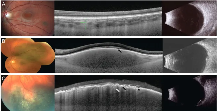
Figure 1. Color fundus photograph (FP), EDI-OCT and BUS of choroidal nevi. (A) FP showing pigmented choroidal nevus.
EDI-OCT showing no drusen, RPE thinning, and thinning of choriocapillaris (CC). (B) FP showing pigmented choroidal nevus.
EDI-OCT showing RPE thinning (black arrow), and normal CC. (C) FP showing pigmented choroidal nevus with overlying drusen.
EDI-OCT showing drusen, trace subretinal fluid (arrowhead), RPE thinning (black arrow), and marked thinning of CC (white arrow).
적 분석은 Statistical Package for the Social Sciences 16.0version (SPSS inc., Chicago, IL)을 이용하여 Student’s ttest로 맥락막 모반의 두께를 비교 분석하였다. p<0.05인 경우를 통계학적으로 유의한 것으로 하였다. EDI-OCT 소 견을 통해 모반의 최대 두께를 측정하였고, 맥락막, 망막과 망막색소상피의 변화를 살펴보았다. 맥락막 모반의 두께를 EDI-OCT와 초음파를 통하여 측정하고 비교하였다.
결 과
43명 43안의 맥락막 모반 환자에서 남자는 23명(53%), 여자는 20명(47%)이었으며, 평균 연령은 58세(35-79 세), 평균 시력은 20/25였다. 맥락막 모반의 EDI SD-OCT 특징적 소견으로 choroidal shadowing, choriocapillary thin- ning과같은 맥락막의 변화가 37안(86%), 망막색소상피의 위축이나 소실과 같은 변화는 27안(63%), 망막하액은 9안 (21%)에서 관찰되었다(Fig. 1).
초음파로 측정된 맥락막 모반의 두께는 1295 μm (780- 2400 μm), EDI-OCT 검사에서는 817 μm (120-1850 μm) 로측정되었다. 두 검사상 측정값의 평균 차이는 475 μm (27-1319 μm)로 맥락막 모반의 두께 측정에 있어서 초음 파가 EDI SD-OCT에 비해 통계학적으로 의미 있게 두껍 게 측정되었다(p<0.05).
고 찰
맥락막 모반은 주로 양성이지만 드물게 커지거나 악성으 로 진행할 가능성이 있기 때문에 종양의 크기를 정확히 측 정하고 크기의 변화를 추적 관찰하며, 망막과 망막색소상피 등 주위 조직에 미치는 영향을 관찰하는 것이 필요하다. SD- OCT의 해상도는 3-4 μm인 반면, TD-OCT는 10 μm,초 음파는 50-200 μm이다.6따라서 TD-OCT를 이용하면 임 상적인 안저 소견에서 관찰되기 어려운 망막, 망막색소상피 등의 변화는 관찰되나, 낮은 해상도로 인해 망막색소상피, 맥락막의 병변을 관찰하기 어렵고 따라서 다른 맥락막 종 양과 감별 진단할 수 있는 유용성이 떨어진다고 알려졌
다.4,7-10그러나 EDI SD-OCT 영상은 단층 이미지를 보다
정확하게 얻을 수 있게 되었고, 망막의 여러 세포층, 망막색 소상피층 및 맥락막모세혈관의 변화 등을 볼 수 있으며, 종 양 자체의 모양과 혈관조직은 물론 주변 조직의 변화에 대 한 정보를 제공할 수 있다. 크기가 작아서 초음파 소견만으 로 진단하기가 어렵거나 크기의 변화를 추적관찰하기 힘든 후극부의 작은 맥락막 종양이나, 맥락막 종양의 활동성을 알 수 있는 주된 예측인자로 보고된 망막하액의 변화 또한 안저 소견이나 초음파보다는 EDI SD-OCT가 진단적 유용 성을 가진다.11-13Shah et al14은 3 mm 이하의 맥락막 모반 에서 최적화된 EDI 이미지를 104안 중 51안(49%)에서 얻
A
B
C
www.ophthalmology.org 389 - 권의용 : 맥락막 모반의 빛간섭단층촬영 소견 -
었다고 보고하며 고령이나 여성 환자인 경우, 병변이 황반 중심으로부터 멀리 떨어진 경우, 크기가 너무 클 때 기술적 으로 최적화된 이미지를 얻기 어렵다고 하였다. 또한 EDI- OCT의 특징적 소견으로 맥락막모세혈관의 위축, 망막색 소상피층, 시세포층의 변화, 망막내 세포층의 변화의 빈 도순으로 보고하였다.14이는 본 연구에서 보고한 choroidal shadowing, choriocapillary thinning과 같은 맥락막의 변화 와 망막색포상피의 이상 및 망막하액이 관찰되는 결과와 부합되었다. 본 연구에서 초음파로 측정된 맥락막 모반의 두께는 1295 μm (780-2400 μm), EDI-OCT 검사에서는 817 μm (120-1850 μm)이었고, 두 검사상 측정값의 평균 차이는 475 μm (27-1319 μm)로 맥락막 모반의 두께 측 정에 있어서 초음파가 EDI SD-OCT에 비해 통계학적으로 의미 있게 두껍게 측정되었으며, 이는 EDI-OCT 측정값에 비하면 평균 58%의 차이를 보인다. 맥락막 흑색종의 초음 파 소견과 조직병리학적 측정을 비교한 보고에 따르면 10%의 경우에서 초음파가 두껍게 측정된다는 보고가 있 다.15 맥락막 모반의 두께와 크기를 측정하는데 있어서 초 음파는 여전히 중요한 역할을 하고 있으나, EDI SD-OCT 를 이용하면 높은 해상도로 인해 종양의 안쪽과 바깥쪽의 경계를 명확히 파악하고 두께를 정확히 측정하게 되어 초 음파의 경우처럼 불가피하게 망막이나 공막이 포함되어 두 껍게 측정되는 경우가 없기 때문에 더 얇고 정확한 결과를 보인다. EDI SD-OCT 검사는 후극부에 가까이 위치하고 2 mm 이하의 비교적 두께가 얇은 맥락막 모반에서 초음파보 다 정확하게 두께를 측정할 수 있으며, 또한 2 mm 이상의 큰 병변일지라도 병변의 위치를 파악하는 데 도움을 줄 뿐 아니라, 망막과 맥락막을 비롯한 여러 주변 조직의 특징적 인 변화에 대한 정보를 제공한다.
결론적으로 EDI SD-OCT는 종양을 진단하고, 두께를 정확히 측정하여 종양의 진행 및 재발을 일찍 발견할 수 있 게 하며, 종양으로 인한 주변 조직에 미치는 영향에 대한 정보를 제공하여 치료 방향을 제시해줄 수 있다.
REFERENCES
1) Ng CH, Wang JJ, Mitchell P, et al. Prevalence and characteristics of choroidal nevi in an Asian vs white population. Arch Ophthalmol 2009;127:314-9.
2) Singh AD, Kalyani P, Topham A. Estimating the risk of malignant transformation of a choroidal nevus. Ophthalmology 2005;112:1784-9.
3) Shields CL, Cater J, Shields JA, et al. Combination of clinical fac- tors predictive of growth of small choroidal melanocytic tumors.
Arch Ophthalmol 2000;118:360-4.
4) Sakata LM, Deleon-Ortega J, Sakata V, Girkin CA. Optical coher- ence tomography of the retina and optic nerve - a review. Clin Experiment Ophthalmol 2009;37:90-9.
5) Spaide RF, Koizumi H, Pozzoni MC. Enhanced depth imaging spectral-domain optical coherence tomography. Am J Ophthalmol 2008;146:496-500.
6) Kiernan DF, Mieler WF, Hariprasad SM. Spectral-domain optical coherence tomography: a comparison of modern high-resolution retinal imaging systems. Am J Ophthalmol 2010;149:18-31.
7) Shields CL, Materin MA, Shields JA. Review of optical coherence tomography for intraocular tumors. Curr Opin Ophthalmol 2005;
16:141-54.
8) Shields CL, Mashayekhi A, Materin MA, et al. Optical coherence tomography of choroidal nevus in 120 patients. Retina 2005;25:
243-52.
9) Muscat S, Parks S, Kemp E, Keating D. Secondary retinal changes associated with choroidal naevi and melanomas documented by optical coherence tomography. Br J Ophthalmol 2004;88:120-4.
10) Schaudig U, Hassenstein A, Bernd A, et al. Limitations of imaging choroidal tumors in vivo by optical coherence tomography.
Graefes Arch Clin Exp Ophthalmol 1998;236:588-92.
11) Shields CL, Furuta M, Berman EL, et al. Choroidal nevus trans- formation into melanoma: analysis of 2514 consecutive cases.
Arch Ophthalmol 2009;127:981-7.
12) Espinoza G, Rosenblatt B, Harbour JW. Optical coherence tomog- raphy in the evaluation of retinal changes associated with suspicious choroidal melanocytic tumors. Am J Ophthalmol 2004;137:90-5.
13) Materin MA, Raducu R, Bianciotto C, Shields CL. Fundus auto- fluorescence and optical coherence tomography findings in choroidal melanocytic lesions. Middle East Afr J Ophthalmol 2010;17:201-6.
14) Shah SU, Kaliki S, Shields CL, et al. Enhanced depth imaging opti- cal coherence tomography of choroidal nevus in 104 cases. Op- hthalmology 2012;119:1066-72.
15) Collaborative Ocular Melanoma Study Group. Comparison of clinical, echographic, and histopathological measurements from eyes with medium-sized choroidal melanoma in the collaborative ocular melanoma study: COMS report no. 21. Arch Ophthalmol 2003;121:1163-71.
www.ophthalmology.org 390
=ABSTRACT=
Enhanced Depth Imaging Optical Coherence Tomography of Choroidal Nevus : Comparison to B-Scan Ultrasonography
Eui Yong Kweon, MD
Department of Ophthalmology, Chonbuk National University Medical School, Jeonju, Korea
Purpose: To evaluate the characteristics of choroidal nevus using the enhanced depth imaging spectral domain optical co- herence tomography (EDI SD-OCT), with a comparison to the B scan ultrasound (BUS) findings.
Methods: Medical records of 124 eyes of 124 choroidal nevus patients were reviewed retrospectively. All patients under- went fundus photography (FP), EDI SD-OCT, and BUS.
Results: Of 124 eyes with choroidal nevus examined by EDI SD-OCT, 43 eyes (35%) displayed good images to study. The most common EDI-OCT imaging features included choroidal shadowing, choriocapillary thinning, retinal pigment epithelial changes, and overlying subretinal fluid. The mean nevus thickness was 817 μm (120-1850 μm) by EDI-OCT compared 1295 μm (780-2400 μm) by BUS. The mean difference in the tumor thickness between two techniques was 475 μm (27-1319 μm) (p < 0.05).
Conclusions: These results have suggested that imaging of choroidal nevus with EDI-OCT shows superior measurement of its characteristics compared with ultrasonography. The clinical utility of this modality is emerging. EDI-OCT is useful in distinguishing suspicious nevi from other chorioretinal lesions, detecting tumor re-growth along the treatment margin, and demonstrating retinal or choroid tumor location.
J Korean Ophthalmol Soc 2014;55(3):387-390
Key Words: Choroidal Nevus, Enhanced Depth Imaging Optical Coherent Tomography, Ultrasonography
Address reprint requests to Eui Yong Kweon, MD
Department of Ophthalmology, Chonbuk National University Hospital
#20 Geonji-ro, Deokjin-gu, Jeonju 561-712, Korea
Tel: 82-63-250-1960, Fax: 82-63-250-1960, E-mail: key@jbnu.ac.kr
- 대한안과학회지 2014년 제 55 권 제 3 호 -