CASE REPORT
IV형 담관 낭종에서 발생한 간내담관암 1예
김석훈, 김형욱, 강대환, 김민대, 이진호, 이재형, 김봉갑, 박종환
부산대학교 의학전문대학원 내과학교실
A Case of Intrahepatic Cholangiocarcinoma Associated with Type IV Choledochal Cyst
Suk Hun Kim, Hyung Wook Kim, Dae Hwan Kang, Min Dae Kim, Jin Ho Lee, Jae Hyung Lee, Bong Gap Kim and Jong Hwan Park Department of Internal Medicine, Pusan National University School of Medicine, Yangsan, Korea
Anomalous union of the pancreaticobiliary duct (AUPBD) is a congenital anomaly that is defined as a junction of the bile duct and pancreatic duct outside the duodenal wall. This anomaly results in a loss of normal sphincteric mechanisms at the pancreaticobiliary junction. As a result, regurgitation of pancreatic juice into the biliary system develops and causes chol- edochal cysts, choledocholithiasis, cholangitis, pancreatitis and malignancy of the biliary tract. Gallbladder cancer or common bile duct cancer associated with AUPBD and choledochal cysts have been frequently reported. But, intrahepatic chol- angiocarcinoma associated with this condition has been only rarely reported. Here, we report a case of intrahepatic chol- angiocarcinoma associated with AUPBD and choledochal cyst. (Korean J Gastroenterol 2012;60:123-127)
Key Words: Anomalous union of pancreaticobiliary duct; Choledochal cyst; Intrahepatic cholangiocarcinoma
Received June 9, 2011. Revised August 19, 2011. Accepted August 19, 2011.
CC This is an open access article distributed under the terms of the Creative Commons Attribution Non-Commercial License (http://creativecommons.org/licenses/
by-nc/3.0) which permits unrestricted non-commercial use, distribution, and reproduction in any medium, provided the original work is properly cited.
교신저자: 김형욱, 626-787, 양산시 물급읍 금오로 20, 양산부산대학교병원 소화기내과
Correspondence to: Hyung Wook Kim, Department of Internal Medicine, Pusan National University Yangsan Hospital, 20 Geumo-ro, Mulgeum-eup, Yangsan 626-787, Korea. Tel: +82-55-360-1534, Fax: +82-55-360-1536, E-mail: [email protected]
Financial support: None. Conflict of interest: None.
서 론
췌담관 합류이상은 십이지장 벽 밖에서 담관과 췌관이 합 류하는 선천성 기형으로, 췌액의 담도내 역류가 발생하여 담 관낭종, 담관염, 담관 결석, 췌장염, 그리고 담낭암, 담관암이 발생하는 것으로 알려져 있다.1 악성 변화에는 담즙과 췌액의 상호 역류가 중요한 인자로 알려져 있고, lysolecithin 등이 세포막에 심한 손상을 주며, 발암과정에 K-ras oncogene과 p53 suppressor gene의 변이가 관여한다고 알려져 있다.2 이 러한 현상은 담즙과 췌액이 지속적으로 저류되는 담낭과 총담 관에 영향을 미쳐 담낭암과 총담관암이 주로 발생하게 되지만 상대적으로 지속적인 저류가 적은 간내담관에서의 담관암 발 생은 드물게 보고되고 있다.3,4 이에 저자들은 췌담관 합류이 상과 담관낭종을 동반한 간내담관암 1예를 경험하여 보고하 는 바이다.
증 례
39세 여자가 4일간의 발열, 우상복부 통증으로 응급실로 왔다. 과거력, 가족력, 사회력에서 특이소견은 없었다. 신체검 사에서 37.7oC의 발열과 복부 압통이 있었고 반발통은 없었 다. 내원 당시 백혈구 5,500/mm3, 혈색소 10.2 g/dL, 혈소판 188,000/mm3이었고, CRP는 5.9 mg/dL로 증가되어 있었다.
생화학검사에서 AST 274 IU/L, ALT 263 IU/L, total bilir- ubin 1.7 mg/dL, direct bilirubin 1.2 mg/dL, ALP 675 U/L, GGT 411 U/L, LDH 570 IU/L로 증가되어 있었고, amylase 221 IU/L, lipase 54 U/L이었다. 종양표지자 검사에서 CEA 는 1.4 ng/mL로 정상범위였지만, CA 19-9은 139.7 U/mL로 증가된 소견이 보였다. 복부 전산화단층촬영에서 양측 간내담 관과 총담관의 확장이 관찰되었고, 좌측 간내담관에서 유두상 돌기형태의 거대 종괴가 관찰되었으며 우측 간내담관에서도
Fig. 1. Abdominal CT findings. Abdominal CT scan showed papillary projected mass (arrow) with ductal dilation in the left intrahepatic duct (A) and ductal dilation of the right intrahepatic duct and the common bile duct (B), right intrahepatic solid mass (arrow) (C). Also abdominal CT showed both kidneys with large, multiple cysts.
역시 작은 유두상 종양이 관찰되었다(Fig. 1). 또한 양측 신장 은 커져있고 많은 수의 낭종이 있었으며, 가족력이 있어 상염 색체 우성 다낭신(autosomal dominant polycystic kidney
에서 저 신호강도, T2 강조영상에서 고 신호강도를 보였으며, 조영 증강 영상에서 점차적인 증강을 나타내었다. 그리고 양 측 신장과 간 내에 다양한 크기의 무수히 많은 낭종들이 관찰 되었다(Fig. 2). 내시경적 역행성 담췌관조영술에서 총담관의 확장이 심해 정확히 판단하기는 힘드나 췌담관의 합류부 이상 으로 췌관이 담관에 유입되는 소견이 관찰되었고, 좌측 간내 담관에 유두상 종괴가 관찰되었다. 총담관의 심한 확장으로 인해 총담관 내에 뚜렷한 종괴를 관찰하기 어려웠고, 간문부 담관의 협착 소견이 의심되었지만 췌담관 합류이상으로 인해 생검 겸자의 진입이 어려워 조직검사는 시행하지 못하였다.
내시경 경비적 담즙배액술을 시행하였고, 배액된 담즙으로 시 행한 검사에서 amylase 27,512 IU/L, lipase 25,340 U/L로 증가되어 있어 췌액의 담관 내로의 역류를 확인할 수 있었다.
유치된 경비적 담즙배액관으로 다음 날 조영술을 시행하였다.
배액관이 총담관 방향으로 이동하여 양측 간내담관의 정확한 관찰이 힘들었지만 우측 간내담관의 작은 유두상 종괴는 관찰 할 수 있었다(Fig. 3). 양전자방출 전산화단층촬영에서 좌측 간내담관에 과대사를 보이는 종괴(SUVmax 14.9)가 관찰되었 고 복부 및 골반 부위의 림프절 전이소견은 관찰되지 않았다 (Fig. 4). 좌측 간내담관에서 초음파 유도 조직생검이 행해졌 고 병리조직검사 결과에서 담관 주위 침윤이 있는 중등도 분 화형 관내 담관형 담관암으로 진단되었다(Fig. 5). 환자는 좌 측 간내담관에 주된 종괴가 있었으나 ERCP, CT와 MRCP에 서 우측 간내담관에서도 종괴가 관찰되어, 근치적 절제가 어 려운 상태로 판단하고 항암치료를 시행하였다. Gemcitabin 과 oxaliplatin 병합요법으로 9차례 항암치료를 받았으며, 10 번째 항암치료 중에 oxaliplatin으로 인한 신경병증으로 약물 을 중단하고, gemcitabin 단독으로 치료를 받았다. 이후에 두 차례 carboplatin과 5-fluorouracil로 항암치료를 받았으나 폐렴이 발생하여 결국 사망하였다.
고 찰
췌담관 합류이상(anomalous union of pancreaticobiliary duct)은 십이지장 벽 밖에서 췌관과 담관의 합류가 이루어지 는 드문 선천성 기형으로서 서양인보다 동양인에서 더 흔하게 발견이 되며, 담관 낭종, 담관염, 담관 결석, 담관암, 췌장염을 유발한다고 알려져 있다.5 이러한 기형은 오디 괄약근 상부에 서 췌관과 담관이 합류하게 되며, 합류방법에 따라 Kimura
Fig. 2. MRCP findings. (A) MRCP showed dilation of the common bile duct and anomalous union of pan- creaticobiliary duct (arrow). (B) The additional small, papillary mass (white arrow) was also seen in the right intra- hepatic duct (IHD) with large projected solid mass (black arrow) in left IHD.
Fig. 3. ERCP findings. (A) It showed fusiform dilation of the common bile duct and papillary mass in the left intrahepatic duct (IHD) (arrow). (B) Endoscopic naso biliary drainage tubography showed small, papillary mass in the right IHD (arrow).
Fig. 4. PET-CT findings. It showed hypermetabolic mass (SUVmax 14.9) in the left intrahepatic duct, suggesting malignant mass.
type의 두 가지로 분류된다. 췌담관형(type 1)은 주췌관이 총 담관으로 합류하게 되고, 담췌관형(type 2)은 총담관이 주췌 관에 합류하는 형태이다.6 진단적 방법으로는 복부 초음파, 전
산화단층촬영, 내시경 역행성 담췌관조영술, 자기공명 췌담관 조영술 등이 있고, 복부 초음파에서 3 mm 이상의 담낭벽의 비후와 8 mm 이상의 담관 확장이 진단에 도움을 줄 수 있 다.6,7 직접적인 췌담관 조영술에서 췌담관 공통관의 길이가 15 mm 이상이면 진단이 가능하지만 공통관의 길이를 정확히 측정하기 어렵고 담관의 확장이 심하거나 췌담관 합류가 복잡 한 경우는 진단이 어려울 수 있다. 이러한 상황에서 가장 도움 이 되는 검사법은 좀더 정확히 해부학적 변화를 관찰할 수 있는 자기공명 췌담관조영술이 될 수 있고, 내시경 경비 담즙 배액술을 시행하여 담관 내 췌장 효소치를 측정하여 비정상적 으로 증가된 소견을 확인하면 담관 내 췌액의 존재를 확인하 게 되므로 진단에 도움을 줄 수 있다.8 이 증례는 내시경 역행 성 담췌관조영술과 자기공명 췌담관조영술을 볼 때 Kimura type 2로 관찰된다. Kimura 등9의 보고에 따르면 췌담관형 (type 1)은 경도의 간외담관 확장과 경한 증상을 보이며, 상대 적으로 고령에서 나타난다. 반면, 담췌관형(type 2)은 비교적 젊은 나이에서 발생하며, 간외담관 확장이 두드러지고 황달, 복통, 복부 종괴가 종종 중한 증상으로 나타난다.
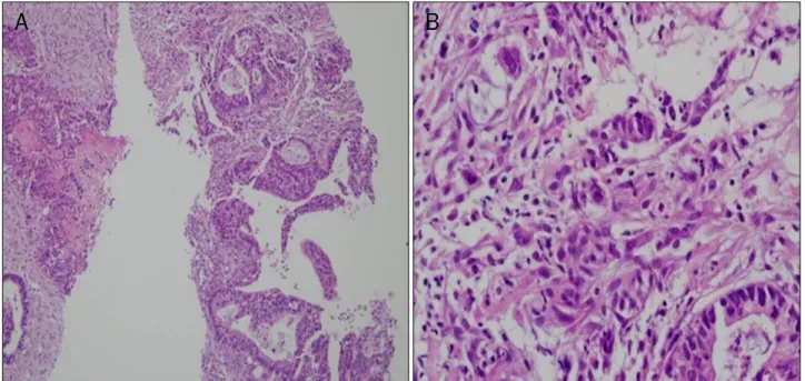
Fig. 5. Moderately differentiated adenocarcinoma was seen (H&E). (A) ×100, (B) ×400.
췌담관 합류이상은 담관 낭종과 깊은 관련이 있는데, 자유 로운 췌액의 담관내 역류로 인해 만성염증이 담관 내에서 지 속적으로 발생하고 점막의 파괴, 섬유화가 초래되어 결국 담 관 내압의 상승을 야기하므로 담관의 낭종성 확장이 발생하게 된다.10 담관 낭종은 Todani 등11에 의해 분류되었는데 1형은 단일로 간외담관에 있는 경우, 2형은 간외 게실이 있는 경우, 3형은 총담관류로 십이지장 벽 내에 간외담관으로 한정된 경 우, 4형은 간외와 간내에 발생한 IV A형과 다발성의 간외 낭 종이 있는 IV B형으로 나누어지고, 5형(Caroli 증후군)은 다 발성의 간내낭종으로 정의된다.11-13 담관 낭종의 유형별로 type I형과 type IV형이 췌담관 합류이상을 가장 많이 동반한 다고 보고되고 있으며, Bakka 등14은 담관 낭종 환자의 40-85%에서 췌담관 합류이상을 동반한다고 보고하였다.
췌담관 합류이상은 담관계암과 연관이 많은데 Sandoh 등15은 담낭암에서 췌담관 합류이상의 빈도가 담관암에서의 빈도보다 훨씬 높다고 보고하였고 특히 췌담관형의 합류이상 이 담낭암으로의 진행을 촉진시킨다고 보고하였다. 기전으로 는 췌장액 내의 trypsin이 phospholipase A2를 활성화시키 게 되고 phospholipase A2는 강력한 점막 파괴 작용을 하게 되며 lecithin을 lysolecithin으로 변화시키게 되는데 이 또한 강력하게 세포 점막에 손상을 주게 된다.16 유전자 변이도 관 계한다고 보고되고 있는데, 췌담관 합류이상이 있는 담낭암 환자에서 췌담관 합류이상이 없는 담낭암 환자에서보다 K-ras 변이와 p-53 변이가 높게 나타났다.17 Hasumi 등18은 담관의 낭종형 확장이 있는 췌담관 합류이상의 환자에서 담관 암의 빈도를 담췌관형에서 7.3%, 췌담관형에서 11.3%로 보고
하였으며, 담관의 낭종형 확장이 없는 췌담관 합류이상의 환 자에서 담관암의 빈도는 담췌관형에서 2.6%, 췌담관형에서 3.2%로 보고하였다. 이번 증례에서는 낭종형의 확장이 있고, 췌담관형으로 빈도가 높은 군에 속하였다.
췌담관 합류이상의 치료는 담낭암을 예방하기 위해 담낭절 제술을 시행하며 총담관 확장을 동반하는 경우는 총담관 절제 및 간공장 문합술을 추가로 시행한다. 일부에서는 췌관 확장 을 동반하는 경우 pylorus preserving pancreaticoduode- nectomy나 Whipple 수술을 주장하지만 좀 더 많은 연구가 필요하다.17
담관 낭종 수술 후에 간내담관암의 발생의 증례가 드물게 보고 되었지만, 본 증례는 췌담관 합류 이상 및 IV형 담관 낭 종을 동반한 간내담관암으로 매우 드문 증례이며 진단 당시 근치적 수술이 어려운 상태로 발견되어 전암성 병변인 췌담관 합류이상의 중요성을 상기시킨다.3,4 이 질환의 조기 진단과 적 절한 치료방법에 대한 더 많은 연구가 필요하다고 생각된다.
REFERENCES
1. Song HK, Kim MH, Myung SJ, et al. Choledochal cyst associated the with anomalous union of pancreaticobiliary duct (AUPBD) has a more grave clinical course than choledochal cyst alone.
Korean J Intern Med 1999;14:1-8.
2. Matsumoto Y, Fujii H, Itakura J, Matsuda M, Nobukawa B, Suda K. Recent advances in pancreaticobiliary maljunction. J Hepato- biliary Pancreat Surg 2002;9:45-54.
3. Shimamura K, Kurosaki I, Sato D, et al. Intrahepatic chol- angiocarcinoma arising 34 years after excision of a type IV-A
congenital choledochal cyst: report of a case. Surg Today 2009;
39:247-251.
4. Goto N, Yasuda I, Uematsu T, et al. Intrahepatic cholangiocar- cinoma arising 10 years after the excision of congenital extra- hepatic biliary dilation. J Gastroenterol 2001;36:856-862.
5. Nomura T, Shirai Y, Wakai T, Yokoyama N, Sakata J, Hatakeyama K. Narrow portion of the terminal choledochus is a cause of up- stream biliary dilatation in patients with anomalous union of the pancreatic and biliary ducts. World J Gastroenterol 2005;11:
6503-6507.
6. Misra SP, Dwivedi M. Pancreaticobiliary ductal union. Gut 1990;31:1144-1149.
7. Kamisawa T, Takuma K, Itokawa F, Itoi T. Endoscopic diagnosis of pancreaticobiliary maljunction. World J Gastrointest Endosc 2011;3:1-5.
8. Min YI, Lee SK, Kim MH, et al. Choledochal cyst and anomalous union of pancreaticobiliary duct in the adult. Korean J Gastroint- est Endosc 1996;16:41-48.
9. Kimura K, Ohto M, Ono T, et al. Congenital cystic dilatation of the common bile duct: relationship to anomalous pancreatico- biliary ductal union. AJR Am J Roentgenol 1977;128:571-577.
10. Babbitt DP, Starshak RJ, Clemett AR. Choledochal cyst: a con- cept of etiology. Am J Roentgenol Radium Ther Nucl Med 1973;119:57-62.
11. Todani T, Watanabe Y, Narusue M, Tabuchi K, Okajima K.
Congenital bile duct cysts: Classification, operative procedures,
and review of thirty-seven cases including cancer arising from choledochal cyst. Am J Surg 1977;134:263-269.
12. Jesudason SR, Govil S, Mathai V, Kuruvilla R, Muthusami JC.
Choledochal cysts in adults. Ann R Coll Surg Engl 1997;79:
410-413.
13. Lee HK, Park SJ, Yi BH, Lee AL, Moon JH, Chang YW. Imaging fea- tures of adult choledochal cysts: a pictorial review. Korean J Radiol 2009;10:71-80.
14. Bakka A, Bergan A, Søreide O. Bile duct cysts in adults. Pitfalls in diagnosis and management. Scand J Gastroenterol 1991;26:
197-206.
15. Sandoh N, Shirai Y, Hatakeyama K. Incidence of anomalous un- ion of the pancreaticobiliary ductal system in biliary cancer.
Hepatogastroenterology 1997;44:1580-1583.
16. Tsuchida A, Itoi T. Carcinogenesis and chemoprevention of bili- ary tract cancer in pancreaticobiliary maljunction. World J Gastrointest Oncol 2010;2:130-135.
17. Matsubara T, Sakurai Y, Zhi LZ, Miura H, Ochiai M, Funabiki T.
K-ras and p53 gene mutations in noncancerous biliary lesions of patients with pancreaticobiliary maljunction. J Hepatobiliary Pancreat Surg 2002;9:312-321.
18. Hasumi A, Matsui H, Sugioka A, et al. Precancerous conditions of biliary tract cancer in patients with pancreaticobiliary mal- junction: reappraisal of nationwide survey in Japan. J Hepatobili- ary Pancreat Surg 2000;7:551-555.