대한소화기학회지 2000;35:400 - 404
14)
서 론
결절성 다발동맥염(polyarteritis nodosa, PAN)은 1866년 Kussmaul과 Maier에 의해 처음 기술된 질환 으로 중, 소 근육형 동맥의 괴사성 혈관염으로 전신 의 장기를 다양하게 침범하는 질환이다.1,2 신장, 심
접수: 1999년 5월 14일, 승인: 1999년 7월 25일 연락처: 윤용범, 110-744, 서울특별시 종로구 연건동 28
서울대학교 의과대학 내과학교실 Tel: (02) 760-3346, Fax: (02) 765-8265
장, 간, 위장관, 근육, 피부, 말초신경, 관절, 뇌혈관, 골, 고환과 자궁 등 거의 전 장기의 혈관에서 발병 가능하며 이들 동맥에서 염증과 섬유소양 괴사 (fibrinoid necrosis)를 일으켜 다양한 임상상을 나타 낸다. 위장관 침범의 빈도는 다양하게 보고되고 있 는데 발열, 복통, 전신 무력감, 구역, 구토 등을 일으 키며 드물게 위장관 출혈이나 천공 같은 중한 합병 증을 일으킨다.1,3 췌담관계의 증례 보고는 대부분이 급성 담낭염에 대한 것이고4-7 이는 국내에서도 보고 된 바 있다.8 그러나 담관이나 췌장을 침범하여 임 상증상을 일으킨 경우는 매우 드물다.9-12 저자들은
총담관협착을 동반한 결절성 다발동맥염 1예
서울대학교 의과대학 내과학교실, 간연구소, 외과학교실*, 병리학교실†
이우진・장유현・김용태・윤용범・김정룡・김선회*・김우호†
A B e n i g n B i li a r y S t r i c t u r e Co m p li c a t i n g Is o la t e d P o ly a rt e r i t i s N o d o s a
Wo o J i n Le e , M.D., Yo o H y u n J a n g , M.D., Yo n g -Ta e Ki m , M.D., Yo n g B u m Yo o n , M .D., Ch u n g Yo n g Ki m , M .D.,
S u n Wh e Ki m , M.D.* a n d Woo H o Kim , M.D.†
Departments of Internal Medicine, Surgery*, and Pathology†, Liver Research Institute, Seoul National University College of Medicine, Seoul, Korea
Polyarteritis nodosa, a disease of the vasculitis of small- and medium-sized arteries, has diverse symptoms and signs. Gastrointestinal involvement is common, but localized involvement of the biliary tree or pancreas is very rare. We report a case of localized necrotizing arteritis involving the arteries around the pancreas and the common bile duct. The patient had jaundice by common bile duct stricture without any pancreatic pathology, and had no underlying abnormal laboratory findings which suggested systemic vasculitis. The arteritis was found incidentally in surgically resected specimens. To our knowledge, only a few cases of localized polyarteritis nodosa causing biliary tree stricture have been reported. It shoud be prudently judged through a long-term follow-up whether this lesion represents an early manifestation of a systemic vasculitis syndrome or a limited form of polyarteritis nodosa. (Kor J Gastroenterol 2000;35:400 - 404)
Key Words: Polyarteritis nodosa, Biliary stricture, Jaundice
이우진 외 6인. 총담관협착을 동반한 결절성 다발동맥염 1예 401
췌장 주위 동맥의 PAN으로 인한 총담관협착 1예를 경험하였기에 문헌 고찰과 함께 보고하는 바이다.
증 례
23세 남자 환자가 내원 1개월 전부터 발생된 황 달을 주소로 내원하였다. 내원 당시 식후 심와부 동 통과 함께 한 달 동안 약 4 kg의 체중 감소 등을 호 소하였으나 발열, 오한 등은 없었으며, 과거력에도 특이 사항은 없었다.
신체검사에서 혈압은 110/60 mmHg, 맥박 64회/
분, 호흡 18회/분, 체온 36.6℃였다. 공막에 황달 소 견이, 복부진찰에서 경도의 우상복부 압통이 관찰되 었다.
검사실 소견으로 말초혈액검사에서 백혈구 5,100/
mm3(호산구 2%), 혈색소 14.8 g/dL, 혈소판 221,000/
mm3, 적혈구침강속도 8 mm/hr이었다. 생화학검사 에서는 총단백 6.6 g/dL, 알부민 3.9 g/dL, 총빌리루 빈 23.4 mg/dL, 직접빌리루빈 10.0 mg/dL, 알칼리성 포스파타제 273 U/L, AST 89 U/L, ALT 123 U/L, γ-GT 50 U/L, 혈중요소질소 9 mg/dL, 크레아티닌 1.1 mg/dL, 총콜레스테롤 134 mg/dL, 공복 혈당 66 mg/dL이었다. 요검사에서는 빌리루빈 3+이었다. B 형 간염바이러스 표면항원 음성, B형 간염바이러스 항체 양성, C형 간염바이러스 항체 음성이었고, C반 응단백 0.2 mg/dL이었으며, 항핵항체(antinuclear antibody, ANA), 류마토이드양 인자, 항중성구세포 질항체(antineutrophil cytoplasmic antibody, ANCA) 모두 음성이었다. 혈청보체는 C3 115 mg/dL, C4 29 mg/dL, 혈청면역글로부린치는 IgG 1,415 mg/dL, IgM 202 mg/dL, IgA 246 mg/dL로 모두 정상 범위 이었다.
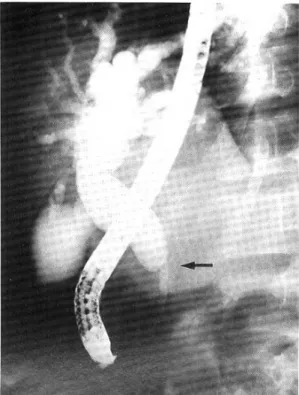
복부 초음파검사와 복부 전산화단층촬영에서 원 위부 총담관의 폐쇄로 폐쇄 상부의 총담관과 양측 간내담관이 심하게 확장되어 있었으나 폐쇄 부위에 종괴는 보이지 않았다(Fig. 1). 내시경적 역행성 담 관조영술에서 원위부 총담관이 갑자기 좁아져 있고 그 상부의 총담관과 양측 간내담관이 심하게 확장 되어 있었다(Fig. 2). 환자의 폐쇄성 황달을 치료하 기 위해 내시경적 경비담관배액술을 시행한 후 협착
의 원인 규명을 위해 시험적 개복술을 시행하였다.
수술시 근위부 총담관 확장, 원위부 총담관의 작은 궤양 및 협착, 부분적 담관벽 비후 등이 관찰되고
Fig. 1. Abdominal CT finding in a patient with common bile duct stricture complicating polyarteritis nodosa.
Abrupt narrowing at the distal common bile duct with proximal dilation (arrow) is observed without definite mass or stone.
Fig. 2. Endoscopic retrograde cholangiographic finding.
Band-like narrowing of the distal common bile duct (arrow) is present.
402 The Korean Journal of Gastroenterology : Vol. 35, No. 3, 2000
담석이나 명확한 종괴 등은 발견되지 않았으나(Fig.
3), 악성 질환을 배제할 수 없어 근치적 절제술인 유 문 보존 췌십이지장절제술(pylorus preserving pan- creaticoduodenectomy, PPPD)을 시행하였다.
병리조직 소견에서 췌장 주변의 중, 소동맥혈관 에 괴사성 염증 소견을 보이나 종양세포는 관찰되 지 않았다. 췌장조직은 정상 소견이었다(Fig. 4).
환자는 수술 후 동통 및 황달이 호전되어 스테로 이드와 cyclophosphamide를 복용하면서 현재까지 외래로 추적관찰 중에 있다.
고 찰
본 증례는 췌장에는 병변이 없이 췌장 주위 동맥 의 국한성 괴사성 혈관염으로 인한 총담관협착으로 황달이 진행하여 내원한 23세 남자의 증례이다.
PAN은 중, 소 근육형 동맥의 혈관염으로 몇몇 질 환과 감별 진단이 필요하다. 현미경적 다발혈관염 (microscopic polyangiitis, MPA)과는 몇 가지 점에서 감별이 되는데, MPA는 중, 소 근육형 동맥을 침범 할 수도 있으나 주로 세동맥, 세정맥, 모세혈관을 침 범하고, PAN에서 관찰되는 신혈관성 고혈압, 신혈 관경색과 미세동맥류(microaneurysm)가 없이 급속 진행성 사구체신염(rapid progressive glomerulone- phritis, RPGN)을 흔히 일으킨다. PAN과 달리 폐출 혈을 일으킬 수 있고, 재발이 더 흔하며 ANCA 양 성률이 더 높은 반면에 B형 간염바이러스 양성률이 낮고 혈관조영술상 미세동맥류가 관찰되지 않는 다.1,2 본 증례는 중, 소 근육형 동맥의 혈관에만 염 증이 관찰되고 더 작은 혈관에는 혈관염이 없어 MPA와 감별이 된다. Churg-Strauss 증후군도 중, 소 혈관의 염증이 관찰되나 천식 및 알레르기성 비염 환자에서 호발하며 말초혈액 호산구증다증과 혈관 내 호산구 침윤이 관찰되므로 PAN과는 감별이 된 다.1,2 본 증례는 PAN의 진단 기준 중에 4 kg의 체 중 감소, 전신 무력감, 조직학적 소견 등 세 가지가 일치하여 정의상 PAN으로 진단내릴 수 있다.1 그 동안 부검 예에서는 PAN의 간 침범은 비교적 흔하 게 보고되고 있으며 담관계 침범은 10-25%로 보고 되고 있고13 췌장의 침범은 35%에서 보고된 바 있 다.14 또한 혈관조영술에서 미세동맥류가 이들의 혈 Fig. 3. Gross finding of the resected specimen after
PPPD operation. An 1.0×0.5 cm sized ulcer crater (arrow) is seen at the narrowed portion of the distal common bile duct.
Fig. 4. Histologic finding of the common bile duct and the peripancreatic artery. (A) The epithelium of the common bile duct is ulcerated and many inflammatory cells are infiltrated in the wall of common bile duct. (B) Necrotizing inflammation is noticed in the medium-sized artery around the pancreas (H&E stain, ×100).
Lee, et al. A Benign Biliary Stricture Complicating Isolated Polyarteritis Nodosa 403
관에서 관찰되는 경우는 많다. 그러나 실제 임상적 으로 증상을 일으켜서 증례가 보고된 것은 급성 담 낭염을 제외하고는 드물다.
담관에 PAN의 침범으로 증례 보고된 것은 매우 드문데, 황달이 발생하였던 60세 남자에서 총담관에 심한 협착과 비후가 관찰되었고 총담관의 협착은 T 자 도관을 설치한 후 담낭절제술을 시행하고 절제 된 담낭과 담낭관에서 PAN과 합당한 조직 소견이 발견된 보고가 있다.9 또한 40년 전에 담낭절제술을 받은 뒤, 황달은 없이 발열과 복통이 발생하였던 80 세 여자에서 좌측 간내 담관에 협착이 발견되어 절 제술과 간-공장문합술을 시행하였을 때, 절제 조직 에서 PAN과 합당한 조직 소견이 관찰되었던 보고10 가 있으며 본 증례와 임상상이 비슷하다. 간외담관 암과 PAN이 연관되어 발생된 증례도 보고되었는데 11 이 경우에는 PAN을 먼저 진단받은 후 황달이 발 생하였고, 수술 소견에서 간외담관암이 확인되어 PAN과 연관성이 있음이 시사되나 PAN이 담관에만 국한된 것이 아니며 또한 담관에서 혈관염이 조직 학적으로 증명된 것도 아니었다.
PAN에 의한 총담관협착의 또다른 원인으로 췌장 병변에 의한 이차적 총담관협착이 초래될 수 있다.
췌장에만 국한된 PAN과 이로 인한 총담관협착은 1 예가 보고되었는데12 황달이 없이 심와부 동통이 발 생하였던 44세 남자에서 췌두부의 부종과 총담관의 심한 협착이 관찰되어 췌장암 의심하에 Whipple 수 술을 하였을 때, 절제된 췌장에서 PAN과 합당한 조 직 소견이 관찰되었다. 본 증례에서처럼 심와부 동 통 이외에는 검사 소견에서 PAN에서 잘 관찰되는 백혈구 증가, 적혈구침강속도나 CRP 증가 및 혈청 면역글로부린치 상승의 소견 등이 관찰되지 않았다.
이는 전신적으로 침범한 PAN과는 달리 국한성 괴 사성 혈관염으로 발현하기 때문이라고 생각된다.
췌장에서 PAN은 부검 예에서 보면 췌장의 혈관 을 침범하여 경색, 미만성 섬유화와 만성 췌장염, 출 혈성 췌장염을 일으킨다.15 임상적으로는 췌장 가성 낭,16,17 췌장 농양,18 급성 췌장염,19 췌장 내 출혈20 등이 보고된 바 있는데 이들은 다른 병변으로 PAN 을 먼저 진단받은 후 췌장의 병변이 다음에 나타난 경우로 췌장에만 침범이 국한된 경우가 아니며 췌
장에서는 혈관염이 조직학적으로 증명된 것도 아니 었다.
PAN은 충수돌기, 담낭, 장, 자궁, 유방, 고환, 부 고환, 신장, 골격 근육, 말초신경계, 피부 등에 국한 성으로 침범할 수 있다. 이 경우 예후가 좋으며 스 테로이드치료에 반응을 잘하고 저절로 호전되기도 한다. 그러나 재발이 흔하고 수 년 후에 전신적으로 진행할 수도 있다. 이렇게 국한성 괴사성 혈관염은 조직학적으로 PAN과 일치하지만 다른 임상상을 보 여 PAN의 초기 임상상인지 아니면 다른 국소성 질 환인지에 대해서는 아직 명확하지 않다.
본 증례에서는 파터 팽대부나 췌장에는 이상 소 견이 없었고 담관궤양과 협착 그리고 췌장 주위에 있는 중, 소동맥에 염증만 관찰되었던 증례이다. 이 들 동맥은 담관조직에도 혈류를 공급하는 혈관으로 생각되며, 담관의 허혈성 괴사로 인해 궤양과 염증 성 부종, 담관벽의 비후로 총담관에 협착이 발생하 여 황달이 발생하였다고 생각된다. 처음에는 총담관 의 악성 종양을 의심하여 근치적 절제술인 PPPD를 시행받았으나 양성 협착이므로 절제술과 간-공장문 합술만으로 완치가 가능했으리라고 생각된다. 그러 나 다시 고찰해 보더라도 악성 종양을 수술 이전이 나 수술 당시에 완전히 배제할 수 있는 진단 방법이 아직 없기 때문에 PPPD는 어쩔 수 없이 시행될 수 밖에 없으리라 여겨진다.
전신적인 PAN의 예후는 스테로이드와 cyclo- phosphamide 병합요법으로 크게 향상되어 B형 간염 과 관련이 없는 PAN의 경우 10년 생존율이 80%에 이르는 것으로 알려져 있다.1 더구나 본 증례처럼 B 형 간염바이러스에 의한 간 손상이 없고 고혈압이 나 신장, 심장, 신경계 등의 침범이 없이 국한성으로 침범한 경우는 추후 재발이나 전신적인 침범이 없 다면 더욱 예후가 좋으리라고 기대된다.
참 고 문 헌
1. Lhote F, Cohen P, Guillevin L. Polyarteritis no- dosa, microscopic angiitis and Churg-Strauss synd- rome. Lupus 1998;7:238-258.
2. Jennette JC, Falk RJ, Andrassy K, et al. Nomen-
404 대한소화기학회지 : 제 35 권 제 3 호 2000
clature of systemic vasculitides. Proposal of an international consensus conference. Arthritis Rheum 1994;37:187-192.
3. Le Thi Huong Du, Wechsler B, Guillevin L, et al.
Digestive manifestations of periarteritis nodosa in a series of 120 cases. Gastroenterol Clin Biol 1985;
9:697-703.
4. Schwartz IS, Mendelow N, Winkler L. Polyarteritis nodosa presenting as acute cholecystitis. Am J Clin Pathol 1966;45:468-471.
5. Remigio P, Zaino E. Polyarteritis of the gallbladder.
Surgery 1970;67:427-431.
6. Bohrod MD, Bodon OR. Isolated polyarteritis no- dosa of the gallbladder. Am Surg 1970;36:681-685.
7. Livolsi VA, Perzid KH, Porter M. Polyarteritis nodosa of the gallbladder, presenting as acute chole- cystitis. Gastroenterology 1973;65:115-123.
8. 조미경, 윤돈영, 함상수 등. 담낭의 결절성 다발 동맥 염 1예. 대한내과학회지 1992;42:826-831.
9. Dillard BM, Black WC. Polyarteritis nodosa of the gallbladder and bile ducts. Am Surg 1970;36:423- 427.
10. Barquist E, Goldstein N, Zinner MJ. Polyarteritis nodosa presenting as a biliary stricture. Surgery 1991;109:16-19.
11. Hatzis GS, Papachristodoulou A, Delladetsima IK, Moutsopoulos HM. Polyarteritis nodosa associated with cholangiocarcinoma. Lupus 1998;7:301-306.
12. Ito M, Sano K, Inaba H, Hotchi M. Localized necrotizing arteritis. A report of a two cases involv-
ing the gallbladder and pancreas. Arch Pathol Lab Med 1991;115:780-783.
13. Rerdbord HE, McCornack LJ, O' Duffy JD. Necro- tizing angiitis: findings at autopsy in twenty-seven cases. Clev Clin Q 1965;32:191-204.
14. Nuzum WJ Jr, Nuzum WJ Sr. Polyarteritis nodosa.
Statistical review of one hundred seventy five cases from literature and report of a typical case. Arch Intern Med 1954;94:942-955.
15. McKay JW, Baggenstoss AH, Wollaeger EE. In- farcts of the pancreas. Gastroenterology 1958;35:
256-264.
16. Bocanegra T, Vasey FB, Espinoza LR, Germain BF. Pancreatic pseudocyst. A complication of necrotizing vasculitis (polyarteritis nodosa). Arch Intern Med 1980;140:1359-1361.
17. Ghosh AK, Sahkuja V, Malik N, Kathuria P, Chugh KS. Pancreatic pseudocyst. A rare complication of polyarteritis nodosa. Dig Dis Sci 1993;38:1347- 1350.
18. Hadary A, Haskel Y, Nissan S. Pancreatic abscess complicating periarteritis nodosa. Am J Gastroen- terol 1986;81:501.
19. Pellegrini CA, Paloyan D, Acosta JM, Skinner DB.
Acute pancreatitis of rare causation. Surg Gynecol Obstet 1977;144:899-902.
20. Jarrousse B, Schlemmer B, Gossot, et al. Periar- teritis nodosa complicated by intrapancreatic vascular rupture. Gastroenterol Clin Biol 1988;12:661-663.