which were successfully treated by curative resection. A 60-year-old male patient with perihilar cholangiocarcinoma was decompressed with single percutaneous transhepatic biliary drainage. Two days after right portal vein embolization, the patient suffered from paralytic ileus with marked abdominal distension. Imaging study revealed that marked fluid collection around the liver and whole abdomen, suggesting intrahepatic bile duct rupture. With abdominal drainage and biliary decompression for 2 weeks, the biliary rupture was controlled. To enhance the safety of right hepatectomy, additional right hepatic vein embolization was performed. The patient underwent routine surgical procedures for right hepatectomy, caudate lobectomy and bile duct resection, and recovered uneventfully and discharged 18 days after surgery. This is the first report of a case of spontaneous rupture of intrahepatic bile duct in a patient with perihilar cholangiocarcinoma. (Korean J Hepatobiliary Pancreat Surg 2013;17:42-47)
Key Words: Intrahepatic bile duct; Liver rupture; Perihilar cholangiocarcinoma; Curative resection
Received: February 10, 2013; Revised: February 21, 2013; Accepted: February 25, 2013 Corresponding author: Shin Hwang
Department of Surgery, Asan Medical Center, University of Ulsan College of Medicine, 388-1, Poongnap-dong, Songpa-gu, Seoul 138-736, Korea Tel: +82-2-3010-3930, Fax: +82-2-3010-6701, E-mail: [email protected]
Copyright Ⓒ 2013 by The Korean Association of Hepato-Biliary-Pancreatic Surgery Korean Journal of Hepato-Biliary-Pancreatic Surgery ∙ ISSN: 1738-6349
INTRODUCTION
Occlusion of perihilar bile duct leads to dilatation of the intrahepatic bile ducts. When slow gradual occlusion happens as like in perihilar cholangiocarcinoma, such di- latation becomes insidious thus showing diffuse dilatation of the involved intrahepatic bile ducts. In contrast, if rapid gradual occlusion is associated with friable liver paren- chyma such as rapidly regenerated remnant livers or parti- al liver grafts following living donor liver transplantation, such pressure increase in the intrahepatic bile duct can in- duce intrahepatic biloma formation.1-4 In any situation, spontaneous rupture of the intrahepatic duct or biloma has not been reported yet in literature.
We herein present a case of spontaneous rupture of in- trahepatic bile duct in a patient with perihilar chol- angiocarcinoma following portal vein embolization, which were successfully treated by curative resection.
CASE
A 60-year-old male patient was referred to our insti- tution under the diagnosis of perihilar cholangiocarcinoma.
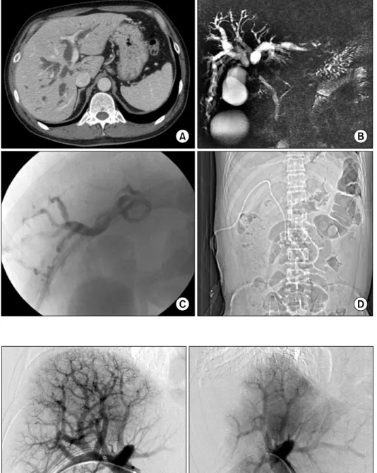
Computed tomography (CT) findings were compatible to those of hilar cholangiocarcinoma Bismuth-Corlette type IIIa with encasement of the right hepatic artery (Fig. 1).
A percutaneous transhepatic biliary drainage (PTBD) tube was inserted into the right hepatic duct, and crossed over the hilar stenotic portion and then passed into the ampulla of Vater for probably simultaneous internal drainage.
Since we were not accustomed to such placement of PTBD tube and total bilirubin level decreased gradually like in the usual perihilar cholangiocarcinoma cases, we did not perform any other procedure for biliary drainage.
In order to induce regeneration of the future remnant left liver, we performed right portal vein embolization (Fig. 2). Two days after this procedure, the patient suf- fered from paralytic ileus with marked abdominal dis-
Fig. 1. Imaging finding of a pa- tient with perihilar cholangiocar- cinoma. (A) Initial computed to- mography image; (B) Initial mag- netic resonance cholangiography;
(C) A percutaneous transhepatic biliary drainage (PTBD) tube was inserted into the right hepatic duct; (D) The PTBD tube was crossed over the hilar stenotic portion and then passed into the ampulla of Vater.
Fig. 2. Preoperative right portal vein embolization with ipsilateral approach (A) and post-emboliza- tion direct portogram (B).
tension (Fig. 3). Emergency CT scan revealed that marked fluid collection around the liver and whole abdomen, thus percutaneous drainage was performed with insertion of a pigtail catheter over the left liver. The pigtail drainage output was bilious in nature, thus leading the diagnosis of intrahepatic bile duct rupture. Follow-up CT scan showed residual fluid collection in the other area of the abdomen, thus another pigtail catheter was inserted into the pelvis. At this time, we recognized that the underlying
cause of intrahepatic bile duct rupture was ineffective drainage of the left hepatic duct with overproduction of bile after right portal vein embolization. Therefore, a PTBD tube was inserted into the left liver (Fig. 4).
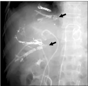
To enhance the safety of right hepatectomy, we per- formed additional right hepatic vein embolization to facil- itate left liver regeneration (Fig. 5).
During laparotomy, we found that the ruptured area was the diaphragmatic side of the segment II because the
Fig. 3. Computed tomography image showing diffuse fluid collection in the abdomen and pelvis. (A) Perihepatic fluid collection;
(B) A percutaneous transhepatic biliary drainage (PTBD) was placed into the right hepatic duct; (C) A large amount of fluid was collected in the pelvis.
Fig. 4. Insertion of a new percutaneous transhepatic biliary drainage tube into the left hepatic duct.
Fig. 5. Embolization of the right hepatic vein and inferior right hepatic veins (arrows).
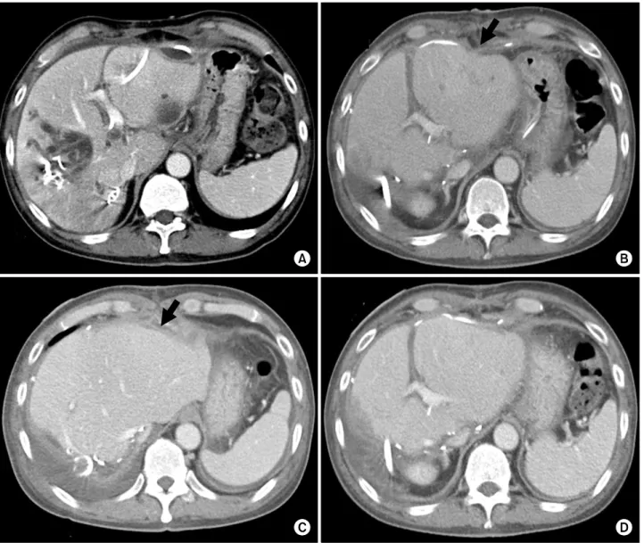
Fig. 6. Perioperative changes of computed tomography (CT) findings. (A) Liver CT taken 2 days before surgery; (B) Liver CT taken 5 days after surgery. An arrow indicates the dimpled site of liver rupture; (C) Liver CT taken 12 days after surgery.
An arrow indicates the dimpled site of liver rupture; (D) Liver CT taken 12 days after surgery shows uneventful regeneration of the remnant left liver.
superficial liver parenchyma at that area was definitely torn out. After confirmation of respectability with limited dissection of the hepatoduodenal ligament, we carried out routine surgical procedures for right hepatectomy, caudate lobectomy and bile duct resection and reconstruction.
Intraabdominal biloma debris and abscess pockets were removed. Multiple abdominal drains were inserted to evacuate residual fluid in the abdomen and pelvis. The pa- tient recovered uneventfully and discharged 18 days after surgery (Fig. 6).
Pathological reports showed that the tumor was a 2.1 cm-sized cholangiocarcinoma with extension to perifi- bromuscular connective tissue and positive lymphovas-
cular invasion (Fig. 7). There was no lymph node metas- tasis. No adjuvant treatment was carried out.
DISCUSSION
The underlying cause of intrahepatic bile duct rupture in this case was ineffective drainage of the left hepatic duct superimposed to overproduction of bile from the left liver following right portal vein embolization. To our knowledge, spontaneous rupture of the intrahepatic duct or biloma at any cause has not been reported yet in literature.
In this case, there was no possibility of iatrogenic intra-
Fig. 7. Gross photograph of the resected right liver and bile duct.
hepatic duct injury because right portal vein embolization was performed through an ipsilateral approach, thus mak- ing no touch to the left liver.
For preoperative evaluation of bile duct involvement in perihilar cholangiocarcinoma, frequent serial surveillance of all dilated intrahepatic ducts is very beneficial for ef- fective biliary decompression. We strongly suggest per- forming dynamic liver CT scan a few days before surgery to finally evaluate the liver status.
Biliary obstruction due to malignancies of the hep- atobiliary system often requires biliary drainage through percutaneous or endoscopic approach. PTBD has been widely used for a long time, but has various drawbacks including procedure-related complications and tumor spread along the PTBD tract, leading to the adoption of endoscopic biliary drainage as the primary method.1-5 Endoscopic biliary drainage including endoscopic na- so-biliary drainage for external drainage and endoscopic retrograde biliary drainage for internal drainage has been shown effective for biliary decompression of distal bile duct cancer, reducing the need for PTBD. In patients with perihilar cholangiocarcinoma, however, endoscopic na- so-biliary drainage is often infeasible or ineffective due to the complexity of the biliary obstruction patterns. Thus, PTBD is still frequently performed in patients with peri- hilar cholangiocarcinoma, with complementary application of endoscopic naso-biliary drainage in patients with less advanced cancers.6,7
ume to total liver volume increased further after hepatic vein embolization without occurrence of any procedure- related complications. We are sure that preoperative se- quential application of sequential portal vein emboliza- tion-ipsilateral hepatic vein embolization is safe and effec- tive in facilitating contralateral liver regeneration by in- ducing more severe liver damage than portal vein emboli- zation alone.8-10
This is the first report of a case of spontaneous rupture of intrahepatic bile duct following portal vein emboliza- tion in a patient with perihilar cholangiocarcinoma.
REFERENCES
1. Takahashi Y, Nagino M, Nishio H, et al. Percutaneous trans- hepatic biliary drainage catheter tract recurrence in cholangio- carcinoma. Br J Surg 2010;97:1860-1866.
2. Maguchi H, Takahashi K, Katanuma A, et al. Preoperative biliary drainage for hilar cholangiocarcinoma. J Hepatobiliary Pancreat Surg 2007;14:441-446.
3. Arakura N, Takayama M, Ozaki Y, et al. Efficacy of preoperative endoscopic nasobiliary drainage for hilar cholangiocarcinoma. J Hepatobiliary Pancreat Surg 2009;16:473-477.
4. Lee SG, Lee YJ, Park KM, et al. One hundred and eleven liver resections for hilar bile duct cancer. J Hepatobiliary Pancreat Surg 2000;7:135-141.
5. Lee SG, Song GW, Hwang S, et al. Surgical treatment of hilar cholangiocarcinoma in the new era: the Asan experience. J Hepa- tobiliary Pancreat Sci 2010;17:476-489.
6. Hwang S, Song GW, Ha TY, et al. Reappraisal of percutaneous transhepatic biliary drainage tract recurrence after resection of perihilar bile duct cancer. World J Surg 2012;36:379-385.
7. Kim TH, Lee SK, Han JH, et al. The role of endoscopic retro- grade cholangiography for biliary stricture after adult living do- nor liver transplantation: technical aspect and outcome. Scand J Gastroenterol 2011;46:188-196.
8. Hwang S, Lee SG, Ko GY, et al. Sequential preoperative ipsi- lateral hepatic vein embolization after portal vein embolization to induce further liver regeneration in patients with hepatobiliary malignancy. Ann Surg 2009;249:608-616.
9. Hwang S. Right hepatectomy in a patient with hepatocellular car- cinoma after induction of hepatic parenchymal atrophy through
subsequent portal and hepatic vein embolizations. Korean J Gastroenterol 2011;58:162-165.
10. Ko GY, Hwang S, Sung KB, et al. Interventional oncology: new options for interstitial treatments and intravascular approaches:
right hepatic vein embolization after right portal vein emboliza- tion for inducing hypertrophy of the future liver remnant. J Hepatobiliary Pancreat Sci 2010;17:410-412.