Korean J Gastroenterol Vol. 61 No. 1, 46-49 http://dx.doi.org/10.4166/kjg.2013.61.1.46 pISSN 1598-9992 eISSN 2233-6869
CASE REPORT
Korean J Gastroenterol, Vol. 61 No. 1, January 2013 www.kjg.or.kr
간세포암에서 담관 침범으로 다량 출혈을 일으킨 혈액담즙증 1예
이경은, 김창욱, 김민주, 박진희, 조규민, 장정원, 이영석, 이창돈
가톨릭대학교 의과대학 내과학교실
Massive Bleeding Hemobilia Occurred in Patient with Hepatocellular Carcinoma
Kyung Eun Lee, Chang Wook Kim, Min Ju Kim, Jinhee Park, Gu Min Cho, Jeong Won Jang, Young Sok Lee and Chang Don Lee Department of Internal Medicine, The Catholic University of Korea College of Medicine, Seoul, Korea
Massive bleeding hemobilia occurs rarely in patients with hepatocellular carcinoma (HCC) without any invasive procedure.
Upper gastrointestinal bleeding in patient with cirrhosis and abdominal pain with progressive jaundice in patient with HCC were usually thought as variceal bleeding and HCC progression respectively. We experienced recently massive bleeding hemobilia in patient with HCC who was a 73-year old man and showed sudden abdominal pain, jaundice and hematochezia. He had alcoholic cirrhosis and history of variceal bleeding. One year ago, he was diagnosed as HCC and treated with transarterial chemoembolization periodically. Sudden right upper abdominal pain occurred then subsided with onset of hemotochezia.
Computed tomography showed bile duct thrombosis spreading in the intrahepatic and extrahepatic ducts, while an ampulla of vater bleeding was observed during duodenoscopy. Hemobilia could be one of the causes of massive bleeding in patients with cirrhosis and HCC especially when they had sudden abdominal pain and abrupt elevation of bilirubin. (Korean J Gastroenterol 2013;61:46-49)
Key Words: Hemobilia; Carcinoma, hepatocellular; Gastrointestinal hemorrhage; Cholestasis
Received February 2, 2012. Revised April 16, 2012. Accepted April 19, 2012.
CC This is an open access article distributed under the terms of the Creative Commons Attribution Non-Commercial License (http://creativecommons.org/licenses/
by-nc/3.0) which permits unrestricted non-commercial use, distribution, and reproduction in any medium, provided the original work is properly cited.
교신저자: 김창욱, 480-717, 의정부시 천보로 271, 가톨릭대학교 의정부성모병원 내과
Correspondence to: Chang Wook Kim, Department of Internal Medicine, The Catholic University of Korea, Uijeongbu St. Mary's Hospital, 271 Cheonbo-ro, Uijeongbu 480-717, Korea. Tel: +82-31-820-3997, Fax: +82-31-847-2719, E-mail: [email protected]
Financial support: None. Conflict of interest: None.
서 론
상부위장관 출혈의 흔한 원인은 소화성 궤양 출혈과 식도 정맥류 출혈 등이고, 혈액담즙증은 매우 드문 원인이다.1 혈액 답즙증은 혈관과 담도계 사이에 연결이 생기면서 상부위장관 출혈을 일으키는 경우로 다른 상부위장관 출혈과 진단 및 치 료에 차이가 있어, 빠른 진단을 위해 반드시 의심을 하는 것이 중요하다. 전형적 임상 삼증후는 담도 산통(biliary colic pain), 폐쇄성 황달, 상부위장관 출혈로서 이와 같은 증상이 있을 경 우 반드시 혈액담즙증을 고려해 보아야 한다.2 혈액담즙증의 원인은 과거에는 외상에 의한 경우가 가장 많았으나 최근에는 의인성(iatrogenic) 원인이 대부분이며,2,3 간세포암의 담관 침 범 및 특별한 시술과 관련없이 발생한 혈액담즙증의 경우는
매우 드물고, 이 중 상부위장의 다량출혈 양상으로 지혈치료 및 수혈이 필요했던 예는 더욱 드물다.
간경변이 있는 경우 상부위장관 출혈의 가장 흔한 원인은 정맥류 출혈이다. 특히, 과거 정맥류 출혈을 진단받은 경우 재출혈의 가능성이 높으므로 정맥류 출혈의 과거력이 있는 간 경변 환자가 상부위장관 출혈로 내원한 경우 정맥류 출혈을 가장 먼저 의심한다. 한편, 간세포암 환자에서 발생하는 황달 과 복통은 대개 간세포암의 진행과 연관된 간기능 이상 및 암성 통증으로 생각한다. 이번 증례는 식도정맥류 출혈 과거 력이 있는 간경변 및 간세포암 환자에서 급성 복통 및 다량의 상부위장관 출혈이 발생한 경우로, 진단 초 암성 통증 및 식도 정맥류 재출혈로 오인되었던 혈액담즙증의 예로서, 간세포암 에서 수술적 치료 또는 시술과 관련없이 다량의 혈액담즙증이
Lee KE, et al. Massive Bleeding Hemobilia by Hepatocellular Carcinoma
47
Vol. 61 No. 1, January 2013
Fig. 1. Abdominal CT scan showed intrahepatic and extrahepatic bile duct thrombosis in the patients with hepatocellular carcinoma and cirrhosis. (A) Contrast enhanced axial CT scans showed the dilated intrahepatic bile duct filled with intraluminal hyperattenuated material, bile duct thrombus (arrows). (B) Contrast enhanced coronal CT scans showed the dilated extahepatic bile duct filled with intraluminal hyperattenuated materials, bile duct thrombus (arrows). (C) Contrast enhanced axial CT scan during arterial phase showed multiple arterial enhancing nodules near the bile duct (arrows).
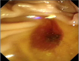
Fig. 2. Bleeding from ampulla of Vater was observed during duoden- oscopy.
발생하는 경우는 매우 드물다. 이에 저자들은 문헌 고찰과 함 께 보고하는 바이다.
증 례
73세 남자 환자가 갑작스럽게 심한 상복부 통증이 있다가 혈변과 흑색변을 보였다. 환자는 과거 알코올성 간경변 및 식 도정맥류 출혈이 있었던 환자로 내원 약 1년 전 간세포암을 진단받고 이후 경동맥 화학색전술을 주기적으로 시행받았다.
내원 약 5개월 전 4차 경동맥 화학색전술을 시행받고 이후 경과 관찰하던 중 내원 전일 갑작스러운 우상복부 동통이 발 생하였으며, 내원일 통증은 호전되었으나 혈변 및 흑색변이 나타나 내원하였다. 내원 당시 고혈압약을 복용하고 있었고, 내원 4개월 전에 동기능부전증후군(sick sinus syndrome)으 로 심박동기를 삽입하였다. 약 50년간 거의 매일 소주 반 병 정도 음주하였으나 약 2년 전부터 금주하였다. 흡연력은 약 10갑년으로 약 2년 전부터 금연하였다.
환자의 키는 153 cm, 몸무게는 55.3 kg이었으며, 내원 당 시 생체징후는 혈압 100/60 mmHg, 맥박 64회/분, 호흡수 20 회/분, 체온 36.0oC였다. 환자는 만성 병색을 보였고 의식은 명료하였다. 두경부에서 공막에 경도의 황달을 보였고 흉부 청진 결과 이상 없었다. 복부는 복수로 팽창된 상태였고 우상 복부에 압통과 반발통이 있었다. 말초혈액검사에서 백혈구 18,100/μL, 혈색소 7.1 gm/dL, 혈소판 41,000/μL이었으며, 생 화학검사에서 혈당 110 mg/dL, 혈중 요소질소 20.7 mg/dL, 혈청 크레아티닌 1.08 mg/dL, 총단백 5.5 g/dL, 알부민 2.5 g/dL, AST 305 U/L, ALT 189 U/L, ALP 805 IU/L, GGT 166 U/L, 총 빌리루빈 10.7 mg/dL, 직접 빌리루빈 6.4 mg/dL, 나트륨 142 mEq/L, 칼륨 4.4 mEq/L, 프로트롬빈 시간은 12.9 초(70.9%, INR 1.21)였다. 직장수지검사에서 흑색변 양성이었
고 내원 전일 시각통증척도(visual analogue scale) 7점 정도 의 간헐적인 우상복부 급경련통(colicky pain)이 있었고 이후 종이컵 4컵 분량의 혈변과 흑색변 소견을 보였다. 혈색소는 7.1 gm/dL로 이전 검사(10.4 gm/dL)보다 감소하였고 총 빌리 루빈은 10.7 mg/dL로 이전 검사(3.99 mg/dL)에 비해 갑자기 증가하였다. 환자는 혈변을 보이면서 우상복부 통증은 사라졌 다고 한다. 식도정맥류 재출혈로 추정하여 시행한 위내시경에 서 식도 및 위 정맥류에서 출혈 병소를 찾지 못하였다. 직장경 에서도 흑색변 소견은 보였으나 출혈 병소는 관찰되지 않았 다. 복부 전산화단층촬영에서 간내담관과 총간관 내부에 전반 적으로 조영전 고밀도 음영의 혈액으로 의심되는 물질이 차있 으면서 확장된 양상을 보였고(Fig. 1A, B), 동맥기 사진에서 간세포암으로 보이는 다발성 조영증강 병변들이 담도 주변에 산재해 있는 것이 확인되었다(Fig. 1C). 간세포암의 간내담도
48
이경은 등. 간세포암에서 담관 침범으로 다량 출혈을 일으킨 혈액담즙증 1예The Korean Journal of Gastroenterology
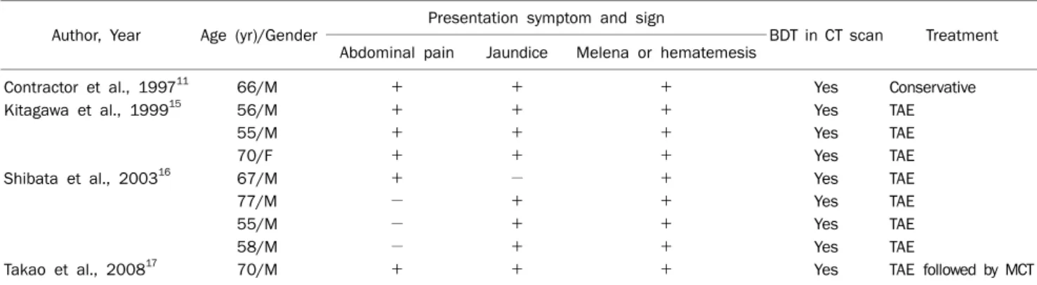
Table 1. Clinical and Radiologic Manifestations of the Patients with HCC and Massive Bleeding Hemobilia
Author, Year Age (yr)/Gender Presentation symptom and sign
BDT in CT scan Treatment Abdominal pain Jaundice Melena or hematemesis
Contractor et al., 199711 66/M + + + Yes Conservative
Kitagawa et al., 199915 56/M + + + Yes TAE
55/M + + + Yes TAE
70/F + + + Yes TAE
Shibata et al., 200316 67/M + − + Yes TAE
77/M − + + Yes TAE
55/M − + + Yes TAE
58/M − + + Yes TAE
Takao et al., 200817 70/M + + + Yes TAE followed by MCT
HCC, hepatocellular carcinoma; BDT, bile duct thrombi; TAE, transcatheter hepatic arterial embolization; MCT, microwave coagulation treat- ment; M, male; F, female.
침범에 의한 혈액담즙증 의심 하에 측시경을 시행하였고 십이 지장 유두부 입구 흡인 시 혈액이 담즙과 혼합된 양상으로 나왔다(Fig. 2). 충전 적혈구, 신선혈장 및 혈소판 수혈 및 보 존적 치료로서 지혈되었으며, 측시경 시 시행한 흡인 이후 황 달 소견도 호전되어 경과 관찰 중이다.
고 찰
1948년 Sandblom4이 처음으로 기술한 혈액담즙증은 혈관 과 담도계 사이의 연결이 생기면서 발생하는 질환으로 전형적 으로 임상 삼증후 소견을 보인다. 즉 담도 산통, 폐쇄성 황달, 상부위장관 출혈이 있을 경우에는 혈액담즙증을 반드시 고려 해야 한다. 혈액담즙증은 드문 질환이지만 전형적인 증상이 있기 때문에 진단하기 위해서는 우선 의심을 하는 것이 무엇 보다 중요하다. 이번 증례에서처럼 전형적인 임상 삼증후 소 견이 다 보이는 경우는 전체 혈액담즙증의 약 22-40%로 보고 되고 있으며, 출혈과 함께 담도 산통은 약 70%, 황달은 약 60%에서 나타난다.2,3 과거에는 외상에 의한 경우가 가장 많 았으나 최근에는 경피경간담도배액술, 간조직검사, 담낭절제 술과 같은 의인성(iatrogenic) 원인이 가장 많고 그 밖에도 드 물게는 담관암과 같은 종양에 의해서도 유발이 된다.2,3
Kojiro 등5은 부검(238예) 및 외과절제(21예)를 시행한 간세 포암을 대상으로 한 연구에서 담도계 침윤이 24예(9.2%)에서 발견되었으며, 이 중 5예(1.9%)에서 혈액담즙증 소견을 보였 고, 이 중 1예(0.4%)에서 다량의 상부위장출혈 양상을 보였음 을 보고하였다. Shibata 등6은 경동맥 색전술을 시행받은 간 세포암 548예 중 4예(0.7%)가 다량의 상부위장출혈 양상을 보이는 혈액담즙증이었음을 보고하였다. 이러한 두 연구가 전 체 간세포암 환자를 대상으로 하지 않고 특정 검사를 시행받 은 환자를 대상으로 한 것을 감안하면 전체 간세포암에서 다 량의 상부위장출혈 양상을 보이는 혈액담즙증의 빈도수는
0.4%나 0.7%보다도 더 낮을 것으로 추정되며, 또한 최근 영 상의학 및 진단기술의 발전으로 보다 초기에 간세포암이 진단 되는 것을 감안하면 다량출혈 혈액담즙증의 빈도수는 더 낮아 질 것이다. 지금까지 전세계적으로 시술과 관련없이 간세포암 에서 발생한 혈액담즙증에 대한 보고는 약 26예7-17가 있었으며 국내 보고는 1예10가 있었다. 이중 상부위장의 다량출혈 양상으 로 지혈치료 및 수혈이 필요했던 예는 모두 9예였다.6,11,15,17 이 들은 대부분 복통, 황달 및 출혈 등 혈액담즙증의 임상 삼증후 소견이 뚜렷하였고, 특히 영상검사에서 모두 담도혈전(bile duct thrombosis) 소견을 나타내었다(Table 1).
간세포암에서 시술과 관련없이 혈액담즙증이 발생하는 이 유는 종양세포가 담관을 침범하고 담도 내로 파열되어 발생하 는 것으로 보고 있다.2,17 간세포암의 담관침범 양상은 세 가지 로 분류되는데, 암이 연속적으로 자라면서 담관을 침윤하는 경우, 이후 종양이 괴사되면서 일부가 근위부 담관에서 떨어 져 나와 원위부로 이동하는 경우, 또 간세포암에서 암세포를 함유한 출혈이 담도를 통해 흘러나가며 담도에 혈액원주 (blood cast)를 형성하는 경우이다.18,19 이 중 다량의 상부위 장관출혈 양상의 혈액담즙증은 마지막 경우로 생각되며, 따라 서 이러한 다량출혈 양상일 경우 담도에 채워진 혈액원주가 담도혈전 소견으로 영상검사에서 나타나는 것이다.
진단은 측시경을 이용하여 십이지장 유두부에서 출혈 소견 이 있는지 보고 내시경 역행성 담도췌관 조영술로 담관 내에 혈병(blood clot) 여부를 볼 수 있다. 복부초음파와 전산화단 층촬영술은 출혈의 증거를 찾는데 유용한 검사방법이며, 출혈 병소를 찾기 위해 Tc-99m 표지 적혈구 스캔(technetium- tagged red blood cell scan)및 선택적 동맥 조영술이 이용될 수 있다. 이번 증례에서는 병소를 찾지 못하는 상부위장관 출 혈에서 전형적인 혈액담즙증 증상을 보였고, 복부 전산화단층 촬영에서 담관 내 혈전 소견이 있어 측시경으로 혈액담즙증을 진단한 경우이다.
Lee KE, et al. Massive Bleeding Hemobilia by Hepatocellular Carcinoma
49
Vol. 61 No. 1, January 2013
혈액담즙증의 치료는 출혈을 멈추고 담관폐쇄를 풀어주는 것이다. 현재는 혈관색전술이 치료의 근간인데, 75%에서 지혈 성공률을 보였으며 반면에 부작용은 적었다.20 혈관색전술 실 패 시 수술적 치료를 고려한다. 이번 환자 경우처럼 간세포암 환자에서 혈액담즙증이 발생하였을 때 효과적인 치료는 정립 되지 않았다. 간절제술이 효과적인 치료이겠으나 거의 대부분 이 진행된 간세포암이기 때문에 수술의 적응증이 되지 못하 며, 선택적 경도관간동맥색전술(selective transcatheter he- patic arterial embolization)이 파열된 간세포암에 의한 출혈 에 유용하다는 증례와15 간기능 저하, 특히 고빌리루빈혈증과 문맥 침범(portal vein invasion) 소견을 갖는 간세포암 환자 에서 경도관간동맥색전술 시행 후 극초단파열치료(microwave ablation)가 혈액담즙증의 재발을 막아줬다는 증례가 있다.17 이번 증례는 식도정맥류 출혈 과거력이 있는 간경변 및 간세 포암 환자에서 발생한 급성 복통 및 다량의 상부위장관 출혈 의 경우로, 이는 정맥류 출혈 및 간세포암 진행에 의한 황달과 복통으로 오인될 수 있지만, 전형적인 임상 삼증후를 보이면 서 다량의 상부위장관 출혈로 수혈이 필요했던 혈액담즙증 환 자였다. 간세포암에서 수술적 치료 또는 시술과 관련없이 다 량의 혈액담즙증이 발생하는 경우는 매우 드물다. 이에 저자 들은 문헌 고찰과 함께 보고하는 바이다.
REFERENCES
1. Dallal HJ, Palmer KR. ABC of the upper gastrointestinal tract:
Upper gastrointestinal haemorrhage. BMJ 2001;323:1115- 1117.
2. Green MH, Duell RM, Johnson CD, Jamieson NV. Haemobilia. Br J Surg 2001;88:773-786.
3. Merrell SW, Schneider PD. Hemobilia--evolution of current diag- nosis and treatment. West J Med 1991;155:621-625.
4. Sandblom P. Hemorrhage into the biliary tract following trauma;
traumatic hemobilia. Surgery 1948;24:571-586.
5. Kojiro M, Kawabata K, Kawano Y, Shirai F, Takemoto N, Nakashi- ma T. Hepatocellular carcinoma presenting as intrabile duct tu- mor growth: a clinicopathologic study of 24 cases. Cancer 1982;
49:2144-2147.
6. Shibata T, Hattori K, Hirai H, Fujii H, Aoyama T, Seuhiro S. Rectus abdominis myocutaneous flap after unsuccessful delayed ster- nal closure. Ann Thorac Surg 2003;76:956-958.
7. Afroudakis A, Bhuta SM, Ranganath KA, Kaplowitz N. Obstruc- tive jaundice caused by hepatocellular carcinoma. Report of three cases. Am J Dig Dis 1978;23:609-617.
8. Cajot O, Descamps C, Navez B, Lacremans D, Druez P. Hemobilia disclosing very small hepatocellular carcinoma ruptured into the biliary ducts. Gastroenterol Clin Biol 1997;21:426-429.
9. Casagrande A, Liscidini P, Pepoli R. Obstructive icterus caused by a biliary thrombus due to hemobilia caused by a hepa- tocarcinoma. Case observation. Minerva Chir 1985;40:571- 576.
10. Cho SJ, Ryu JK, Myung SJ, et al. A case of hepatocellular carcino- ma invading intrahepatic duct complicated by hemobilia.
Korean J Gastrointest Endosc 2005;31:278-281.
11. Contractor QQ, Boujemla M, ul Haque I. Hepatoma cast obstruct- ing common bile duct: not always a terminal event. Indian J Gastroenterol 1997;16:121.
12. Hamy A, Paineau J, Savigny B, et al. Tumoral rupture in the intra- hepatic biliary tracts: a rare manifestation of hepatocellular carcinoma. J Chir (Paris) 1997;134:128-132.
13. Hsu CL, Wang CH, Chen RJ, Chen TC. Hepatocellular carcinoma presenting as jaundice, hemobilia, and acute pancreatitis: a case report. Changgeng Yi Xue Za Zhi 1998:21:232-236.
14. Johns WA, Zimmerman A. Biliary obstruction due to hemobilia caused by liver cell carcinoma. Ann Surg 1961;153:706-710.
15. Kitagawa K, Yamakado K, Nakatsuka A, et al. Selective trans- catheter hepatic arterial chemoembolization for hemobilia from hepatocellular carcinoma: report of three cases. J Vasc Interv Radiol 1999;10:1357-1360.
16. Shibata T, Sagoh T, Maetani Y, et al. Transcatheter arterial emboli- zation for bleeding from bile duct tumor thrombi of hepatocellular carcinoma. Hepatogastroenterology 2003;50:1119-1123.
17. Takao Y, Yoshida H, Mamada Y, Taniai N, Bando K, Tajiri T.
Transcatheter hepatic arterial embolization followed by micro- wave ablation for hemobilia from hepatocellular carcinoma. J Nippon Med Sch 2008;75:284-288.
18. Qin LX, Tang ZY. Hepatocellular carcinoma with obstructive jaun- dice: diagnosis, treatment and prognosis. World J Gastroenterol 2003;9:385-391.
19. Xiangji L, Weifeng T, Bin Y, et al. Surgery of hepatocellular carci- noma complicated with cancer thrombi in bile duct: efficacy for criteria for different therapy modalities. Langenbecks Arch Surg 2009;394:1033-1039.
20. Srivastava DN, Sharma S, Pal S, et al. Transcatheter arterial em- bolization in the management of hemobilia. Abdom Imaging 2006;31:439-448.