Korean J Gastroenterol Vol. 61 No. 1, 50-53 http://dx.doi.org/10.4166/kjg.2013.61.1.50 pISSN 1598-9992 eISSN 2233-6869
CASE REPORT
Korean J Gastroenterol, Vol. 61 No. 1, January 2013 www.kjg.or.kr
총담관으로 전이되어 폐쇄성 황달을 유발한 폐선암
차인혜, 김진남, 김유선, 류수형, 문정섭, 이혜경1
인제대학교 의과대학 서울백병원 내과학교실, 병리학교실1
Metastatic Common Bile Duct Cancer from Pulmonary Adenocarcinoma Presenting as Obstructive Jaundice
In Hye Cha, Jin Nam Kim, You Sun Kim, Soo Hyung Ryu, Jeong Seop Moon and Hye Kyung Lee1
Departments of Internal Medicine and Pathology1, Seoul Paik Hospital, Inje University College of Medicine, Seoul, Korea
We report an extremely rare case of metastatic common bile duct cancer from pulmonary adenocarcinoma presenting as obstructive jaundice. The patient was a 76-year-old male, who presented with generalized weakness and right upper quadrant pain. Plain chest X-ray noted multiple small nodules in both lung fields. Abdominal computed tomography scan showed a stricture of the mid common bile duct along with ductal wall enhancement. Endoscopic retrograde cholangiography revealed a concentric, abrupt narrowing of the mid-common bile duct suggestive of primary bile duct cancer. However, pathology comfirmed metastatic common bile duct cancer arising from pulmonary adenocarcinoma with immunohistochemical study with thyroid transcriptional factor-1 (TTF-1). (Korean J Gastroenterol 2013;61:50-53)
Key Words: Common bile duct neoplasms; Neoplasm metastasis; Lung adenocarcinoma; Obstructive jaundice; TTF-1
Received February 17, 2012. Revised April 2, 2012. Accepted April 3, 2012.
CC This is an open access article distributed under the terms of the Creative Commons Attribution Non-Commercial License (http://creativecommons.org/licenses/
by-nc/3.0) which permits unrestricted non-commercial use, distribution, and reproduction in any medium, provided the original work is properly cited.
교신저자: 김진남, 100-032, 서울시 중구 저동 2가 85번지, 인제대학교 의과대학 서울백병원 소화기내과학교실
Correspondence to: Jin Nam Kim, Division of Gastroenterology, Department of Internal Medicine, Seoul Paik Hospital, Inje University College of Medicine, 85 Jeo-dong 2-ga, Jung-gu, Seoul 100-032, Korea. Tel: +82-2-2270-0012, Fax: +82-2-2270-0257, E-mail: [email protected]
Financial support: None. Conflict of interest: None.
INTRODUCTION
Malignant distal biliary obstruction is mostly caused by pri- mary bile duct cancer or metastasis of other tumors to the peripancreatic, periportal, peribiliary lymph nodes, and head of the pancreas.1 Common malignant tumors that could cause metastatic distal biliary obstruction are stomach can- cer and colon cancer, but lung cancer rarely causes it.1-3 Furthermore, biliary obstruction associated with metastasis to the common bile duct is even rarer in non-biliary cancers, especially in lung cancer.
CASE REPORT
A 76-year-old man presented with generalized weakness, poor oral intake and right upper quadrant pain of 2 months’
duration. He was a never smoker. Since he had a history of Billroth-II operation for stomach cancer 12 years ago, surveil- lance esophagogastroduodenoscopy and screening colono- scopy were performed to rule out gastrointestinal cause of patient’s symptoms. However, there was no evidence of stomach cancer recurrence and only one 1.2 cm sized lateral spreading tumor was found on the cecum, which was re- moved with snare electrocautery. On physical examination, the patient was chronic ill-looking and showed right upper quadrant tenderness. Laboratory evaluation revealed ane-
Cha IH, et al. Metastatic CBD Cancer from Pulmonary Adenocarcinoma Presenting as Obstructive Jaundice 51
Vol. 61 No. 1, January 2013 Fig. 1. Plain chest X-ray. Multiple small nodules were found in both
lung parenchyma.
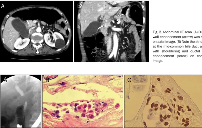
Fig. 2. Abdominal CT scan. (A) Ductal wall enhancement (arrow) was seen on axial image. (B) Note the stricture at the mid-common bile duct along with shouldering and ductal wall enhancement (arrow) on coronal image.
Fig. 3. Imaging and histologic findings of the common bile duct. (A) Endoscopic retrograde cholangiography revealed a concentric, abrupt narrowing of the mid-common bile duct. (B) Biopsy exhibited clusters of malignant columnar cells with prominent nucleoli and intracytoplasmic mucin vacuoles (H&E, ×400). (C) Adenocarcinoma cells demonstrated strong nuclear immunoreactivity for thyroid transcriptional factor-1 (×400).
mia with hemoglobin of 10.5 g/dL (normal 12.0-16.0), ob- structive jaundice with total bilirubin of 6.8 mg/dL (0.2-1.3), direct bilirubin of 4.5 mg/dL (0-0.4), alkaline phosphatase of
970 IU/L (39-117), aspartate aminotransferase of 237 IU/L (0-31), and alanine aminotransferase of 278 IU/L (0-31), and CEA of 10.2 ng/mL (0-5), CA19-9 of 2.7 U/mL (0-37). Multiple small nodules were found in both lung fields on plain chest X-ray (Fig. 1). Abdominal computed tomography scan showed a ductal wall enhancement on cross-sectional view, and a stricture of the mid-common bile duct along with shouldering on coronal view (Fig. 2). Chest computed tomography scan demonstrated obstructive pneumonia caused by lympha- denopathy compressing the bronchus of the left lower lobe additionally. Endoscopic retrograde cholangiography reveal- ed a concentric, abrupt narrowing of the mid-common bile duct suggestive of primary bile duct cancer (Fig. 3A). However, bi- opsy specimen obtained at the strictured bile duct revealed clusters of columnar epithelial cells with immunoreactivity for Cytokeratin 7 and prominent nucleoli with strong im- munoreactivity for thyroid transcriptional factor-1 (TTF-1) (Fig. 3B, C). The morphologic features and this immunohisto- chemical characteristic could make the diagnosis of meta- static adenocarcinoma from the lung. Additionally broncho-
52 차인혜 등. 총담관으로 전이되어 폐쇄성 황달을 유발한 폐선암
The Korean Journal of Gastroenterology
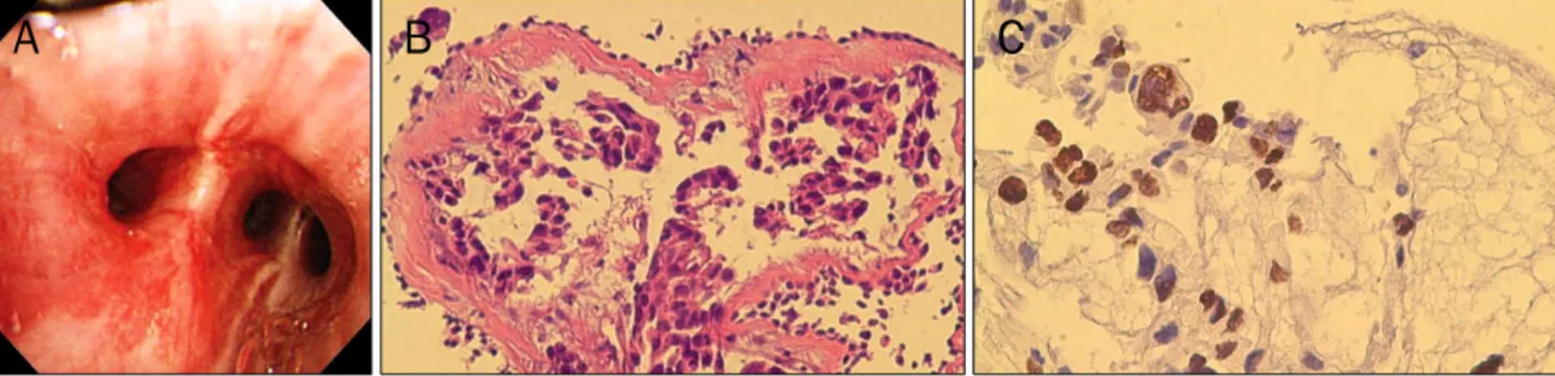
Fig. 4. Bronchoscopic findings and bronchial biopsy specimen. (A) Bronchoscopy showed hyperemic mucosal change, finding suspicious of cancer cell infiltration. (B) Biopsy revealed haphazardly infiltrating nests of adenocarcinoma cells with intracytoplasmic mucin production (H&E,
×200). (C) Adenocarcinoma cells were also reactive for thyroid transcriptional factor-1 (×400).
scopy showed hyperemic mucosal change, finding suspi- cious of cancer cell infiltration (Fig. 4A). Biopsy also showed invasive nests of adenocarcinoma cells, which were strong positive for TTF-1 (Fig. 4B, C). Thus, the diagnosis of meta- static common bile duct cancer from pulmonary ad- enocarcinoma could be made. PET scan revealed metastasis to the bran, L2 lumbar spine, and left supraclavicular lymph node additionally. The patient was referred to the oncologist to undergo systemic chemotherapy after we inserted un-cov- ered biliary metal stent.
DISCUSSION
Patients with primary bile duct cancers typically present with painless jaundice resulting from hilar or distal biliary strictures or with an intrahepatic mass causing abdominal pain. Common non-biliary malignancy that can cause distal biliary obstruction are stomach cancer and colon cancer.
Biliary obstruction caused by lung cancer is rare and mostly due to the compression of the bile duct by metastasis to the surrounding the lymph nodes or to the pancreas head.
More than half of all lung cancer patients present with ad- vanced stage at the time of diagnosis. Extrathoracic meta- stasis is found at autopsy in more than 50% of patients with squamous carcinoma, 80% of patients with adenocarci- noma, and more than 95% of patients with small cell lung cancer. The most frequent site of extrathoracic spread of lung cancer is to the liver and adrenal gland.1 Patients with liver metastases may present with anorexia, weight loss, hep- atomegaly, and right upper quadrant pain. However, liver dys- function or biliary obstructions are rare. Metastasis to com- mon bile duct-induced jaundice is very rare in lung cancer
and has not been reported in pulmonary adenocarcinoma in the English literature so far.
TTF-1 is a 38 kDa homeodomain-containing nuclear pro- tein that plays a role in transcriptional activation during em- bryogenesis in the thyroid, diencephalon, and respiratory epithelium.4-6 TTF-1 has been demonstrated to be expressed specifically in the lung or thyroid neoplasm.7,8 TTF-1 ex- pression varies according to the subtype of lung cancers.
TTF-1 is expressed in 26% to 76% of adenocarcinomas, in 0%
to 38% of squamous cell carcinomas, in 40% of large cell car- cinomas, in 40% to 75% of large cell neuroendocrine carcino- mas, and in 81% to 100% of small cell carcinomas.8-10 Thus, it could serve as a reliable marker of primary lung cancer.11 Roh and Hong12 reported that TTF-1 was expressed in 69%
of metastatic lung cancers in the cervical lymph nodes and had a specificity of 95% and a sensitivity of 69% for meta- static lung cancer.
When plain chest X-ray reveals multiple lung nodules and abdominal computed tomography scan shows solitary com- mon bile duct stricture with shouldering and ductal wall en- hancement without evidence of other intra-abdominal meta- stasis as is with the present case, it is necessary to differ- entiate between double primary cancer and common bile duct cancer with multiple lung metastasis. To make the diag- nosis in our case, biopsy was performed at the strictured bile duct, but histopathology revealed adenocarcinoma cells with no biliary differentiation, i.e., no immunoreactivity for CA19-9.
Since multiple pulmonary nodules were present, immunohis- tochemical study with TTF-1 was performed and it revealed strong positivity. Bronchoscopic biopsy also showed ad- enocarcinoma cells with strong immunoreactivity for TTF-1.
Thus, we could confirm the diagnosis of metastatic common
Cha IH, et al. Metastatic CBD Cancer from Pulmonary Adenocarcinoma Presenting as Obstructive Jaundice 53
Vol. 61 No. 1, January 2013
bile duct cancer arising from pulmonary adenocarcinoma.
Considering the patient’s age, generalized condition, and ad- vanced stage of malignancy irrespective of primary site, im- munohistochemical study for localizing primary origin of bile duct stricture might not affect treatment modality and prog- nosis of the patient. However, If treatment modality such as surgery improves prognosis according to the bile duct histol- ogy, we should make every effort to identify the primary origin in patient with bile duct stricture and other lesion including lung. In summary, when it is necessary to differentiate be- tween primary bile duct cancer and metastatic bile duct can- cer from pulmonary adenocarcinoma, immunohistochem- ical study of the biopsy samples from bile duct with TTF-1 can be useful in differential diagnosis.
REFERENCES
1. Smith HJ. Extrahepatic bile duct obstruction in primary carcino- ma of the lung: incidence, diagnosis, and non-operative treatment. J Natl Med Assoc 1980;72:215-220.
2. Moon SG, Han JK, Kim TK, Kim AY, Kim TJ, Choi BI. Biliary ob- struction in metastatic disease: thin-section helical CT findings.
Abdom Imaging 2003;28:45-52.
3. Lee YJ, Kim SH, Lee JY, et al. Differential CT features of intra- ductal biliary metastasis and double primary intraductal poly- poid cholangiocarcinoma in patients with a history of extra- biliary malignancy. AJR Am J Roentgenol 2009;193:1061- 1069.
4. Guazzi S, Price M, De Felice M, Damante G, Mattei MG, Di Lauro R. Thyroid nuclear factor 1 (TTF-1) contains a homeodomain and displays a novel DNA binding specificity. EMBO J 1990;9:3631- 3639.
5. Lazzaro D, Price M, de Felice M, Di Lauro R. The transcription fac- tor TTF-1 is expressed at the onset of thyroid and lung morpho- genesis and in restricted regions of the foetal brain. Develop- ment 1991;113:1093-1104.
6. Bohinski RJ, Huffman JA, Whitsett JA, Lattier DL. Cis-active ele- ments controlling lung cell-specific expression of human pulmo- nary surfactant protein B gene. J Biol Chem 1993;268:11160- 11166.
7. Fabbro D, Di Loreto C, Beltrami CA, Belfiore A, Di Lauro R, Damante G. Expression of thyroid-specific transcription factors TTF-1 and PAX-8 in human thyroid neoplasms. Cancer Res 1994;
54:4744-4749.
8. Fabbro D, Di Loreto C, Stamerra O, Beltrami CA, Lonigro R, Damante G. TTF-1 gene expression in human lung tumours. Eur J Cancer 1996;32A:512-517.
9. Folpe AL, Gown AM, Lamps LW, et al. Thyroid transcription fac- tor-1: immunohistochemical evaluation in pulmonary neuro- endocrine tumors. Mod Pathol 1999;12:5-8.
10. Kaufmann O, Dietel M. Expression of thyroid transcription fac- tor-1 in pulmonary and extrapulmonary small cell carcinomas and other neuroendocrine carcinomas of various primary sites.
Histopathology 2000;36:415-420.
11. Dennis JL, Hvidsten TR, Wit EC, et al. Markers of adeno- carcinoma characteristic of the site of origin: development of a diagnostic algorithm. Clin Cancer Res 2005;11:3766-3772.
12. Roh MS, Hong SH. Utility of thyroid transcription factor-1 and cy- tokeratin 20 in identifying the origin of metastatic carcinomas of cervical lymph nodes. J Korean Med Sci 2002;17:512-517.