CASE REPORT
내시경역행담췌관조영술 후 발생한 복강 가성동맥류 파열
박태영, 류수형, 문정섭
인제대학교 서울백병원 내과
Visceral Artery Pseudoaneurysm Rupture after Endoscopic Retrograde Cholangiopancreatography
Tae Young Park, Soo Hyung Ryu and Jeong Seop Moon
Department of Internal Medicine, Inje University Seoul Paik Hospital, Seoul, Korea
A visceral artery pseudoaneurysm after ERCP is a rare adverse event that is potentially life-threatening. Most cases reported pre- viously originated from the peripancreatic arteries, including the splenic artery, gastroduodenal artery, or pancreaticoduodenal artery. The mechanism of the occurrence of visceral artery pseudoaneurysms after ERCP has not been elucidated until now.
Recently, a pseudoaneurysm rupture originating from the superior mesenteric artery after ERCP was observed in a patient with- out a history of pancreatitis. This paper reports this case with a review of the relevant literature. (Korean J Gastroenterol 2020;75:162-166)
Key Words: Endoscopic retrograde cholangiopancreatography; Pseudoaneurysm; Rupture
Received October 2, 2019. Revised December 4, 2019. Accepted January 5, 2020.
CC This is an open access article distributed under the terms of the Creative Commons Attribution Non-Commercial License (http://creativecommons.org/licenses/
by-nc/4.0) which permits unrestricted non-commercial use, distribution, and reproduction in any medium, provided the original work is properly cited.
Copyright © 2020. Korean Society of Gastroenterology.
교신저자: 박태영, 04551, 서울시 중구 마른내로 9, 인제대학교 서울백병원 내과
Correspondence to: Tae Young Park, Department of Internal Medicine, Inje University Seoul Paik Hospital, 9 Mareunnae-ro, Jung-gu, Seoul 04551, Korea. Tel:
+82-2-2270-0001, Fax: +82-2-2170-0001, E-mail: [email protected], ORCID: https://orcid.org/0000-0002-3767-504X Financial support: None. Conflict of interest: None.
INTRODUCTION
A visceral artery pseudoaneurysm is a rare vascular compli- cation caused by peripancreatic vascular erosion or vascular wall component disruption induced by pancreatic enzymes from acute or chronic pancreatitis, necrotizing pancreatitis, pancreatic tumor, vascular anomaly, and trauma.1 To date, most reported visceral artery pseudoaneurysms have been associated with acute, chronic, or necrotizing pancreatitis.1,2 The splenic artery, gastroduodenal artery, and pan- creaticoduodenal artery are frequently reported locations of pseudoaneurysms.3 A visceral artery pseudoaneurysm after ERCP without pancreatitis is extremely rare.4 A pseudoaneur- ysm after ERCP usually occurs in the gastroduodenal artery
or pancreaticoduodenal artery, which supplies blood to the major papillary area. The rupture of a pseudoaneurysm from the pancreaticoduodenal artery or gastroduodenal artery is potentially life-threatening.5,6 This paper reports a case of pseudoaneurysm rupture originating from the superior mes- enteric artery (SMA), which is a very rare location, with a re- view of the literature.
CASE REPORT
A 55-year-old man presented with severe abdominal pain and visited the emergency room. He had a prior medical his- tory of diabetes that had not been managed. He was a current smoker and a social drinker. The abdominal pain was asso-
Fig. 1. Diffuse GB wall edema with pericholecystic fluid collection and subtle calcification in the CBD were noted in the abdominal CT scan. GB, gallbladder; CBD, common bile duct; CT, computed tomography.
A B
Fig. 2. Endoscopic retrograde cholangiopancreatography. (A) A filling defect was noted in the distal CBD during cholangiography. (B) After EST, a round stone was removed using a retrieval balloon catheter. CBD, common bile duct; EST, endoscopic sphincterotomy.
ciated with fever and chills. An examination revealed a body temperature of 38.3℃ with his other vital signs unremarkable.
Tenderness was noted in the epigastric area, but there was no rebound tenderness. Icteric sclera was noted.
The laboratory findings revealed the following: leukocytosis of 23,220/mm3, hemoglobin level of 14.7 g/dL, total bilirubin of 8.0 mg/dL, an AST level of 316 IU/L, ALT level of 350 IU/L, ALP level of 317 IU/L, GGT level of 1,191 IU/L, and hemoglo- bin A1c level of 8.6%. An abdominal CT scan revealed diffuse gallbladder wall edema with pericholecystic fluid collection and subtle calcification in the distal common bile duct (CBD) (Fig. 1). MRCP failed due to claustrophobia.
Intravenous third-generation cephalosporin was administered
to control the acute cholangitis, and therapeutic ERCP was performed for CBD stone removal and biliary drainage. Written informed consent for ERCP was obtained from the patient before the procedure. ERCP was performed under conscious sedation using midazolam and propofol. After selective cannu- lation of the CBD, the suppurative discharge was drained through the papilla, but the patient showed a severe para- doxical response. Therefore, the procedure was stopped, and the endoscope was withdrawn for the safety of the patient.
After stabilizing the paradoxical activity, ERCP was performed without sedation. On cholangiography, a filling defect was not- ed in the distal CBD (Fig. 2A). The endoscopic sphincterotomy (EST) was performed until the hooding fold of the ampulla of vater using a pull type sphincterotome (UltratomeTM XL;
Boston Scientific Co., Natick, MA, USA). The mode of electro- cautery used in EST was ENDO CUT I mode (upmax, 550 Vp;
effect, 2; cut duration, 2; cut interval, 2) (ERBE®; Erbe Elektromedizin GmbH, Tuebingen, Germany). A papillary bal- loon dilator or papillary large balloon dilator was not used in sphincter management. After EST, a round-shaped stone was removed using a retrieval balloon catheter (Fig. 2B). Six Fr endoscopic nasobiliary drainage was then performed.
After ERCP, there was no evidence of adverse events, such as pancreatitis, bleeding, or bowel perforation. After 1 week, the liver function test result normalized, and the patient was transferred to the department of general surgery for a laparo- scopic cholecystectomy. On the day of the elective operation, the patient complained of sudden abdominal distension and dizziness before the operation, and the procedure was cancelled. The vital signs showed 80/60 mmHg, and the he-
Fig. 3. 2.5 cm sized heart-shaped contrast leakage was noted inside the hematoma on enhanced abdominal CT. CT, computed tomo- graphy.
Fig. 5. No contrast leakage from the embolized first jejunal branch of the SMA was observed on enhanced abdominal CT. SMA, superior mesenteric artery; CT, computed tomography.
Fig. 4. Contrast leakage (arrowhead) from the right side wall of the first jejunal branch originating from the SMA was noted on angiography. SMA, superior mesenteric artery.
moglobin level was 7.5 g/dL. No symptoms of gastrointestinal bleeding, such as hematemesis, melena, or hematochezia, were noted. On an abdomen CT scan, a 12×9 cm sized well-capsulated hematoma was observed in the retroperitoneal area, and 2.5 cm heart-shaped contrast leakage was noted inside the hematoma (Fig. 3). As a result, the rupture of a visceral artery pseudoaneurysm was diagnosed. Angiography was performed to evaluate and manage the pseudoaneurysm.
On angiography, contrast leakage was confirmed at the right side wall of the first jejunal branch originating from the SMA, and embolization was performed using 0.5 mL of Histoacryl®
(N-butyl-2-cyanoacrylate; Braun, Melsungen, Germany) (Fig. 4).
Four weeks after angioembolization, the size of the retro- peritoneal hematoma was decreased to 8×7 cm, and the Histoacryl® (N-butyl-2-cyanoacrylate; Braun) and lipiodol ag- glomerate were noted inside the pseudoaneurysm. No con- trast leakage from the embolized first jejunal branch of the SMA was noted on enhanced abdominal CT (Fig. 5). The pa- tient was discharged without additional adverse events. The Institutional Review Board of the Inje University Seoul Paik Hospital approved this case report (IRB No. PAIK 2019-02-002).
DISCUSSION
Visceral artery pseudoaneurysm can be caused by vascular wall damage from pancreatitis, malignancy, vascular malfor- mation, or trauma.1,2 A visceral artery pseudoaneurysm is a rare adverse event, but it can be potentially life-threatening.
A visceral artery pseudoaneurysm is clinically associated with pancreatitis, and most cases originate from the peripancreatic arteries, including the splenic artery, gastroduodenal artery, or pancreaticoduodenal artery.3 The mortality of visceral ar- tery pseudoaneurysm rupture has been reported to be 20%
in cases caused by chronic pancreatitis and up to 60% in acute pancreatitis.7 The mortality can be higher in the rupture of pseudoaneurysms originating from the pancreaticoduodenal artery than in the rupture of pseudoaneurysms originating from the splenic artery or gastroduodenal artery.7
Among cases previously reported, a visceral artery pseu- doaneurysm after ERCP without the association of acute or
Table 1. Clinical Features of Visceral Artery Pseudoaneurysm Rupture after ERCP
Study Sex/
age
Underlying
disease Anatomy Indication for ERCP
Details of procedure
Post-ERCP pancreatitis
Location of
pseudoaneurysm Treatment Al-Jeroudi et al.
(2001)8
F/76 None Normal Pancreas cancer Precut sphinc- terotomy
No Pancreaticoduod- enal artery
Embolization
Gaduputi et al.
(2013)4
M/74 Chronic hep- atitis C
Billroth II gas- trectomy
CBD stone Unintended PD cannulation
Yes Gastroduodenal artery
Embolization
Kurita et al.
(2015)5
F/71 Chronic re- nal failure
Normal CBD stone EPLBD No Gastroduodenal
artery
Embolization
Priya et al. (2016)6 M/64 None Normal Acute cholangitis with CBD stone
EST, plastic stent
No Gastroduodenal artery
Embolization
M/72 None Normal Periampullary
mass with jaun- dice
EST, plastic stent
No Gastroduodenal artery
Embolization
Mohapatra et al.
(2017)11
M/53 None Normal Acute cholangitis with CBD stone
EST, plastic stent
No SMA Embolization
El Hajj et al.
(2017)10
F/55 None Normal Acute cholangitis with CBD stone
EST, plastic stent
No Right hepatic ar- tery
Stent graft
Ding et al. (2017)9 M/56 Chronic hep- atitis B
Liver trans- plantation
CBD stricture EST, plastic stent
No Left hepatic artery Embolization
Current case M/55 DM Normal Acute cholangitis
with CBD stone
EST, ENBD No SMA Embolization
ERCP, endoscopic retrograde cholangiopancreatography; F, female; M, male; CBD, common bile duct; PD, pancreatic duct; EPLBD, endoscopic papillary large balloon dilation; EST, endoscopic sphincterotomy; SMA, superior mesenteric artery; DM, diabetes mellitus; ENBD, endoscopic nasobiliary drainage.
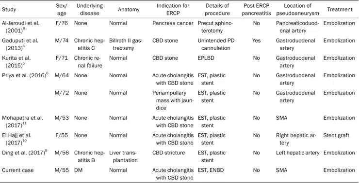
chronic pancreatitis is very rare. Until now, the mechanism of occurrence of visceral artery pseudoaneurysms after ERCP has not been investigated. Several hypotheses have been pro- posed, including direct mechanical injury from the accessories or endoscope, ischemic injury from electric stimulus, and sec- ondary injury from pancreatitis.4,6 The rupture of a visceral artery pseudoaneurysm after ERCP occurs in diverse patients with a range of characteristics, such as gender; middle to advanced age; surgically altered anatomy, including Billroth II gastrectomy and liver transplantation; association with pan- creatitis; sphincter therapy, including EST; endoscopic papil- lary large balloon dilation; and precut sphincterotomy.4-6,8-11 In most cases, fatal hemorrhage occurred, and the clinical presentations were diverse, including gastrointestinal bleed- ing, hemobilia, intra-abdominal hematoma, and hypovolemic shock. In the current case, there were several risks of adverse events according to the previously reported cases. These in- cluded middle age, an indication of ERCP for CBD stone, and the use of EST as sphincter management. Among them, the extent of the sphincterotomy and mode of electrocautery in performing the EST can be a procedure-related risk of this event. Therefore, technical issues should be considered as a cause of this adverse event. Table 1 lists the detailed clinical
features of cases of visceral artery pseudoaneurysm rupture after ERCP.
In conclusion, this paper reports a case of a pseudoaneur- ysm rupture of the SMA after ERCP, which is a very rare loca- tion for a pseudoaneurysm. The pseudoaneurysm rupture of the SMA was managed successfully with angioembolization.
The current case reminds the ERCP endoscopist always to be alert to the occurrence of unexpected and rare but possibly lethal adverse events after ERCP.
REFERENCES
1. Bergert H, Hinterseher I, Kersting S, Leonhardt J, Bloomenthal A, Saeger HD. Management and outcome of hemorrhage due to arterial pseudoaneurysms in pancreatitis. Surgery 2005;137:323-328.
2. de Perrot M, Berney T, Bühler L, Delgadillo X, Mentha G, Morel P.
Management of bleeding pseudoaneurysms in patients with pancreatitis. Br J Surg 1999;86:29-32.
3. Udd M, Leppäniemi AK, Bidel S, Keto P, Roth WD, Haapiainen RK.
Treatment of bleeding pseudoaneurysms in patients with chron- ic pancreatitis. World J Surg 2007;31:504-510.
4. Gaduputi V, Tariq H, Dev A. Visceral arterial aneurysms compli- cating endoscopic retrograde cholangiopancreatography. Case Rep Gastrointest Med 2013;2013:515201.
5. Kurita A, Ito T, Kudo Y, Yazumi S. Rupture of a pseudoaneurysm caused by endoscopic papillary large-balloon dilation. Endoscopy
2015;47 Suppl 1 UCTN:E532-E533.
6. Priya P, Bhattacharyya A, Mohammed S, Gulati S, Ghatak S.
Angiographic management of pseudoaneurysms of gastro- duodenal artery following endoscopic sphincterotomy. Indian J Gastroenterol 2016;35:242-244.
7. Flati G, Salvatori F, Porowska B, et al. Severe hemorrhagic compli- cations in pancreatitis. Ann Ital Chir 1995;66:233-237.
8. Al-Jeroudi A, Belli AM, Shorvon PJ. False aneurysm of the pan- creaticoduodenal artery complicating therapeutic endoscopic
retrograde cholangiopancreatography. Br J Radiol 2001;74:375-377.
9. Ding Z, Tang XL, Lin R, Han C, Liu J. ERCP-related complication is not the only cause of GI bleeding in post-liver transplantation patients: a case report. Medicine (Baltimore) 2017;96:e7716.
10. El Hajj II, Sherman S, Pyko M, Lehman GA. Life threatening hemo- bilia after endoscopic retrograde cholangiopancreatography (ERCP). Dig Liver Dis 2017;49:1336-1337.
11. Mohapatra S, Moradi D, Broder A. A rare cause of post-sphincter- otomy bleeding. Gastroenterology 2017;153:1193-1194.