© 2019 The Korean Ophthalmological Society
This is an Open Access article distributed under the terms of the Creative Commons Attribution Non-Commercial License (http://creativecommons.org/licenses /by-nc/3.0/) which permits unrestricted non-commercial use, distribution, and reproduction in any medium, provided the original work is properly cited.
198
Korean J Ophthalmol Vol.33, No.2, 2019
Macular Hole Formation after In- travitreal Injection of Bevacizum- ab for Diabetic Macular Edema
Dear Editor,
Macular hole (MH) formation after anti-vascular endothelial growth factor therapy (VEGF) is a rare complication. Some cas- es of MH development after intravitreal bevacizumab have been reported, but there has been only one reported case of MH after intravitreal anti-VEGF for treatment of diabetic macular edema (DME). We report a patient who developed an MH after intrav- itreal bevacizumab injection for DME and MH closure after vit- rectomy.
A 56-year-old male presented with non-proliferative diabetic retinopathy in the right eye. The best-corrected visual acuity (BCVA) was 20 / 200 in the right eye and 20 / 30 in the left eye.
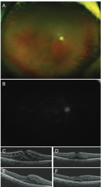
Intraocular pressure was 14 mmHg in the right eye and 15 mmHg in the left eye. Anterior segment findings were normal in the right eye. Fundus examination showed a few retinal hemor- rhages in his right eye (Fig. 1A). Fluorescein angiography showed a diffuse petaloid pattern of leakage around the central fovea (Fig. 1B). Optical coherence tomography (OCT) revealed a thin epiretinal membrane, serous macular detachment and intra- retinal edema that was located at the outer retina (Fig. 1C). After giving informed consent, the patient received an intravitreal 1.25 mg bevacizumab injection with a 30-gauge needle. Two weeks after injection, the BCVA in his right eye improved to 20 / 100.
OCT revealed the formation of a full thickness MH and decreas- ing intraretinal edema (Fig. 1D). Four weeks after injection, the MH was still open. The patient underwent vitrectomy combined with cataract surgery and intraocular lens implantation. Internal limiting membrane peeling and gas tamponade with 20% sulfur hexafluoride were successfully performed. One month after sur- gery, OCT confirmed successful closure of the MH, but the se- rous macular detachment still remained (Fig. 1E). The BCVA in his right eye was 20 / 100. The patient refused further treatment due to economic reasons. Six months after vitrectomy, the se- rous macular detachment remained unchanged (Fig. 1F).
The responsible factors for MH formation after intravitreal Korean J Ophthalmol 2019;33(2):198-199 https://doi.org/10.3341/kjo.2018.0079
Fig. 1. Ocular findings at initial presentation. (A) Fundus examina- tion showed a few retinal hemorrhages. (B) Fluorescein angiography revealed a diffuse petaloid pattern of leakage around the central fovea. Serial changes in optical coherence tomography (OCT) image.
(C) Before treatment, OCT revealed a thin epiretinal membrane, serous macular detachment, and intraretinal edema that was located at the outer retina. (D) Two weeks after the injection of bevacizum- ab, OCT showed the formation of a full thickness macular hole and decreasing intraretinal edema. (E) One month after vitrectomy, the macular hole was closed, but the serous macular detachment re- mained. (F) Six months after vitrectomy, the serous macular detach- ment remained unchanged
a
B
C E
D
F
199 anti-VEGF have been assumed to exist at the retinal pigment ep-
ithelium, retinal surface, and in the vitreous. Several potential mechanisms have been implicated to explain this process. In- duction of vitreous incarceration following anti-VEGF injections could enhance vitreoretinal traction and subsequently MH de- velopment [1]. Chemical compounds introduced into the vitreous cavity and structural modification of the vitreous body follow- ing anti-VEGF therapy could also trigger incomplete posterior vitreous detachment (PVD), vitreomacular traction, and subse- quent MH formation [2]. Grigoropoulos et al. [3] hypothesized that intravitreal injections can increase vitreomacular traction due to globe deformation during needle insertion and vitreous incarceration at the insertion site following treatment. This was proposed to cause vitreous syneresis and increase vitreofoveal traction leading to incomplete PVD, resulting in focal sites of traction on the retinal surface and MH formation. In our case, the PVD itself might not have been a causative factor for MH formation, because the PVD was induced with active aspiration during surgery. Liquefaction necrosis of the Müller cells and ad- jacent neural cells due to persistent ischemia leads to cystoid macular edema, a known cause of MH formation [4]. We postu- lated that intravitreal bevacizumab injection might have had an indirect role in the development of MH formation by favoring the rupture of distended Müller cells and intraretinal cysts. In this case, the coalescence and breakdown of large intraretinal cysts after bevacizumab injection in the presence of serous mac- ular detachment could have caused MH. In addition, contraction of the thin epiretinal membrane and increased vitreomacular traction caused by the intravitreal injection could have contrib- uted to the formation of MH.
In conclusion, we report a case of MH formation after intrav- itreal bevacizumab injection for treatment of DME. Although the occurrence of MH after intravitreal bevacizumab injection is uncommon, physicians should be well acquainted with this complication.
Yun Ji Lee, Moosang Kim
Department of Ophthalmology, Kangwon National University School of Medicine, Chuncheon, Korea
E-mail (Moosang Kim): [email protected]
Conflict of Interest
No potential conflict of interest relevant to this article was re- ported.
Acknowledgements
This study was supported by 2017 Research Grant from Kangwon National University (520170439).
References
1. Querques G, Souied EH, Soubrane G. Macular hole follow- ing intravitreal ranibizumab injection for choroidal neovas- cular membrane caused by age-related macular degenera- tion. Acta Ophthalmol 2009;87:235-7.
2. Raiji VR, Eliott D, Sadda SR. Macular hole overlying pig- ment epithelial detachment after intravitreal injection with ranibizumab. Retin Cases Brief Rep 2013;7:91-4.
3. Grigoropoulos V, Emfietzoglou J, Nikolaidis P, et al. Full- thickness macular hole after intravitreal injection of ranibi- zumab in a patient with retinal pigment epithelium detach- ment and tear. Eur J Ophthalmol 2010;20:469-72.
4. Kim M, Lee P, Kim Y, et al. Effect of intravitreal bevacizum- ab based on optical coherence tomography patterns of diabetic macular edema. Ophthalmologica 2011;226:138-44.