망막앞막이 동반된 당뇨황반부종에서 유리체내 덱사메타손 임플란트 삽입술의 단기 치료 효과
Short-term Effect of Intravitreal Dexamethasone Implant on Diabetic Macular Edema with Epiretinal Membrane
노동현, 안장환, 이준엽, 사공민
Donghyoun Noh, Jang Hwan Ahn, Junyeop Lee, Min Sagong
영남대학교 의과대학 안과학교실
Department of Ophthalmology, Yeungnam University College of Medicine, Daegu, Korea
Purpose: To assess the short term effect of an intravitreal dexamethasone implant on diabetic macular edema (DME) with epiretinal membrane (ERM).
Methods: We conducted a retrospective study including 39 eyes of 39 patients with DME who underwent intravitreal dexamethasone implant injection and were able to be observed for more than 6 months. Patients were divided into two groups according to presence or absence of ERM from March 2015 to June 2016.
Results: Thirteen DME eyes with ERM and 26 DME eyes without ERM were enrolled. The classification of ERM was grade 0 in 5 (38.5%) eyes, grade 1 in 7 eyes (53.8%) and grade 2 in 1 eye (7.7%). There was no difference in best corrected visual acuity (BCVA) and central macular thickness (CMT) between the DME with ERM group and the DME without ERM group at baseline. After the injection, there was no difference in mean BCVA and mean CMT between the two groups at 1, 3, and 6 months. DME without ERM group showed a signifi- cant decrease in CMT compared to baseline at 1, 3, and 6 months (p < 0.001, p = 0.003, p = 0.001, respectively). The DME with ERM group showed a significant decrease in CMT compared to baseline at 1 and 3 months, but no difference at 6 months (p = 0.048, p = 0.024, p = 0.275, respectively).
Conclusions: Intravitreal dexamethasone implantation may be a useful treatment modality for patients with DME with ERM, but the duration of the anatomical improvement may be shorter than that for patients with DME without ERM.
Keywords: Best corrected visual acuity; Central macular thickness; Dexamethasone implant; Diabetic macular edema; Epiretinal membrane
서론
당뇨황반부종은 당뇨를 가지고 있는 환자에서 가장 흔한 시력
저하의 원인으로 알려져 있다[1]. 당뇨황반부종의 원인은 아직까 지 완전하게 밝혀지지는 않았지만, 망막혈관내피세포와 혈관주 위세포의 소실로 인한 혈관내피세포 치밀이음부(tight junction)
Address reprint requests to Min Sagong, MD, PhD
Department of Ophthalmology, Yeungnam University College of Medicine, #170 Hyeonchung-ro, Nam-gu, Daegu 42415, Korea
Tel: 82-53-620-3443, Fax: 82-53-626-5936 E-mail: [email protected]
*This study was presented as a poster at the 117th Annual Meeting of the Korean Ophthalmological Society 2017.
Received: 2017. 8. 1 Revised: 2017. 9. 22 Accepted: 2017. 9. 22
의 손상으로 인한 혈액망막장벽의 손상[2]과, 조직의 저산소증 으로 인하여 발생하는 혈관내피세포성장인자와 케모카인, 인 터루킨-6, 인터루킨-8, 프로스타글란딘과 같은 염증을 유발하 는 사이토카인의 발현 증가[3,4]가 당뇨황반부종의 원인으로 알 려져 있다.
당뇨황반부종의 치료에서 꾸준히 시행되어 온 국소레이저 치 료는 Early Treatment Diabetic Retinopathy Study (ETDRS) 연 구 결과에 따르면 3년 동안의 경과관찰에서 중등도 이상의 시 력 감소를 보인 환자를 절반 정도 줄일 수 있지만 약 12%의 환 자에서는 여전히 중등도 이상 시력 감소가 지속되는 한계가 있 었다[5,6]. 최근에는 유리체내 항혈관내피세포성장인자 주사가 당뇨황반부종의 주된 치료법으로 사용되면서 더 좋은 결과를 보고하고 있다[7-9]. 추가적으로 유리체내 스테로이드 주입술 은 혈관내피세포성장인자와 염증성 사이토카인의 발현을 막고 [10], 백혈구 울혈을 억제시키며[11], 혈관내피세포 치밀이음부의 방벽 역할을 증가시켜[12], 최대교정시력 향상과 해부학적 호전 을 보이는 것으로 알려져 있다[9,13,14]. 특히, 덱사메타손 임플 란트(Ozurdex®, Allergan, Irvine, CA, USA)는 한 번의 삽입술 로 6개월까지 유리체내에 존재하며[15], 21.5-22.2%의 환자에서 3줄 이상의 시력 상승을 보고하고 있다[13,16]. 이러한 덱사메타 손 임플란트는 항혈관내피세포성장인자치료에 비해 긴 작용 시 간과 함께 유사한 치료 효과를 보여주었고[17,18], 항혈관내피세 포성장인자 치료에 반응하지 않는 난치성(refractory) 당뇨황반 부종 환자에게도 효과가 있는 것으로 알려지면서[19,20], 당뇨황 반부종 환자에서 그 적응증을 넓혀 가고 있다.
당뇨황반부종에서 유리체황반경계면 이상은 흔히 발견되는데 약 27-34% 정도에서 망막전막이 동반되는 것으로 보고되고 있 다[21,22]. 유리체황반견인은 염증반응과 혈관내피세포성장인자 의 지속적인 발현을 유발하는 것으로 알려져 있다[23,24]. 유리 체와 망막조직에서 발현되어 당뇨망막병증의 병태 생리에 중요 한 역할을 하는 염증성 사이토카인 인터루킨-6는[25] 망막 혈관 신생의 가장 중요한 일차 매개체인 혈관내피세포성장인자의 발 현을 유도하는 것으로 보고되고 있으며[26,27], 혈관내피세포성 장인자와 그 수용체와 함께 당뇨망막병증 환자의 혈관성 망막 앞막과 무혈관성 망막앞막에 위치하여 염증을 증가시키고 지속 적인 당뇨황반부종을 유발하는 것으로 알려져 있다[22,28]. 망 막앞막을 동반한 당뇨황반부종 환자는 더 많은 횟수의 항혈관 내피세포성장인자 주사를 필요로 하지만, 망막앞막이 물리적 장벽으로 작용하여 약물의 투과를 감소시키는 것으로 보고되 고 있다[29]. 당뇨황반부종에서 유리체황반경계면 이상이 동반 되는 경우 항혈관내피세포성장인자에 대한 치료 효과는 감소 될 수 밖에 없다[29-31]. 하지만 아직까지 망막앞막을 동반한 당 뇨황반부종 환자에서 유리체내 덱사메타손 임플란트 삽입술의 치료 효과를 비교한 연구는 없었다. 그러므로 본 연구에서는 당 뇨황반부종 환자에서 망막앞막 동반 유무에 따른 유리체내 덱
사메타손 임플란트의 단기 치료 효과를 비교해 보고자 하였다.
대상과 방법
2015년 3월부터 2016년 6월까지 형광안저혈관조영술(fluorescein angiography)에서 미만성 누출을 보이는 당뇨황반부종으로 진 단 받고 유리체내 덱사메타손 임플란트 삽입술 1회 시행 후 6 개월 이상 경과관찰이 가능하였던 당뇨황반부종 환자를 대상 으로 의무기록을 후향적으로 분석하였다. 백내장 수술을 제외 한 안내 수술력이나 천공 외상의 과거력, 당뇨황반부종을 제외 한 망막질환을 가진 경우, 6개월 미만의 경과관찰 기간을 가진 경우, 유리체내 덱사메타손 임플란트 삽입술을 시행 받고 6개월 이내 항혈관내피세포성장인자 주입술, 국소레이저 등의 치료를 받은 경우 등은 제외하였다. 본 연구는 영남대학교병원 임상연 구심의위원회(Institutional review board, IRB)의 승인을 받아 진 행하였으며 헬싱키 선언(Declaration of Helsinki)을 준수하였다.
당뇨황반부종은 안저검사 및 스펙트럼영역 빛간섭단층촬영 (Spectralis optical coherence tomography®, Heidelberg Engineering, Heidelberg, Germany)을 통하여 판단했다. 안저검사에서 황반중 심에서 1/2 유두지름 원 안에서 1 유두 크기 이상의 망막이 두 꺼워지며, 낭포성(cystoid) 또는 확산(diffuse) 부종이 관찰되고, 빛간섭단층촬영에서 중심황반두께가 300 μm 이상인 경우로 정 의하였다. 빛간섭단층촬영에서 얇은 고반사막이 신경망막 앞에 관찰되는 경우 망막앞막이 존재하는 것으로 정의하였다[32]. 망 막앞막은 안저검사 소견을 Gass 분류법[33]을 이용하여 셀로판 황반병증을 grade 0, 내측 망막의 주름을 만드는 주름셀로판황 반병증을 grade 1, 혈관 및 망막전층의 왜곡을 동반하는 황반 주름을 grade 2로 분류하였다.
모든 환자는 유리체내 덱사메타손 임플란트 삽입술 전 최대 교정시력을 측정하고, 세극등현미경검사, 안저검사, 빛간섭단층 촬영을 시행하였다. 주사 후 1개월, 3개월 그리고 6개월째 최대 교정시력을 측정하고 빛간섭단층촬영을 시행하였다. 최대교정 시력은 한천석 시력표를 이용하여 측정했으며, 통계분석을 위하 여 로그마 시력(Logarithm of the minimal angle of resolution, logMAR)으로 전환하였다.
통계분석을 위하여 SPSS Statistics ver. 20 (IBM Corp., Armonk, NY, USA) 소프트웨어를 이용하였다. 두 군 간의 최대 교정시력 및 중심황반두께를 비교하기 위하여 Mann-Whitney U-test를 사용하였으며, 유리체내 덱사메타손 임플란트 삽입술 전후의 최대교정시력 및 중심황반두께의 변화를 비교하기 위하 여 Wilcoxon signed-rank test를 사용하였다. 두 군 사이의 당 뇨망막병증 정도와 황반허혈 유무를 비교하기 위하여 Fisher’s exact test를 사용하였다. 모든 통계는 p-value가 0.05 미만인 경 우를 통계학적으로 유의한 것으로 간주했다.
결과
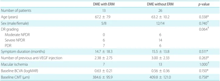
당뇨황반부종으로 유리체내 덱사메타손 임플란트 삽입술을 시 행 받은 환자는 총 92명이었으며, 6개월 이내의 경과관찰을 보 인 45명, 첫 번째 삽입술 후 6개월 이전 다른 치료를 시행 받 은 8명을 제외하고, 총 39명 39안을 대상으로 연구를 진행하였 다. 망막앞막을 동반한 당뇨황반부종 환자는 13명 13안이었으 며, 망막앞막을 동반하지 않은 당뇨황반부종 환자는 26명 26안 이었다. 망막앞막을 동반한 당뇨황반부종 환자의 평균 나이는 67.2 ± 7.9세였고, 5명이 남자, 8명이 여자였다. 망막앞막을 동 반하지 않는 당뇨황반부종 환자는 63.2 ± 10.2세였고, 12명이 남자, 14명이 여자였다. 나이와 성별은 두 군 사이에 차이를 보 이지 않았다(p=0.338, p=0.740). 망막앞막을 동반한 당뇨황반부 종군은 중증(Severe) 비증식당뇨망막병증이 6안, 증식성당뇨망 막병증이 7안이 포함되었으며, 망막앞막을 동반하지 않은 당 뇨황반부종군은 중등도(Moderate) 비증식성당뇨망막병증 6안, 중증 비증식성당뇨망막병증 14안, 증식성당뇨망막병증이 6안이 포함되었다. 두 군 사이의 당뇨망막병증의 분포에는 차이를 보 이지 않았다(p=0.064). 당뇨황반부종의 증상기간은 망막앞막을 동반한 당뇨황반부종군에서 14.7 ± 18.3달, 망막앞막을 동반하 지 않은 당뇨황반부종군은 15.5 ± 13.8달이었고, 두 군 사이의 차이는 없었다(p=0.511). 유리체내 덱사메타손 임플란트 삽입술 이전 유리체내 항혈관내피성장인자 주입술을 시행 받은 횟수는 망막앞막을 동반한 당뇨황반부종군에서 2.38 ± 2.75회, 망막 앞막을 동반하지 않은 당뇨황반부종군에서 3.00 ± 2.33회로 두
군 간의 유의한 차이는 없었다(p=0.263). 형광안저혈관조영술에 서 황반허혈이 관찰되는 경우는 망막앞막을 동반한 당뇨황반부 종군에서 7안, 망막앞막을 동반하지 않은 당뇨황반부종군에서 13안으로 두 군 간의 차이를 보이지 않았다(p=1.000). 유리체내 덱사메타손 임플란트 삽입술을 시행 받기 전의 최대교정시력과 중심황반두께는 망막앞막을 동반한 당뇨황반부종군이 0.63 ± 0.21, 384.62 ± 95.88 μm였으며, 망막앞막을 동반하지 않은 당 뇨황반부종군은 0.56 ± 0.36, 409.8 ± 121.03 μm로 두 군 사 이의 차이를 보이지 않았다(p=0.150, p=0.758) (Table 1). 망막앞 막을 동반한 당뇨황반부종군에서 망막앞막의 grade는 grade 0 이 5안(38.5%), grade 1이 7안(53.8%), grade 2는 1안(7.7%)이었다.
유리체내 덱사메타손 임플란트 삽입술 후 최대교정시력은 망 막앞막을 동반한 당뇨황반부종군에서 1개월째 0.66 ± 0.25, 3개월째 0.56 ± 0.31 그리고 6개월째 0.57 ± 0.25로 나타났고, 삽입술 전과 비교하여 유의한 최대교정시력 향상을 보이지 않 았다(p=0.668, p=0.547, p=0.375). 망막앞막을 동반하지 않은 당 뇨황반부종군의 최대교정시력은 1개월째 0.54 ± 0.38, 3개월 째 0.50 ± 0.39, 그리고 6개월째 0.56 ± 0.49로 나타났으며, 망 막앞막을 동반한 당뇨황반부종군과 마찬가지로 삽입술 전과 비교하여 유의한 최대교정시력 향상을 보이지 않았다(p=0.473, p=0.163, p=0.574). 두 군 사이의 최대교정시력을 비교하였을 때 1, 3, 6개월째 모두 차이를 보이지 않았다(p=0.208, p=0.501, p=0.439) (Fig. 1).
유리체내 덱사메타손 임플란트 삽입술 후 중심황반두께는 망막앞막을 동반한 당뇨황반부종군에서 1개월째 309.31 ±
Table 1. Baseline characteristics of diabetic macular edema with or without epiretinal membrane patients
DME with ERM DME without ERM p-value
Number of patients 13 26
Age (years) 67.2 ± 7.9 63.2 ± 10.2 0.338*
Sex (male/female) 5/8 12/14 0.740†
DR grading Moderate NPDR Severe NPDR PDR
0 6 7
6 14
6
0.064†
Symptom duration (months) 14.7 ± 18.3 15.5 ± 13.8 0.511*
Number of previous anti-VEGF injection 2.38 ± 2.75 3.00 ± 2.33 0.263*
Macular ischemia 7 13 1.000†
Baseline BCVA (logMAR) 0.63 ± 0.21 0.56 ± 0.36 0.150*
Baseline CMT (μm) 384.6 ± 95.9 409.8 ± 121.0 0.758*
Values are presented as mean ± standard deviation or n.
DME = diabetic macular edema; ERM = epiretinal membrane; DR = diabetic retinopathy; NPDR = non-proliferative diabetic retinopathy; PDR = proliferative diabetic retinopathy; VEGF = vascular endothelial growth factor; BCVA = best-corrected visual acuity; CMT = central macular thick- ness.
*Mann-Whitney U-test; †Fisher’s exact test.
79.20 μm, 3개월째 294.73 ± 76.59 μm 그리고 6개월째 335.70
± 103.72 μm로 나타났으며, 삽입술 전과 비교하여 1, 3개월 까지는 유의한 중심황반두께 감소를 보였지만 6개월째에는 그 렇지 못했다(p=0.048, p=0.024, p=0.275). 망막앞막을 동반하 지 않은 당뇨황반부종군의 중심황반두께는 1개월째 285.67 ± 74.38 μm, 3개월째 315.42 ± 111.46 μm 그리고 6개월째 339.36
± 131.42 μm로 나타났으며, 삽입술 전과 비교하여 1, 3, 6개 월째 모두 유의한 중심황반두께 감소를 유지하였다(p<0.001, p=0.003, p=0.001). 하지만 두 군 사이의 1, 3, 6개월째 평균 중 심황반두께 비교에서는 모두 차이를 보이지 않았다(p=0.224, p=0.903, p=0.788) (Fig. 2, 3).
고찰
본 연구에서는 당뇨황반부종으로 유리체내 덱사메타손 임플란 트 삽입술을 시행 받은 환자에서 망막앞막이 시력과 해부학적 소견의 변화에 미치는 영향을 비교 분석하였다. 망막앞막을 동 반하지 않은 당뇨황반부종군은 유리체내 덱사메타손 임플란트 삽입술 이후에 6개월까지 유의한 중심황반두께 감소를 유지하 였지만, 망막앞막을 동반한 당뇨황반부종군은 3개월까지 유지 하였던 중심황반두께의 감소를 6개월까지 유지하지 못하였다.
하지만 두 군 모두에서 유의한 최대교정시력의 향상을 보이지 는 않았다.
망막앞막을 동반한 당뇨황반부종군과 망막앞막을 동반하지 않은 당뇨황반부종군 모두에서 주사 후 3개월까지 중심황반두 께가 감소하였다. 이후, 망막앞막을 동반하지 않은 당뇨황반부 종군에서는 6개월까지 중심황반두께가 감소를 유지하였지만, 3개월에 비해 6개월째 중심황반두께가 증가하는 양상을 보였 다. 이는 덱사메타손 임플란트가 시간이 지나감에 따라 유리되 는 약물의 양이 점차 줄어들면서 생기는 변화라고 할 수 있고 [34] 기존의 연구 결과에서 3개월까지 감소하였던 중심황반두께 가 6개월째에 다시 증가하는 톱니 모양의 망막두께 변화를 보 고한 것과 같은 결과를 보여주었다[13,34-36]. 망막앞막을 동 반한 당뇨황반부종군은 3개월까지 중심황반두께 감소를 보였 지만, 6개월째에는 유의한 감소를 보여주지 못하였다. 이는 유 리되는 약물이 양이 점점 줄어들면서 발생하는 변화[34]와 함 께 유리체황반경계면 이상에 의해 발생하는 지속적인 견인력 이 부종을 유발하여 생기는 변화라고 생각할 수 있다[37,38].
유리체황반경계면 이상에 의하여 혈관내피세포성장인자, 인터 루킨-6 등과 같은 혈관의 투과성을 증가시키는 인자들의 발현 이 증가하여 주사의 효과가 6개월까지 지속되지 못하고 부종의 발생하였을 수 있다[22,28]. 그리고 망막앞막이 기계적인 장벽 Figure 1. The mean best-corrected visual acuity (BCVA) over time
after intravitreal dexamethasone implantation. The mean BCVA did not showed significant improvement at all-time points in both groups (diabetic macular edema [DME] with epiretinal membrane [ERM] group, p = 0.668, p = 0.547, p = 0.375; DME without ERM group, p = 0.208, p = 0.501, p = 0.439 by Wilcoxon signed-rank test vs. base- line). There was no difference in BCVA at all-time points between the two groups. Values are presented as mean (standard deviation).
*Mann-Whitney U-test, DME with ERM group vs. DME without ERM group.
Figure 2. The mean central macular thickness (CMT) over time after intravitreal dexamethasone implant. The diabetic macular edema (DME) with epiretinal membrane (ERM) group showed significant CMT improvement at 1 and 3 months but not at 6 months (p = 0.048, p = 0.024, p = 0.275, respectively)*. The DME without ERM group showed signifcant CMT improvement at all-time points (p < 0.001, p = 0.003, p = 0.001, respectively)*. There was no difference in CMT at all-time points between the two groups. Values are presented as mean (standard deviation)†. *Wilcoxon signed-rank test vs. baseline;
†Mann-Whitney U-test, DME with ERM group vs. DME without ERM group.
1 0.9 0.8 0.7 0.6 0.5 0.4 0.3 0.2 0.1 0
0.63 0.66 0.56 0.57
(0.21) (0.25) (0.31) (0.25)
0.56 0.54 0.50 0.56
(0.36) (0.38) (0.39) (0.49)
0.150 0.208 0.501 0.439
0 1 3 6 (month)
p-value*
BCVA (logMAR)
DME with ERM DME without ERM
450 400 350 300 250 200 150 100 50 0
384.62 309.31 294.73 335.70 (95.88) (79.20) (76.59) (103.72) 0.048 0.024 0.275 409.81 285.67 315.42 339.36 (121.03) (74.38) (111.46) (131.42) <0.001 0.003 0.001 0.758 0.224 0.903 0.788
0 1 3 6
p-value† (between 2 groups) p-value*
(vs. baseline)
p-value*
(vs. baseline)
CMT (µm)
DME with ERM DME without ERM (month)
으로 작용하여 약물 전달을 억제하는 것도 한 요인이라고 생각 할 수 있겠다[29].
두 군 모두 이전의 유리체내 항혈관내피세포성장인자 주입술 을 시행 받은 과거력이 있었지만(망막앞막을 동반한 당뇨황반 부종군 2.38 96 ± 2.75회 vs. 망막앞막을 동반하지 않은 당뇨황 반부종군 3.00 ± 2.33회, p = 0.263) 두 군 모두에서 추가적인 해부학적 호전을 관찰할 수 있었다. 유리체내 항혈관내피세포 성장인자 주입술에도 불구하고 당뇨황반부종이 지속되는 난치 성 당뇨황반부종에서도 유리체내 덱사메타손 임플란트 삽입술 후 의미 있는 해부학적 호전을 보였다는 기존의 연구와 같은 결 과를 보여주었다[16,39-41].
두 군 모두에서 유리체내 덱사메타손 임플란트 삽입술 전에 비해 1, 3, 6개월 모두에서 비록 의미 있는 최대교정시력 호전 을 보여주지는 못하였지만 3개월까지 최대교정시력이 증가하다 6개월째 다시 감소하는 양상을 보였다. 이러한 양상은 3개월까 지 최대교정시력이 상승하다 6개월째 상승의 정도가 다소 감소 하는 이전의 연구들과 유사한 결과를 보여주었다[35,36]. 두 군 모두에서 유의한 최대교정시력의 상승을 보여주지 못하였던 것 은 최대교정시력이 20/200–20/50 사이의 환자를 대상으로 시 행한 MEAD 연구[13]나 20/400–20/40 사이의 환자를 대상으 로 한 BEVODEX 연구[18]에 비해 더 좋은 주사 전 시력을 가진 환자들이 많이 포함되었기 때문으로 생각할 수 있다. 망막앞막 을 동반한 당뇨황반부종군과 망막앞막을 동반하지 않은 당뇨 황반부종군 모두 평균 증상기간이 약 15개월로 길었으며, 기존 치료에 내성을 보이던 환자가 망막앞막을 동반한 당뇨황반부종 군에서 84.6% (11/13안), 망막앞막을 동반하지 않은 당뇨황반부 종군에서 80.8% (21/26안)로 많이 포함되었던 것도 한 요인이라 고 할 수 있다. 이는 난치성 당뇨황반부종 환자가 기존의 치료
를 받지 않은 환자(treatment naïve)에 비해 시력 상승 정도가 더 작았다는 기존의 연구 결과와 유사한 결과라고 할 수 있겠다 [41]. ETDRS 시력표를 이용한 시력 측정이 Snellen 시력표를 이 용한 시력 측정에 비해 더 좋은 시력과 정확성을 보인다는 이전 의 보고[42,43]와 같이, 본 연구에서 한천석 시력표를 이용하여 시력을 측정했기 때문에 ETDRS 시력표를 이용한 이전 연구들 에 비해 시력 상승의 정도가 제한되었거나 측정의 정확성이 떨 어졌을 가능성도 있다.
본 연구의 제한점으로는 후향적 연구였고 대상 환자 수가 적 었다는 점 그리고 덱사메타손 임플란트 삽입술 1회 시행 후 6개월 내 재치료가 필요했던 경우가 제외되어 치료 후 시력 예 후가 나쁜 환자들이 더 많이 배제되었을 가능성이 있다. 하지만 6개월 내 재치료를 시행한 경우나 추가적 치료를 시행한 경우 는 망막앞막을 동반한 당뇨황반부종군에서 18.8% (3명 3안), 망 막앞막을 동반하지 않은 당뇨황반부종군에서 16.1% (5명 5안) 로 나타나 차이를 보이지는 않았다. 또한 망막앞막의 심한 정도 에 대한 객관적 분류법의 부재로 당뇨황반부종에 미치는 영향 을 정확히 평가하기 어려웠다는 점이 있겠다. 향후 더 많은 환자 를 대상으로 한 전향적인 비교 연구가 필요할 것으로 생각된다.
결론적으로 심하지 않은 망막앞막을 동반한 당뇨황반부종 환 자에서도 유리체내 덱사메타손 임플란트 삽입술을 시행했을 때 뚜렷한 해부학적 호전을 얻을 수 있었다. 하지만 망막앞막이 동 반되지 않은 당뇨황반부종 환자에 비해 해부학적 호전의 지속 기간이 짧기 때문에 첫 삽입술 후 재치료 간격을 좁히거나 항혈 관내피세포성장인자와의 병합치료, 수술적 치료 등 보다 적극 적인 치료를 고려해야 할 필요가 있겠다.
Figure 3. The case of a 73-year-old female patient who had diabetic macular edema with epiretinal membrane. Optical coherence scans of the same case at baseline (A), 1 month (B), 3 months (C), and 6 months (D) after intravitreal dexamethasone implantation. There was a marked decrease in central macular thickness at 1 and 3 months, but edema recurred at 6 months.
A
C
B
D
Conflicts of interest
The authors have no conflicts to disclose.
References
1. Otani T, Kishi S, Maruyama Y. Patterns of diabetic macular ede- ma with optical coherence tomography. Am J Ophthalmol 1999;127:688-93.
2. Yannuzzi LA, Shakin JL, Fisher YL, Altomonte MA. Peripheral ret- inal detachments and retinal pigment epithelial atrophic tracts secondary to central serous pigment epitheliopathy. Ophthal- mology 1984;91:1554-72.
3. Rechtman E, Harris A, Garzozi HJ, Ciulla TA. Pharmacologic ther- apies for diabetic retinopathy and diabetic macular edema. Clin Ophthalmol 2007;1:383-91.
4. Antcliff RJ, Marshall J. The pathogenesis of edema in diabetic maculopathy. Semin Ophthalmol 1999;14:223-32.
5. Treatment techniques and clinical guidelines for photocoagula- tion of diabetic macular edema. Early Treatment Diabetic Reti- nopathy Study Report Number 2. Early Treatment Diabetic Reti- nopathy Study Research Group. Ophthalmology 1987;94:761-74.
6. Photocoagulation for diabetic macular edema. Early Treatment Diabetic Retinopathy Study report number 1. Early Treatment Diabetic Retinopathy Study research group. Arch Ophthalmol 1985;103:1796-806.
7. Brown DM, Schmidt-Erfurth U, Do DV, et al. Intravitreal afliber- cept for diabetic macular edema: 100-week results from the VISTA and VIVID studies. Ophthalmology 2015;122:2044-52.
8. Mitchell P, Wong TY; Diabetic Macular Edema Treatment Guide- line Working Group. Management paradigms for diabetic mac- ular edema. Am J Ophthalmol 2014;157:505-13.e1-8.
9. Diabetic Retinopathy Clinical Research Netwrok, Elman MJ, Aiello LP, et al. Randomized trial evaluating ranibizumab plus prompt or deferred laser or triamcinolone plus prompt laser for diabetic macular edema. Ophthalmology 2010;117:1064-77.e35.
10. Wang K, Wang Y, Gao L, et al. Dexamethasone inhibits leukocyte accumulation and vascular permeability in retina of streptozo- tocin-induced diabetic rats via reducing vascular endothelial growth factor and intercellular adhesion molecule-1 expression.
Biol Pharm Bull 2008;31:1541-6.
11. Tamura H, Miyamoto K, Kiryu J, et al. Intravitreal injection of cor- ticosteroid attenuates leukostasis and vascular leakage in experi- mental diabetic retina. Invest Ophthalmol Vis Sci 2005;46:1440-4.
12. Antonetti DA, Wolpert EB, DeMaio L, et al. Hydrocortisone de-
creases retinal endothelial cell water and solute flux coincident with increased content and decreased phosphorylation of oc- cludin. J Neurochem 2002;80:667-77.
13. Boyer DS, Yoon YH, Belfort R Jr, et al. Three-year, randomized, sham-controlled trial of dexamethasone intravitreal implant in patients with diabetic macular edema. Ophthalmology 2014;121:1904-14.
14. Gillies MC, Sutter FK, Simpson JM, et al. Intravitreal triamcino- lone for refractory diabetic macular edema: two-year results of a double-masked, placebo-controlled, randomized clinical trial.
Ophthalmology 2006;113:1533-8.
15. Chang-Lin JE, Attar M, Acheampong AA, et al. Pharmacokinetics and pharmacodynamics of a sustained-release dexamethasone intravitreal implant. Invest Ophthalmol Vis Sci 2011;52:80-6.
16. Augustin AJ, Kuppermann BD, Lanzetta P, et al. Dexamethasone intravitreal implant in previously treated patients with diabetic macular edema: subgroup analysis of the MEAD study. BMC Ophthalmol 2015;15:150.
17. Shah SU, Harless A, Bleau L, Maturi RK. Prospective randomized subject-masked study of intravitreal bevacizumab monotherapy versus dexamethasone implant monotherapy in the treatment of persistent diabetic macular edema. Retina 2016;36:1986-96.
18. Gillies MC, Lim LL, Campain A, et al. A randomized clinical trial of intravitreal bevacizumab versus intravitreal dexamethasone for diabetic macular edema: the BEVORDEX study. Ophthalmology 2014;121:2473-81.
19. Totan Y, Güler E, Gürağaç FB. Dexamethasone intravitreal im- plant for chronic diabetic macular edema resistant to intravitreal bevacizumab treatment. Curr Eye Res 2016;41:107-13.
20. Alshahrani ST, Dolz-Marco R, Gallego-Pinazo R, et al. Intravitreal dexamethasone implant for the treatment of refractory macular edema in retinal vascular diseases: results of the KKESH Interna- tional Collaborative Retina Study Group. Retina 2016;36:131-6.
21. Ghazi NG, Ciralsky JB, Shah SM, et al. Optical coherence tomog- raphy findings in persistent diabetic macular edema: the vitreo- macular interface. Am J Ophthalmol 2007;144:747-54.
22. Yamamoto T, Akabane N, Takeuchi S. Vitrectomy for diabetic macular edema: the role of posterior vitreous detachment and epimacular membrane. Am J Ophthalmol 2001;132:369-77.
23. Seko Y, Seko Y, Fujikura H, et al. Induction of vascular endothelial growth factor after application of mechanical stress to retinal pigment epithelium of the rat in vitro. Invest Ophthalmol Vis Sci 1999;40:3287-91.
24. Schepens CL, Avila MP, Jalkh AE, Trempe CL. Role of the vitreous in cystoid macular edema. Surv Ophthalmol 1984;28 Sup-
pl:499-504.
25. Kojima S, Yamada T, Tamai M. Quantitative analysis of interleu- kin-6 in vitreous from patients with proliferative vitreoretinal diseases. Jpn J Ophthalmol 2001;45:40-5.
26. Cohen T, Nahari D, Cerem LW, et al. Interleukin 6 induces the expression of vascular endothelial growth factor. J Biol Chem 1996;271:736-41.
27. Aiello LP, Avery RL, Arrigg PG, et al. Vascular endothelial growth factor in ocular fluid of patients with diabetic retinopathy and other retinal disorders. N Engl J Med 1994;331:1480-7.
28. Chen YS, Hackett SF, Schoenfeld CL, et al. Localisation of vas- cular endothelial growth factor and its receptors to cells of vascular and avascular epiretinal membranes. Br J Ophthalmol 1997;81:919-26.
29. Ercalik NY, Imamoglu S, Kumral ET, et al. Influence of the epiret- inal membrane on ranibizumab therapy outcomes in patients with diabetic macular edema. Arq Bras Oftalmol 2016;79:373-5.
30. Yoon D, Rusu I, Barbazetto I. Reduced effect of anti-vascular en- dothelial growth factor agents on diabetics with vitreomacular interface abnormalities. Int Ophthalmol 2014;34:817-23.
31. Wu PC, Lai CH, Chen CL, Kuo CN. Optical coherence tomograph- ic patterns in diabetic macula edema can predict the effects of intravitreal bevacizumab injection as primary treatment. J Ocul Pharmacol Ther 2012;28:59-64.
32. Wilkins JR, Puliafito CA, Hee MR, et al. Characterization of epireti- nal membranes using optical coherence tomography. Ophthal- mology 1996;103:2142-51.
33. Gass JDM. Stereoscopic atlas of macular diseases: diagnosis and treatment, 4th ed. St. Louis: Mosby, 1997;938-51.
34. Danis RP, Sadda S, Li XY, et al. Anatomical effects of dexametha- sone intravitreal implant in diabetic macular oedema: a pooled
analysis of 3-year phase III trials. Br J Ophthalmol 2016;100:796- 801.
35. Sacconi R, Battaglia Parodi M, Casati S, et al. Dexamethasone implants in diabetic macular edema patients with high visual acuity. Ophthalmic Res 2017;58:125-30.
36. Pacella F, Ferraresi AF, Turchetti P, et al. Intravitreal injection of Ozurdex((R)) implant in patients with persistent diabetic mac- ular edema, with six-month follow-Up. Ophthalmol Eye Dis 2016;8:11-6.
37. Kaiser PK, Riemann CD, Sears JE, Lewis H. Macular traction de- tachment and diabetic macular edema associated with posteri- or hyaloidal traction. Am J Ophthalmol 2001;131:44-9.
38. Lewis H, Abrams GW, Blumenkranz MS, Campo RV. Vitrectomy for diabetic macular traction and edema associated with poste- rior hyaloidal traction. Ophthalmology 1992;99:753-9.
39. Unsal E, Eltutar K, Sultan P, et al. Efficacy and safety of intravitreal dexamethasone implants for treatment of refractory diabetic macular edema. Korean J Ophthalmol 2017;31:115-22.
40. Pacella F, Romano MR, Turchetti P, et al. An eighteen-month follow-up study on the effects of intravitreal dexamethasone implant in diabetic macular edema refractory to anti-VEGF ther- apy. Int J Ophthalmol 2016;9:1427-32.
41. Escobar-Barranco JJ, Pina-Marín B, Fernández-Bonet M. Dexa- methasone implants in patients with naïve or refractory diffuse diabetic macular edema. Ophthalmologica 2015;233:176-85.
42. Kaiser PK. Prospective evaluation of visual acuity assessment: a comparison of snellen versus ETDRS charts in clinical practice (An AOS Thesis). Trans Am Ophthalmol Soc 2009;107:311-24.
43. Shamir RR, Friedman Y, Joskowicz L, et al. Comparison of Snellen and Early Treatment Diabetic Retinopathy Study charts using a computer simulation. Int J Ophthalmol 2016;9:119-23.
망막앞막이 동반된 당뇨황반부종에서 유리체내 덱사메타손 임플란트 삽입술의 단기 치료 효과
목적: 망막앞막에 동반된 당뇨황반부종에서 유리체내 덱사메타손 임플란트 삽일술의 단기 치료 효과를 알아보고자 하였다.
대상과 방법: 2015년 3월부터 2016년 6월까지 당뇨황반부종으로 유리체내 덱사메타손 임플란트 삽입술을 1회 시행 받고 6개월 이상 경과 관찰이 가능하였던 39명(39안)의 환자를 대상으로 망막앞막 동반 여부에 따른 치료 효과를 후향적으로 비교 분석하였다.
결과: 망막앞막이 동반된 당뇨황반부종이 13안이었고 망막앞막이 동반되지 않은 당뇨황반부종이 26안이었다. 당뇨황반부종에 동반된 망막앞막은 grade 0이 5안(38.5%), grade 1이 7안(53.8%), grade 2가 1안(7.7%)이었다. 당뇨황반부종에서 망막앞막이 동반된 군과 동반되지 않은 군 사이의 주사 전 최대교정시력과 평균 중심황반두께는 차이를 보이지 않았고, 주사 후 1, 3, 6개월째 두 군 간의 최대교정시력, 평균 중심황반두께도 차이를 보이지 않았다. 중심황반두께는 망막앞막이 동반되지 않은 군에서 주사 후 1, 3, 6개월째 모두 주사 전에 비하여 유의한 감소를 보였으나(p<0.001, p=0.003, p=0.001), 망막앞막이 동반된 군은 1, 3개월에는 감소를 보였 지만 6개월째는 차이를 보이지 않았다(p=0.048, p=0.024, p=0.275).
결론: 경도의 망막앞막이 동반된 당뇨황반부종에서 유리체내 덱사메타손 임플란트 삽입술은 유용한 치료 방법이 될 수 있으나, 해부학 적 호전의 지속기간이 망막앞막이 동반되지 않은 당뇨황반부종에 비해 더 짧게 나타날 수 있다.
국문초록