ISSN 0378-6471 (Print)⋅ISSN 2092-9374 (Online)
http://dx.doi.org/10.3341/jkos.2016.57.10.1604
Original Article
당뇨황반부종의 형태학적 분류에 따른 유리체강 내 라니비주맙 초기치료 후의 장기 임상결과
Long-term Outcomes of Diabetic Macular Edema Following Initial Intravitreal Ranibizumab Injection Based on Morphologic Pattern
김기영1⋅김응석2⋅곽형우2⋅유승영2
Kiyoung Kim, MD1, Eung Suk Kim, MD, PhD2, Hyung Woo Kwak, MD, PhD2, Seung-Young Yu, MD, PhD2
경희대학교 대학원 의학과 안과학교실1, 경희대학교 의학전문대학원 경희대학교병원 안과학교실2 Division of Ophthalmology, Department of Medicine, Kyung Hee University Graduate School1, Seoul, Korea Department of Ophthalmology, Kyung Hee University Hospital, Kyung Hee University School of Medicine2, Seoul, Korea
Purpose: To evaluate the 3-year visual and morphological outcomes of diabetic macular edema (DME) based on the morpho- logical pattern observed on optical coherence tomography (OCT) after intravitreal ranibizumab injections.
Methods: Thirty-two eyes of 32 patients with DME were classified according to the following OCT features: diffuse retinal thick- ening (DRT), cystoid macular edema (CME), and serous retinal detachment (SRD). All patients received 3 consecutive monthly intravitreal injections of 0.5 mg ranibizumab. After 3 injections, patients received ranibizumab or dexamethasone implantation as needed. The primary outcome was the number of treatments received based on the DME type over 36 months. Best-corrected visual acuity (BCVA), central subfoveal thickness, and macular volume changes were also evaluated.
Results: The eyes were classified as DRT (n = 16), CME (6), or SRD (10). The mean number of injections over 3 years was sig- nificantly different among the groups: DRT (4.25), CME (7.5), SRD (7.6; p = 0.048). The number of patients who did not need ad- ditional treatment after the initial 3 injections was 13 with DRT (81.3%), 2 with CME (33.3%), and 5 with SRD (50%; p = 0.045).
BCVA at 36 months significantly improved from baseline in the DRT group (p = 0.003). The CME group showed the worst BCVA among the groups (p = 0.023). Six patients who received intravitreal dexamethasone implantation showed no significant im- provement of BCVA but significant decrease in macular volume from 12 to 36 months.
Conclusions: Clinical courses varied according to the morphological pattern of DME after intravitreal ranibizumab injection, and patients with DRT maintained visual and anatomical improvement with fewer injections over 36 months. Additional dex- amethasone implantation showed limited effect in reducing macular edema with persistent macular cystic change, but no sig- nificant improvement in visual acuity.
J Korean Ophthalmol Soc 2016;57(10):1604-1612
Keywords: Dexamethasone implantation, Diabetic macular edema, Optical coherence tomography, Ranibizumab
■Received: 2016. 4. 28. ■ Revised: 2016. 7. 27.
■Accepted: 2016. 9. 13.
■Address reprint requests to Seung-Young Yu, MD, PhD Department of Ophthalmology, Kyung Hee University Hospital,
#23 Kyungheedae-ro, Dongdaemun-gu, Seoul 02447, Korea Tel: 82-2-958-8451, Fax: 82-2-966-7340
E-mail: [email protected]
* This study was presented as a narration at the 114th Annual Meeting of the Korean Ophthalmological Society 2015.
ⓒ2016 The Korean Ophthalmological Society
This is an Open Access article distributed under the terms of the Creative Commons Attribution Non-Commercial License (http://creativecommons.org/licenses/by-nc/3.0/) which permits unrestricted non-commercial use, distribution, and reproduction in any medium, provided the original work is properly cited.
당뇨망막병증은 대사성 질환인 당뇨병에 의한 망막의 미 세혈관계의 순환 장애로 발생하며, 이 중 당뇨황반부종은 전 세계적으로 성인의 시력저하를 유발하는 주요 질환 중 의 하나이다.1,2 황반부종은 다양한 망막질환에서 나타날 수 있는 비특이적 증상이며, 일반적으로 통증이 없는 시력저 하와 동반되는데 이는 황반부의 망막 세포 내 또는 외 공간 의 국소적 팽창에서 기인한다.3 황반부종의 형태학적, 지형
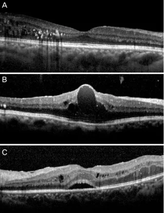
Figure 1. Morphologic patterns of diabetic macular edema by
optical coherence tomography. (A) Diffuse retinal thickening type looks as a sponge-like swelling area with reduced retinal reflectivity. (B) Cystoid macular edema type shows intra-reti- nal cystoid spaces. (C) Serous retinal detachment exhibits ele- vation of retina and fluid is accumulated between retina and retinal pigment epithelium.학적 특징을 결정하는 것은 치료에 대한 서로 다른 반응을 야기하는 질병의 병태생리를 규명하는 데 도움이 된다.
당뇨황반부종에서 빛간섭단층촬영을 통해 얻은 부종의 형태는 다양한 특징을 보이는데, Otani et al4은 이를 미만 성으로 망막이 두꺼워진 황반부종(diffuse retinal thicken- ing, DRT), 낭포황반부종(cystoid macular edema, CME), 장액성 망막박리가 동반된 황반부종(serous retinal detach- ment, SRD)의 3종류로 분류하였다. 각 종류의 발생기전은 서로 다르다고 알려져 있으며, 이에 따라 서로 다른 형태학 적 및 지형학적 특징을 보이게 된다.5,6 이후 많은 연구에서 당뇨황반부종의 형태에 따른 항혈관내피성장인자(anti-vas- cular endothelial growth factor, Anti-VEGF) 및 트리암시놀 론 주입술의 임상적 결과 및 효과가 보고되었다.7-14 당뇨황 반부종에서 항혈관내피성장인자의 약물학적 치료는, 내인 성 매개체에 직접적으로 작용함으로써 혈관 투과성, 신생 혈관형성 및 증식의 기전을 억제하는 효과를 가진다.15 최 근 당뇨망막병증에서 혈관내피세포성장인자 외에 염증반 응을 통한 그 매개체가 되는 싸이토카인(IL-1β, IL-6, IL-8, MCP-1, IP-10)들의 역할이 주목 받고 있는데, 스테로이드 의 광범위한 항염증작용이 염증성 싸이토카인 억제 및 내 피세포의 밀착결합부(tight junction)의 연속성을 강화하여 당뇨황반부종의 치료에 효과가 있음이 보고되었다.16-21 또 한 여러 차례의 항혈관내피성장인자 주입술 등의 치료에도 반응하지 않는 당뇨황반부종에서 덱사메타존 임플란트(Ozu- rdex®, Allergan, Irvine, CA, USA) 치료가 효과를 보인 연구 가 보고되었다.22-25
본 안과학교실에서는 당뇨황반부종에서 빛간섭단층촬영의 형태학적 특징에 따른 유리체강 내 라니비주맙(ranibizumab, Lucentis®, Genentech Inc., San Francisco, CA, USA) 주입 술 후 1년간의 시력과 형태학적 예후를 전향적으로 연구하 여 보고한 바 있다.26 본 연구에서는 당뇨황반부종 환자에 서 빛간섭단층촬영에 따른 형태학적 특징이 유리체강 내 라니비주맙 주입술 후 장기간 시력과 형태학적 예후의 예 측인자가 될 수 있는지를 알아보고, 이에 반응하지 않는 황 반부의 낭포성 변화가 지속되는 경우 시행한 추가 덱사메 타존 임플란트 주입술의 효과에 대해서 알아보고자 하였다.
대상과 방법
본 연구는 본원에서 당뇨황반부종을 진단 받은 환자 55 명의 초기 1년간 전향적, 개방적, 연속적인 케이스 비교연 구 후, 이 중 3년 이상 경과관찰이 가능했던 32명의 의무기 록을 후향적으로 비교 분석한 연구이다. 연구 대상은 다음 과 같은 기준을 만족시키는 환자로 이루어졌다. (a) 18세
이상의 1형 또는 2형 당뇨가 있으며 황반부종으로 인한 시 력장애가 있는 경우, (b) 최대교정시력(best-corrected visual acuity, BCVA)이 스넬렌시력표의 20/160에서 20/32 및 이 에 해당하는 Early Treatment Diabetic Retinopathy Study (ETDRS) 차트를 이용한 시력 39에서 78글자 사이, (c) 중 심와두께가 300 μm 이상인 경우이다. 중요한 제외기준은 (a) 활성화된 안내 염증 또는 감염이 어느 한눈에라도 있는 경우, (b) 조절되지 않는 녹내장이 어느 한눈에라도 있는 경우(예: 치료에도 불구하고 안압이 24 mmHg 이상), (c) 6 개월 내 범망막광응고술을 받았거나 연구 등록 전 3개월 이내에 국소 또는 격자광응고술을 받은 경우, (d) 연구 등 록 전 3개월 이내에 항혈관내피성장인자 치료를 받은 경우, (e) 이전에 유리체절제술 또는 유리체강 내 트리암시놀론 주입술을 시행 받은 경우, (f) 뇌졸중의 과거력, (g) 치료 받 지 않았거나, 치료 후에도 수축기 혈압 160 mmHg 또는 이 완기 혈압 100 mmHg 이상인 경우였다.
초기 중심와두께 및 황반부 부피는 빛간섭단층촬영기
A
B
C
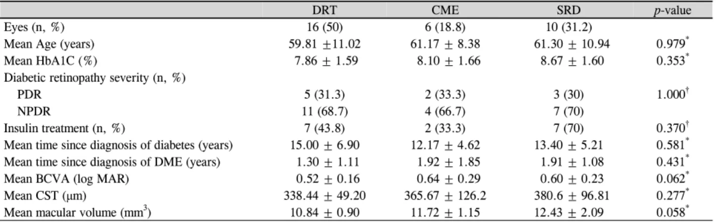
Table 1. Baseline characteristics of eyes and optical coherence tomography findings
DRT CME SRD p-value
Eyes (n, %) 16 (50) 6 (18.8) 10 (31.2)
Mean Age (years) 59.81 ±11.02 61.17 ± 8.38 61.30 ± 10.94 0.979*
Mean HbA1C (%) 7.86 ± 1.59 8.10 ± 1.66 8.67 ± 1.60 0.353*
Diabetic retinopathy severity (n, %)
PDR 5 (31.3) 2 (33.3) 3 (30) 1.000†
NPDR 11 (68.7) 4 (66.7) 7 (70)
Insulin treatment (n, %) 7 (43.8) 2 (33.3) 7 (70) 0.370†
Mean time since diagnosis of diabetes (years) 15.00 ± 6.90 12.17 ± 4.62 13.40 ± 5.21 0.581* Mean time since diagnosis of DME (years) 1.30 ± 1.11 1.92 ± 1.85 1.91 ± 1.08 0.431*
Mean BCVA (log MAR) 0.52 ± 0.16 0.64 ± 0.29 0.60 ± 0.23 0.062*
Mean CST (μm) 338.44 ± 49.20 365.67 ± 126.2 380.6 ± 96.81 0.277*
Mean macular volume (mm3) 10.84 ± 0.90 11.72 ± 1.15 12.43 ± 2.09 0.058*
Values are presented as mean ± SD or n (%) unless otherwise indicated.
DRT = diffuse retinal thickening; CME = cystoid macular edema; SRD = serous retinal detachment; PDR = proliferative diabetic retinop- athy; NPDR = non-proliferative diabetic retinopathy; DME = diabetic macular edema; BCVA = best-corrected visual acuity; log MAR = logarithm of the minimum angle of resolution; CST = central subfoveal thickness.
*Kruskal–Wallis test, statistical significance at 5% level; †Fisher exact test, statistical significance at 5% level.
(Cirrus HD-OCT, Carl Zeiss Meditec, Dublin, CA, USA)의 macular cube 512×128 scans 모드를 이용해 측정하였으며 매달 안저검사와 빛간섭단층촬영을 시행하였다. 모든 환자 는 3회의 연속된 유리체강 내 0.5 mg 라니비주맙 주입술을 1개월 간격으로 받았다. 첫 3회의 주사 후 다음 중 1개 이 상의 기준에 해당하는 환자의 경우 추가 유리체강 내 0.5 mg 라니비주맙 주입술을 시행 받았다. (a) ETDRS 차트를 이용한 최대교정시력의 5글자 이상 감소, (b) 중심와두께 100 μm 이상 증가, (c) 새로 형성된 망막 내 낭종이나 망막 하 삼출물로 인한 최대교정시력의 감소가 연구자에 의해 판단된 경우. 또한 다음 기준 중 어느 한 개 이상 만족하는 경우 치료를 중지하기로 하였다. (a) 최근 2회의 방문에서 시행 받은 유리체강 내 라니비주맙 주입술에도 불구하고, 마지막 연속된 3회 이상의 방문에서 측정한 최대교정시력 이 호전되지 않은 경우, (b) 최근 2회의 연속된 방문에서 ETDRS 차트를 이용한 최대교정시력 값이 84 글자 이상인 경우(Snellen 시력표에서 20/20 해당함). 경과관찰 1년 후부 터는 위의 기준에 따른 추가 유리체강 내 라니비주맙 주입 술에도 불구하고 지속적인 망막 내 낭종으로 인한 시력 저 하가 3개월 이상 지속되었던 환자의 경우 유리체강 내 덱 사메타존 임플란트 주입술을 추가로 시행하였다.
빛간섭단층촬영의 형태학적 특징에 따라 황반부종을 다 음과 같이 세 군으로 분류하였다(Fig. 1). 스펀지 형태의 미 만성으로 망막이 두꺼워진 황반부종과 망막 내 반사도의 감소를 동반한 경우 ‘1군(DRT)’, 망막 내 저반사도의 낭포 성 공간과 고반사도 격막에 의해 분리된 황반부에 낭포성 공간이 있는 경우 ‘2군(CME)’, 망막의 얕은 융기와 광학적 으로 깨끗한 망막과 망막색소상피층 사이의 공간이 있는
경우 ‘3군(SRD)’으로 정의하였다. 미만성으로 망막이 두꺼 워진 황반부종(1군)이 낭포황반부종(2군) 또는 장액성 망막 박리가 동반된 황반부종(3군)과 혼합되었을 경우, 각각 2 군, 3군으로 분류되었으며, 세 가지 형태가 모두 나타날 경 우는 3군으로 분류하였다.
모든 환자에 있어 치료 전과 후의 분석을 위해 paired t-test를 이용하였다. 황반부종의 종류에 따른 치료 전후 변 화 분석을 위해 Wilcoxon signed rank test를 데이터 검정 후에 실시하였고, 황반부종 종류 간의 평균값 비교를 위해 Kruskal–Wallis test 및 Fisher exact test를 실시 후 Bonferroni 방법을 통해 세 군 간의 사후검정을 시행하였다. 치료 전과 후의 시력과 각 측정값들은 Pearson 또는 Spearman corre- lation analysis를 통해 연관성을 검정하였다. 모든 통계학적 분석은 SPSS software version 18.0 (SPSS Inc., Chicago, IL, USA)를 이용했으며 p값이 0.05 미만인 경우 통계학적 유의성이 있다고 간주하였다.
결 과
본 연구에 등록되어 36개월 이상 경과관찰이 이루어진 32명 환자의 초기 측정 값이 Table 1에 나타나 있다. 본 연 구의 12개월 추적관찰 시점에서 처음 등록된 55명 중 6명 의 환자가 누락되었으며, 36개월 추적관찰 시점에서는 총 32명의 환자에서 결과 수집이 이루어졌다. 빛간섭단층촬영 기를 이용한 당뇨황반부종의 분류에서 1군은 16명(50%), 2 군은 6명(18.8%) 그리고 3군은 10명(31.2%)이었다. 세 군 사이의 당뇨 특성치 및 초기 중심와두께(1군=338.4 μm; 2 군=365.7 μm; 3군=380.6 μm; p=0.227), 황반부 부피(1군 =
Table 2. Mean number of intravitreal treatments received over 3 years
DRT CME SRD p-value
Mean number of Ranibizumab treatments (n) (Day 1–Month 36)
4.68 ± 0.95 7.50 ± 2.88 7.60 ± 3.98 0.048*
Mean number of Ranibizumab treatments (n) (Day 1–Month 12)
3.50 ± 0.73 5.16 ± 1.94 4.90 ± 2.28 0.074*
Mean number of Ranibizumab treatments (n) (Month 12– Month 36)
1.19 ± 0.73 2.33 ± 0.49 2.70 ± 0.68 0.037*
Eyes, no additional injections (n) (Month 12–Month 36)
13 (81.3%) 2 (33.3%) 5 (50%) 0.045†
Eyes, received Dexamethasone implantation (n) (Month 12–Month 36)
1 (5.95%) 2 (33.3%) 3 (30%) 0.222†
Mean number of Dexamethasone implantation (n) (Month 12–Month 36)
1 1.5 1.67
Values are presented as mean ± SD or n (%) unless otherwise indicated.
DRT = diffuse retinal thickening; CME = cystoid macular edema; SRD = serous retinal detachment.
*Kruskal–Wallis test, statistical significance at 5% level; †Fisher exact test, statistical significance at 5% level.
Figure 2. Change in the mean best-corrected visual acuity
(BCVA) over time after intravitreal Ranibizumab injection. In diffuse retinal thickening (DRT) type, significant improve- ment of BCVA was maintained from 3 to 36 months. In cys- toid macular edema (CME) type, BCVA significantly im- proved over 12 months, but there was no significant difference with baseline from 18 to 36 months. serous retinal detachment (SRD) type showed significant BCVA improvement at 3 and 18months, which were not maintained over 36 months. SRD type showed poorer BCVA at 6, 12 months, and CME type showed poorer BCVA from 18 to 36 months than the other types. Error bar indicates 95% confidence interval.10.84 mm3; 2군=11.72 mm3; 3군=12.43 mm3; p=0.058)에 는 유의한 차이가 없었다. 1군에서 다른 두 군보다 더 나은 초기 시력(logMAR)을 보였으나 그 차이는 통계적으로 유 의하지는 않았다(1군=0.52; 2군=0.64; 3군=0.60; p=0.062).
Table 2에서 경과관찰 기간에 따른 유리체강 내 주입술 횟수를 정리하였다. 36개월간의 추적관찰에서 유리체강 내 라니비주맙 주입술의 평균횟수는 1군에서 4.68회, 2군에서 7.5회, 3군에서 7.6회로 세 군 간에 유의한 차이가 있었고 (p=0.048), Bonferroni 사후 검정 결과 1군과 2군, 1군과 3 군 사이에서 각각 유의한 차이가 검정되었다(p=0.032, 0.042). 초기 12개월부터 36개월까지 추가 실시한 유리체강 내 라니비주맙 주입술의 평균횟수는 각각 1.19회, 2.33회, 2.7회였고, 이 기간 동안 추가 주입술이 필요하지 않았던 환자는 각각 13명(81.3%), 2명(33.3%), 5명(50%)이었다. 두 항목 모두 세 군 간에 유의한 차이가 있었고(p=0.037, 0.045), Bonferroni 사후 검정결과 모두 1군과 2군(p=0.03, 0.013)과 1군과 3군(p=0.018, 0.03) 사이에서 유의한 차이가 검정되었다.
당뇨황반부종의 종류에 따라 최대교정시력(logMAR)을 평가했을 때 1군에서는 초기 0.53에서 치료 후 12, 24, 36 개월 각각 0.27, 0.18, 0.19로 초기값과 비교한 유의한 시력 호전이 36개월까지 유지되었다(p=0.007, 0.002, and 0.003, respectively). 2군은 초기 0.64로부터 치료 후 12개월에 0.32로 유의한 호전을 보였으나(p=0.018), 치료 후 18, 24, 36개월에는 각각 0.41, 0.50, 0.50으로 초기값과 유의한 차 이를 보이지 않았다(p=0.128, 0.398, 0.398, respectively). 3 군에서는 초기 0.6으로부터 치료 후 3개월에 0.39로 유의한 시력호전이 있었으나(p=0.005), 6개월 후 0.52로 증가하여 초기값과 비교했을 때 유의한 차이가 없었다(p=0.169). 하 지만 18개월째는 0.30으로 유의하게 호전되었고(p=0.004),
36개월까지 시력호전이 유지되었으나 초기값과 유의한 차 이를 보이지는 않았다(p=0.114) (Fig. 2). 세 군 간의 36개 월 치료기간 동안 최대교정시력 평균값을 사후검정을 통해 비교하여 보았을 때, 치료 후 6, 12개월에서 3군이 1군에 비해 유의하게 낮았으며(p=0.005, 0.015), 치료 후 24, 30, 36개월에서 2군이 1군에 비해 유의하게 낮았다(p=0.032, 0.014, 0.011, respectively).
평균 중심와두께 변화를 살펴보면, 1군은 초기 338.4 μm
Figure 3. Change in the mean central subfoveal thickness (CST)
over time after intravitreal Ranibizumab injection. In diffuse retinal thickening (DRT) and serous retinal detachment (SRD) types, CST significantly improved from the baseline through 36 months, except 18 months of SRD type. In cystoid macular ede- ma (CME) type, significant improvement of CST was not main- tained after 6 months. There was no difference in the CST be- tween the individual types. Error bar indicates 95% confidence interval.Figure 4. Change in the mean macular volume over time after
intravitreal Ranibizumab injection. In diffuse retinal thicken- ing (DRT) type, macular volume significantly improved from the baseline until 18 months. In cystoid macular edema (CME) and serous retinal detachment (SRD) types, macular volume significantly deceased at 3 months after treatment, then ag- gravated until 18 months. However both types showed sig- nificant reduction of macular volume at 36 months compared to baseline. Error bar indicates 95% confidence interval.에서 치료 후 12, 24, 36개월 각각 269.3 μm, 298.9 μm, 292.9 μm로 치료 후부터 36개월까지 유의한 호전이 유지 되었다(all p<0.001). 2군은 초기 365.7 μm에서 치료 후 6개 월 281.7 μm로 유의한 감소를 보였으나(p=0.028), 이후부 터 36개월까지는 초기값과 유의한 차이를 보이지 않았다. 3 군에서는 초기 380.6 μm로부터 치료 후 18개월째 317.2 μm 값을 제외한(p=0.285) 12, 24, 36개월에 각각 286.3 μm, 279.2 μm, 270.6 μm로 유의한 감소를 보였다(p=0.022, 0.021, 0.008, respectively). 치료 후 6, 12, 24, 36개월의 평균 중심와두께는 당뇨황반부종의 종류에 따라 유의한 차이를 보이지 않았다 (p=0.845, 0.356, 0.921, 0.665, respectively, Fig. 3).
치료 후 시간에 따른 황반부 부피 변화는, 1군에서 초기 10.8 mm3에서 치료 후 18개월까지 10.53 mm3로 유의하게 감소하였으나(p=0.021), 치료 후 24, 36개월 값은 각각 10.8 mm3, 10.6 mm3로 유의한 차이가 없었다(p=0.092, 0.088). 2 군은 초기 11.7 mm3에서 치료 후 3개월 10.7 mm3로 유의 하게 호전된 후(p=0.018), 18개월까지 11.88 mm3로 악화되 었다가 호전되어 36개월에 10.25 mm3로 유의한 감소를 보 였다(p=0.028). 3군은 초기값 12.43 mm3에서 치료 후 3개 월 11 mm3로 유의하게 호전된 후(p=0.005), 18개월에 12.2 mm3까지 증가하였다가 호전되어, 36개월에 10.9 mm3로 초 기값에 비해 유의한 감소를 보였다(p=0.007) (Fig. 4).
1군 중 1명(5.95%), 2군 중 2명(33.3%), 3군 중 3명(30%)
을 포함한 총 6명의 환자에서 유리체강 내 라니비주맙 주 입술에도 지속적인 망막 내 낭종으로 인한 시력 저하가 판 단되어 추적관찰 12개월 이후부터 유리체강 내 덱사메타존 임플란트 주입술을 추가로 시행하였다. 평균 주입술 횟수 는 각각 1회, 1.5회, 1.67회였다(Table 2). 6명의 환자에서 36개월 관찰 시 최대교정시력은 0.6으로 12개월의 0.48보 다 악화되었으나 통계적으로 유의하지 않았고(p=0.322), 평 균 중심와두께는 12개월 302.5 μm에서 36개월 323.0 μm로 유의한 차이가 없었으며(p=0.833), 황반부 부피는 12개월 12.1 mm3에서 36개월까지 11.38 mm3로 유의한 호전이 있 었다(p=0.001) (Fig. 5).
고 찰
당뇨황반부종은 당뇨망막병증에서 시력저하로 인한 장 기간 치료를 요하는 주요원인이며, 여러 안과적 병적 상태 를 유발할 수 있기 때문에, 당뇨황반부종의 다양한 형태에 대한 연구는 의학적 및 사회경제학적 중요성을 가진다고 할 수 있다. 황반부종은 당뇨망막병증에서 발생하는 여러 병태생리학적 기전의 마지막 공통 형태이며, 이에 대한 효 과적인 치료에 대한 연구는 병인을 밝히는 데 기초를 둔다 고 할 수 있다.
당뇨황반부종은 망막 혈관내피세포의 구조적 변화로 인 한 혈관망막장벽이 손상되어 투과성이 증가하게 되고, 이
Figure 5. Change in the mean best-corrected visual acuity
(BCVA), central subfoveal thickness (CST), and macular vol- ume over time in Dexamethasone implantation added patients.(A) Mean BCVA was aggravated at 36 months (0.6) compared to 12 months, which was not significant (0.48) (p = 0.322).
(B) Mean CST did not showed significant difference between 12 and 36 months (p = 0.833). (C) Mean macular volume sig- nificantly decreased from 12 months (12.1 mm3) to 36 months (11.38 mm3) (p = 0.001). Error bar indicates 95% confidence interval.
에 따른 체액 및 단백물질이 비정상적으로 누출되어 황반 부에 축척되면서 생기게 된다.27 혈관내피성장인자가 이러 한 당뇨황반부종의 발생기전에서 주요한 역할을 하고 있으 며, 당뇨황반부종이 있는 안구의 유리체강 내 혈관내피성 장인자 농도가 높다는 결과가 여러 연구를 통해 밝혀져 있
다.28-30 특히 혈관내피성장인자에 대한 항원결합분절만으로
구성된 단일 클론 형태인 라니비주맙의 유리체강 내 주입
술이 최근 당뇨황반부종의 유용한 치료로 널리 사용되고 있다. 이는 당뇨망막병증에서 혈관 투과성과 신생혈관증식 을 억제하고, 단기간에 황반부종의 감소에 기인한 해부학 적 구조 개선과 유의한 시력 호전이 유지됨이 여러 임상연 구를 통해 보고되었다.31-33
당뇨황반부종에 있어 부종의 형태학적 분류는 빛간섭단 층촬영기를 통해 3개의 주요 형태(미만성으로 망막이 두꺼 워진 황반부종, 낭포황반부종, 장액성 망막박리가 동반된 황반부종)로 나눌 수 있다.4 각 형태의 발생기전은 서로 다 르다고 밝혀져 있는데, 망막 내 혈관의 허혈성 변화로 인한 삼출물이 뮐러세포(Müller cells)를 통한 흡수 장애로 인해 세포 외 공간 축척되면서 망막조직이 미만성으로 두꺼워지게 되며, 낭포황반부종은 뮐러세포의 액화괴사와 이에 동반된 염증매개체들의 작용으로 두 개의 망상층 내 낭포성 공간을 형성하며, 그리고 장액성 망막박리가 동반된 황반부종은 망 막 외 경계막 손상과 삼투압의 증가로 인한 망막하 공간으로 삼출물 이동이 망막신경층의 장액성 박리를 일으킴이 주요 발생기전으로 알려져 있다.5-9 당뇨황반부종의 형태학적 분 류에 따른 서로 다른 치료반응과 효용성에 대한 많은 연구 가 보고되고 있으며, 본 교실에서도 유리체강 내 베바시주 맙(Avastin, Genetech Inc., San Francisco, CA, USA) 주입 술이 미만성으로 망막이 두꺼워진 황반부 부종에서 낭포황 반부종, 장액성 망막박리가 동반된 황반부종보다 효과적임 을 보고한 바 있다.11 이는 본 연구의 결과를 뒷받침하는 소 견이며, 항혈관내피성장인자가 망막 혈관의 과투과성을 감 소시키는 주요한 역할을 하여 미만성으로 망막이 두꺼워진 경우 효과적으로 망막 부종을 감소시킨다고 설명할 수 있 다.28 또한 전향적으로 분석한 55명의 당뇨황반부종 환자 에서 유리체강 내 라니비주맙 주입술 후 12개월 추적 관찰 임상결과에서, 시력 회복과 망막구조의 호전은 세 군 모두 에서 첫 1년 동안 유지되었으며 특히 미만성으로 망막이 두꺼워진 황반부종에서 더 적은 횟수의 주사에 좋은 반응 을 보였다. 하지만 장액성 망막박리가 동반된 황반부종에 서 중심와부근 망막구조는 치료 6개월 후 악화되었다. 또한 광수용체의 연속성 중단은 불량한 시력 예후와 연관성이 있었고 장액성 망막박리가 동반된 황반부종에서 더 빈번하 게 발생하였다.26 이 대상자 중 32명을 36개월까지 연장 추 적 관찰하여 후향적으로 분석한 본 연구 결과에서도 1군에 서는 다른 두 군에 비해 유의하게 적은 횟수의 추가 주사로 12개월부터 36개월까지 시력과 해부학적 구조의 호전이 유 지되었다. 그러나 다른 두 군에서는 36개월까지 유의한 호 전이 유지되지 못했으며, 치료 후 24개월부터는 2군에서 1 군보다 유의하게 낮은 시력 예후를 보였다. 당뇨황반부종 1 군에서 보다 적은 주사횟수로 3년간 시력회복과 망막의 구
A
B
C
조적 회복을 유지했다는 점과 다른 두 군에서 유의한 호전 을 유지하지 못했던 점을 고려할 때, 임상적 상황에서 당뇨 황반부종의 형태학적 특징에 따라 재치료 기준을 조절하여 주사횟수를 줄이고 비용 효율성을 향상시킬 수 있는 추가 적인 연구가 필요할 것으로 생각된다
본 연구의 황반부종 1군은 나머지 두 군보다 초기 최대 교정시력 값이 더 좋았으며 평균 주입술 횟수도 4.68회로 더 적었지만(p=0.048) 각 군 간 환자의 초기 특성 값에는 유의한 차이가 없었다. 따라서 환자의 예후는 황반부종의 형태에 영향을 받았다고 간주할 수 있었다. 대조적으로 2군 에서 시력은 12개월까지 유의하게 호전되었으나 24개월 후 부터 다른 군에 비해 불량한 값을 보였다(Fig. 2). 이는 장 기간 경과관찰 시 뮐러세포의 괴사와 조직위축이 비가역적 으로 진행하여 시력호전 및 망막구조의 회복이 감소하였을 것이라 생각한다. 3군 또한 12개월까지 다른 두 군에 비하 여 예후가 불량하였고 36개월 관찰 시 유의한 호전을 보이 지 못하였는데, 이는 망막 바깥경계막 이상과 망막색소상 피 손상과 관련되어 있으므로 항혈관내피성장인자 치료에 잘 반응하지 않았음을 알 수 있었다.9
낭포황반부종의 경우 그 발생기전에서 혈관내피성장인 자뿐만 아니라 TNFα, IL-1β, Prostaglandin과 같은 다른 염 증 매개체들이 작용한다고 알려져 있어,16-19 만성적인 경과 를 보이는 당뇨황반부종에서 스테로이드의 유리체강 내 주 입술이 항염증 기전을 통해 혈관내피성장인자억제제 치료 보다 효과 있었음이 보고되어 있다.32-36 특히 덱사메타존 임플란트는 유리체강 내 삽입 후 수용성의 부신피질호르몬 인 덱사메타존을 저용량으로 수개월 동안 서서히 방출하여 첫 2개월에 그 농도가 최대치에 이르고, 삽입 후 6개월까지 그 농도와 생화학적 활성이 유지된다고 알려져 있다.37 또 한 최근 난치성 당뇨황반부종에서 덱사메타존 임플란트 사 용이 시력 및 중심와두께의 회복에 효과가 있음을 보고한 연구들이 발표되었다.22-25 이에 기반하여 본 연구에서는 12 개월 이후에도 유리체강 내 라니비주맙 주입술에 호전되지 않는 황반부 낭포성 변화가 있는 경우 덱사메타존 임플란 트 주입술을 시행하였다. 주입술을 시행 받은 군에서 시력 과 중심와두께의 유의한 호전을 보이지 않았지만, 12개월 부터 36개월까지 황반부 부피의 유의한 감소가 유지되었음 을 미루어 볼 때, 덱사메타존 임플란트 주입술의 추가 치료 가 낭포성 변화를 보이는 당뇨황반부종 환자군에서 황반부 종의 완화에 제한적 효과가 있음을 알 수 있었다. 그러나 본 연구에서 덱사메타존 임플란트 주입술의 대상안이 6안 으로 적었고, 그 치료효과를 당뇨황반부종의 형태학적 유 형에 따라 비교하지 못한 한계점이 있어 추가 연구를 통한 치료효과에 대한 결과 제시가 필요하다 생각된다.
본 연구는 이전의 당뇨황반부종의 형태학적 특징에 따른 초기 유리체강 내 라니비주맙 치료 후 전향적인 12개월 경 과관찰 연구를 확장하여 36개월까지의 장기간 예후를 분석 하였다. 기존에 이루어진 당뇨황반부종의 형태학적 특징에 따른 연구는 항혈관내피성장인자 치료 후 12개월 이내의 단기임상결과로 제한점이 있었다. 본 연구에서 제시한 초 기 형태학적 특징에 따른 36개월의 결과는 당뇨황반부종 환자를 치료하고 추적관찰함에 있어, 장기간의 예후를 예 측하고 재치료 프로토콜을 설정하며 임상적 근거로 활용될 수 있음에 그 의의가 있다고 할 수 있다. 또한 유리체강 내 라니비주맙 치료에 불량한 장기예후를 보이는 형태의 당뇨 황반부종의 경우 덱사메타손 임플란트 주입술의 추가 치료 가 망막부종의 완화에 제한적 효과를 얻을 수 있음을 제시 하였다. 그러나 피시험자 수가 충분하지 못했고, 각 군 내 의 치료효과를 비교함에 있어 통계적 정규성이 검증되지 않은 점은 한계였다고 할 수 있다.
결론적으로 미만성으로 망막이 두꺼워진 황반부종에서 는 시력 회복과 망막구조의 호전이 다른 두 군에 비해 적은 횟수의 유리체강 내 라니비주맙 주입술로 3년 동안 유지되 었으며, 81.3%의 환자에서는 경과관찰 1년 이후 추가 라니 비주맙 주입술이 필요하지 않았다. 장액성 망막박리가 동 반된 황반부종 및 낭포황반부종 군에서는 초기 3회 라니비 주맙 주입술 후 호전된 예후 유지를 위해 추가 치료와 임상 적 경과관찰이 더 자주 필요할 것으로 생각되며, 특히 장기 간 경과관찰 시 치료에 잘 반응하지 않는 낭포황반부종에 서는 덱사메타존 임플란트 주입술의 추가가 황반부종의 완 화에 제한적으로 도움이 될 수 있을 것이다.
REFERENCES
1) Ciulla TA, Amador AG, Zinman B. Diabetic retinopathy and dia- betic macular edema: pathophysiology, screening, and novel therapies. Diabetes Care 2003;26:2653-64.
2) Nentwich MM, Ulbig MW. Diabetic retinopathy - ocular compli- cations of diabetes mellitus. World J Diabetes 2015;6:489-99.
3) Tranos PG, Wickremasinghe SS, Stangos NT, et al. Macular edema. Surv Ophthalmol 2004;49:470-90.
4) Otani T, Kishi S, Maruyama Y. Patterns of diabetic macular edema with optical coherence tomography. Am J Ophthalmol 1999;127:
688-93.
5) Scholl S, Augustin A, Loewenstein A, et al. General pathophysiol- ogy of macular edema. Eur J Ophthalmol 2011;21 Suppl 6:S10-9.
6) Yanoff M, Fine BS, Brucker AJ, Eagle RC Jr. Pathology of human cystoid macular edema. Surv Ophthalmol 1984;28 Suppl:505-11.
7) Lim JH, Kim IH, Bae GH, et al. Analysis of optical coherence to- mographic patterns and clinical courses in diabetic macular edema after treatment. J Korean Ophthalmol Soc 2014;55:222-9.
8) Kang SW, Park CY, Ham DI. The correlation between fluorescein
angiographic and optical coherence tomographic features in clin- ically significant diabetic macular edema. Am J Ophthalmol 2004;137:313-22.
9) Shimura M, Yasuda K, Yasuda M, Nakazawa T. Visual outcome af- ter intravitreal bevacizumab depends on the optical coherence to- mographic patterns of patients with diffuse diabetic macular edema.
Retina 2013;33:740-7.
10) Wu PC, Lai CH, Chen CL, Kuo CN. Optical coherence tomo- graphic patterns in diabetic macula edema can predict the effects of intravitreal bevacizumab injection as primary treatment. J Ocul Pharmacol Ther 2012;28:59-64.
11) Kim M, Lee P, Kim Y, et al. Effect of intravitreal bevacizumab based on optical coherence tomography patterns of diabetic mac- ular edema. Ophthalmologica 2011;226:138-44.
12) Kim SH, Park JM. Comparison of intravitreal triamcinolone versus bevacizumab in bilateral diabetic macular edema by optical coher- ence tomography (OCT) patterns. J Korean Ophthalmol Soc 2010;
51:210-9.
13) Kim YG, Yu SY, Kwak HW. The effect of intravitreal triamcinolone acetonide injection according to the diabetic macular edema type. J Korean Ophthalmol Soc 2005;46:84-9.
14) Yoon SC, Lee DY, Nam DH. The effect of intravitreal tri- amcinolone injection according to the OCT patterns of diabetic macular edema. J Korean Ophthalmol Soc 2008;49:1611-8.
15) Das A, McGuire PG, Rangasamy S. Diabetic macular edema: path- ophysiology and novel therapeutic targets. Ophthalmology 2015;
122:1375-94.
16) Dong N, Xu B, Wang B, Chu L. Study of 27 aqueous humor cyto- kines in patients with type 2 diabetes with or without retinopathy.
Mol Vis 2013;19:1734-46.
17) Sohn HJ, Han DH, Kim IT, et al. Changes in aqueous concen- trations of various cytokines after intravitreal triamcinolone versus bevacizumab for diabetic macular edema. Am J Ophthalmol 2011;152:686-94.
18) Funatsu H, Noma H, Mimura T, et al. Association of vitreous in- flammatory factors with diabetic macular edema. Ophthalmology 2009;116:73-9.
19) Yoshimura T, Sonoda KH, Sugahara M, et al. Comprehensive anal- ysis of inflammatory immune mediators in vitreoretinal diseases.
PLoS One 2009;4:e8158.
20) Sarao V, Veritti D, Boscia F, Lanzetta P. Intravitreal steroids for the treatment of retinal diseases. Scientific World Journal 2014;2014:
989501.
21) Felinski EA, Cox AE, Phillips BE, Antonetti DA. Glucocorticoids induce transactivation of tight junction genes occludin and clau- din-5 in retinal endothelial cells via a novel cis-element. Exp Eye Res 2008;86:867-78.
22) Lee SH, Kim SY, Park HS. Short-term results of dexamethasone intravitreal implant in patients with refractory diabetic macular edema. J Korean Ophthalmol Soc 2015;56:1201-7.
23) Lazic R, Lukic M, Boras I, et al. Treatment of anti-vascular endo-
thelial growth factor-resistant diabetic macular edema with dex- amethasone intravitreal implant. Retina 2014;34:719-24.
24) Pacella E, Vestri AR, Muscella R, et al. Preliminary results of an in- travitreal dexamethasone implant (Ozurdex(R)) in patients with persistent diabetic macular edema. Clin Ophthalmol 2013;7:
1423-8.
25) Zalewski D, Raczyńska D, Raczyńska K. Five-month observation of persistent diabetic macular edema after intravitreal injection of Ozurdex implant. Mediators Inflamm 2014;2014:364143.
26) Seo KH, Yu SY, Kim M, Kwak HW. Visual and morphologic out- comes of intravitreal ranibizumab for diabetic macular edema based on optical coherence tomography patterns. Retina 2016;36:
588-95.
27) Antcliff RJ, Marshall J. The pathogenesis of edema in diabetic maculopathy. Semin Ophthalmol 1999;14:223-32.
28) Aiello LP, Bursell SE, Clermont A, et al. Vascular endothelial growth factor-induced retinal permeability is mediated by protein kinase C in vivo and suppressed by an orally effective beta-iso- form-selective inhibitor. Diabetes 1997;46:1473-80.
29) Funatsu H, Yamashita H, Noma H, et al. Increased levels of vas- cular endothelial growth factor and interleukin-6 in the aqueous humor of diabetics with macular edema. Am J Ophthalmol 2002;
133:70-7.
30) Funatsu H, Yamashita H, Ikeda T, et al. Vitreous levels of inter- leukin-6 and vascular endothelial growth factor are related to dia- betic macular edema. Ophthalmology 2003;110:1690-6.
31) Nguyen QD, Shah SM, Khwaja AA, et al. Two-year outcomes of the ranibizumab for edema of the mAcula in diabetes (READ-2) study. Ophthalmology 2010;117:2146-51.
32) Fischer S, Renz D, Schaper W, Karliczek GF. In vitro effects of dexamethasone on hypoxia-induced hyperpermeability and ex- pression of vascular endothelial growth factor. Eur J Pharmacol 2001;411:231-43.
33) Bandi N, Kompella UB. Budesonide reduces vascular endothelial growth factor secretion and expression in airway (Calu-1) and al- veolar (A549) epithelial cells. Eur J Pharmacol 2001;425:109-16.
34) Gillies MC, Sutter FK, Simpson JM, et al. Intravitreal tri- amcinolone for refractory diabetic macular edema: two-year re- sults of a double-masked, placebo-controlled, randomized clinical trial. Ophthalmology 2006;113:1533-8.
35) Shimura M, Nakazawa T, Yasuda K, et al. Comparative therapy evaluation of intravitreal bevacizumab and triamcinolone acetonide on persistent diffuse diabetic macular edema. Am J Ophthalmol 2008;145:854-61.
36) Paccola L, Costa RA, Folgosa MS, et al. Intravitreal triamcinolone versus bevacizumab for treatment of refractory diabetic macular oedema (IBEME study). Br J Ophthalmol 2008;92:76-80.
37) Chang-Lin JE, Attar M, Acheampong AA, et al. Pharmacokinetics and pharmacodynamics of a sustained-release dexamethasone in- travitreal implant. Invest Ophthalmol Vis Sci 2011;52:80-6.
= 국문초록 =
당뇨황반부종의 형태학적 분류에 따른 유리체강 내 라니비주맙 초기치료 후의 장기 임상결과
목적: 당뇨황반부종 환자에서 초기 유리체강 내 라니비주맙 주입술 후 시력 및 해부학적 구조 변화를 빛간섭단층촬영의 형태학적 분류에 따라 살펴보았다.
대상과 방법: 당뇨황반부종을 진단 받은 32명의 총 32안을 대상으로 빛간섭단층촬영의 형태학적 특징에 따라, 1군은 미만성으로 망막 이 두꺼워진 황반부종, 2군은 낭포황반부종, 3군은 장액성 망막박리가 동반된 황반부종으로 분류하였다. 모든 환자는 첫 3달 동안 유리체강 내 0.5 mg 라니비주맙 주입술을 한 달 간격으로 3회 시행 받은 후 기준에 따라 라니비주맙 또는 덱사메타존 임플란트 주입 술을 추가로 시행 받았다. 모든 환자는 36개월 이상의 경과관찰을 통해 유리체강 내 주입술 횟수, 최대교정시력, 중심와두께 및 황반 부 부피의 변화를 분석하였다.
결과: 대상은 1군 16안(50%), 2군 6안(18.8%), 3군 10안(31.2%)으로 분류되었다. 3년간의 평균 주입술 횟수는 1군 4.68회, 2군 7.5회, 3군 7.6회로 세 군 간에 유의한 차이가 있었고(p=0.048), 1군 13명(81.3%), 2군 2명(33.3%), 3군 5명(50%)에서 초기 3회 주입술 후 36개월까지 추가 주입술이 필요하지 않았다(p=0.045). 36개월의 최대교정시력은 1군에서 초기값과 비교하여 유의한 호전이 있었으며 (p=0.003), 2군에서는 다른 두 군에 비해 유의하게 낮은 값을 보였다(p=0.023). 추가 덱사메타존 임플란트 주입술을 시행 받은 6명의 환자에서 36개월의 최대교정시력은 12개월에 비해 유의한 호전이 없었으나 황반부 부피는 유의한 감소를 보였다.
결론: 당뇨황반부종에서 유리체강 내 라니비주맙 주입술 후 임상경과는 형태학적 유형에 따라 서로 다른 양상을 보였으며, 1군에서는 다른 두 군에 비해 적은 횟수의 추가 주입술로 36개월까지 시력과 해부학적 호전이 유지되었다. 황반부의 낭포성 변화가 지속되는 경우 덱사메타존 임플란트 주입술 후 시력의 유의한 호전은 없었으나 황반부종의 완화에 제한적 효과를 보임을 알 수 있었다.
<대한안과학회지 2016;57(10):1604-1612>