pISSN: 0378-6471 eISSN: 2092-9374 http://dx.doi.org/10.3341/jkos.2013.54.9.1475
= 증례보고 =
침술에 의한 안구천공과 시야장애 1예
강현승⋅이동규⋅임수진⋅김형은⋅권오웅 누네안과병원
목적: 침술에 의한 안구천공 및 선형 망막찢김에서 다초점국소망막전위도상 정상 망막기능을 보이나 시야장애가 지속된 증례를 경험하 였기에 이를 보고하고자 한다.
증례요약: 42세 여자가 안와 주변부에 침을 맞은 후 우안 시력저하가 발생하였고, 소량의 유리체 출혈 및 후극부 망막찢김 진단 하에 장벽 레이저 광응고술을 시행받았다. 3개월 후, 시력저하 및 변형시 악화되었고 안저검사상 선형의 망막찢김과 레이저 반흔, 황반부에 망막끌림을 동반한 망막전막이 관찰되었다. 빛간섭단층촬영에서 망막신경섬유조직을 포함한 전층 망막찢김이 관찰되었다. 다초점국소 망막전위도는 정상 소견을 보였으나 자동시야검사상 이측 시야결손이 관찰되었다. 망막전막 제거술을 시행하여 교정시력이 1.0으로 회복되었고 변형시도 일부 호전되었으나, 시야결손은 지속되었다.
결론: 주사침에 의한 안구천공은 주로 외상에 의해 발생하며 흔하지 않은 합병증으로, 그에 대한 다양한 안과 검사를 보고한 경우는 드물다. 국내에서는 아직 보고된 바 없는 침술에 의한 안구천공 및 망막찢김에서 정상 망막기능을 보임과 동시에 망막신경섬유의 절단 에 의한 시야장애가 지속된 증례를 경험하였기에 이를 보고하는 바이다.
<대한안과학회지 2013;54(9):1475-1479>
■Received: 2013. 1. 4. ■ Revised: 2013. 4. 8.
■Accepted: 2013. 6. 25.
■Address reprint requests to Oh Woong Kwon, MD, PhD Nune Eye Hospital, #404 Seolleung-ro, Gangnam-gu, Seoul 135-841, Korea
Tel: 82-2-2086-7766, Fax: 82-2-2086-7894 E-mail: [email protected]
* This study was presented as a poster at the 108th Annual Meeting of the Korean Ophthalmological Society 2012.
각 나라 및 인종에 따른 안질환에서의 보완대체의학 이용 율에 대한 학술조사 및 체계적 고찰은 아직 이루어진 바 없 지만, 미국에서 3차병원에 내원하는 녹내장 환자 중 5%가 보완대체의학의 도움을 받고 있으며, 그 중 한방침술이 2%
를 차지한다고 보고하였다.1,2 최근 침술이 안질환에서 자율 신경계와 면역계를 조절하면서 치료효과를 보인다는 연구들 이 발표됨에 따라 안과 영역에서도 건성안, 안검경련, 녹내 장, 약시 등에서 침술의 이용율이 점차 증가하고 있는 추세 이다.3-8또한 안과 영역 내에서도 근원추내 마취 및 눈둘레 마취 시 주사바늘에 의한 안구천공의 발생빈도는 2,000- 12,000예 중에 1예의 비율로, 근시안에서는 140예 중에 1예 로 위험성이 증가한다.9저자들은 아직 국내 보고가 없는 침 구술에 의해 발생한 안구 천공 및 선형 망막찢김을 보고하 고, 다초점국소망막전위도상 정상 망막 기능을 보이나 시야 장애가 지속된 1예를 경험하여 이를 보고하고자 한다.
증례보고
6년전 양안 굴절교정수술 외 특이 과거력 없는 42세 여자 환자가 안검경련으로 무허가 침술소에서 안와주변부에 침을 맞은 후 발생한 우안 시력저하 및 통증을 주소로 내원하였다.
내원 당시 우안의 교정시력은 0.08, 안압은 15 mmHg였다. 초 음파 검사상 극소량의 유리체출혈이 관찰되었고 혈관궁내를 가로지르는 선형의 망막찢김이 중심와에서 1/2 DD 이측으로 근접해 있었다. 장벽 레이저 광응고술을 시행받고 3개월 후 우안 변형시와 지속되는 시력저하를 주소로 다시 내원하였을 때 우안 교정 시력은 0.9, 안압은 14 mmHg로 측정되었다.
안저검사상 침으로 인한 직접적인 외상으로 보이는 선상의 망막찢김과 레이저 반흔이 후극부에서 관찰되었고, 망막 주름을동반한 망막전막이 관찰되었다(Fig. 1A). 빛간섭단층 촬영(Spectralis HRA SD-OCT; Heidelberg Engineering, Heidelberg, Germany)에서는 망막신경섬유층을 포함한선형 의 전층 망막찢김을 확인할 수 있었다(Fig. 1C). 다초점국소 망막전위도 검사에서 정상 소견을 보였고, 자동시야 검사상 우안 이측 시야결손이 관찰되었다(Fig. 1B, 3). 25-gauge 유리체절제술과 망막전막 및 내경계막 제거술을 시행하였으 며, 수술 후 우안 교정시력은 1.0, 안압은 10 mmHg로 측정 되었고 변형시도 일부 호전되었다. 안저 검사 및 빛간섭단층 촬영에서 망막전막의 소실이 관찰되었으나 자동시야검사상 우안 이측 시야결손은 지속되었다(Fig. 2A-C).
Figure 1. Preoperative examination. (A) At initial presentation, fundus photograph revealed distortion of blood vessels,
marked retinal wrinkling and striae with epiretinal membrane. (B) Humphrey automated perimetry demonstrated atypical temporal hemianopsia of the right eye. (C) Spectralis Domain Optical Coherence Tomography (SD-OCT) showed retinal nerve fiber layer defect with a full-thickness posterior wall hole as well as epiretinal membrane and macular pucker.고 찰
안구천공은 주로 외상에 의해서 발생하는 것으로 알려져 있으며, 이외에도 백내장수술, 섬유주절제술, 전방인공수정 체 삽입술 등과 같은 안내수술을 위한 구후마취의 합병증으 로도 생길 수가 있다.10주사침으로 인해 안구천공이 발생하 게 되면 해당부위 망막찢김, 시야장애, 저안압증, 망막 적반 사 감소, 유리체출혈 등이 발생하게 되어 시력저하가 유발되 며 영구적인 시력 손상이 생길 수도 있다.4 본 증례와 같이 망막열상만 있거나 유리체출혈이 적으면 레이저치료나 냉동 치료를 하며 때로는 경과관찰만 한다. 수술하지 않은 유리체 출혈은 증식유리체망막병증, 망막박리로 진행할 수 있으며, 공막천공에 대한 처치는 복잡하고 수주의 기간이 요구되므
로 예방에 주의를 기울여야 한다.4
침구술에 의한 안구천공에 대해 보고된 증례로, 국외에 서는 Fielden et al11이 녹내장 치료를 위해 침술 치료 도 중 발생한 안구천공을 23-gauge 유리체절제술과 눈속 레 이저 광응고술 및 액체공기-가스교환술을 통해 치료함으 로써 시력과 망막열상을 안정화 시켰다고 보고하였다. 최 근 건성안을 비롯하여 많은 안과적 질환에서 침구술이 시 행되고 있고, 침을 놓는 부위가 안와연에 밀접하게 맞닿아 있어 안구천공을 포함한 합병증의 발생률이 증가할 가능 성이 높다. 침구술을 눈 및 안와 주변부에서 시행할 시 안 구천공에 따른 시력상실 가능성이 높기 때문에 시술 후 면 밀히 관찰하고 추후에 발생할 수 있는 합병증에 대해 주의 해야 할 것이다.
A B
C
Figure 2. Postoperative examination. (A) Fundus photograph reveals improved distortions after the operation and yet re-
mained retinal tear. (B) On visual field test, temporal hemianopsia remains the same. (C) Spectralis Domain Optical Coherence Tomography (SD-OCT) reveals persistent retinal nerve fiber disconnection with the epiretinal membrane removed.본 증례를 정리해보면, 주사침이 안구를 뚫을 때 중심와를 비껴가면서 시력의 저하는 미미했으나 레이저 치료 후 회 복 과정 중에 망막전막이 발생하였고 변형시까지 초래되었 다. 수술 후 시력은 외상 전 시력으로 회복되었으며 변형시 도 일부 호전되었다. 망막 기능도 정상이었으나 망막신경 섬유층의 절단에 의한 시야장애는 지속되었다. 본 증례에 서처럼 외상으로 인한 전층 망막찢김이 있을 때 망막 기능 은 정상이나 시야 손상만을 보이는 사례는 보고된 바 없다.
하지만 유사한 예로 황반원공 수술 후 망막기능은 정상적 으로 유지되면서 시야장애가 발생했던 사례가 보고되어 있 다. Oh et al12은 황반원공 수술 후 나타나는 시야장애의 원 인은 기존에 생각되어왔던 허혈적 원인이 아닌 망막신경섬 유층에 직접적인 물리적 손상이 가해져서 발생한다고 주장 하였다. 또한 Cullinane and Cleary13은 황반원공 수술시
황반을 벗어나 시신경 주위까지 후유리체막을 제거하였을 때 이러한 시야장애가 발생할 가능성이 높다고 보고하였 다. 본 증례에서 발생한 시야장애 또한 망막신경섬유층의 물리적 절단에 의한 것으로 추정되며, 그 정확한 기전에 대 해서는 앞으로 많은 연구가 필요하다.
본 증례에서 안저 검사상 변형시의 원인으로 의심할 수 있는 황반부 영역의 망막전막과 중심와를 비껴간 선형의 망막찢김이 후극부에서 관찰되었다. 그 외 색각 및 형광안 저촬영, 망막전위도는 정상소견을 보였으며, 빛간섭단층촬 영에서 망막신경섬유층의 절단이 시야장애의 원인을 설명 해주고 있다. 국내에서는 아직 보고된 바 없는 침술에 의한 안구천공 및 망막찢김에서 이차적으로 발생한 망막전막에 의한 변형시를 유리체절제술로 성공적으로 치료하였고, 정 상 망막기능을 보임과 동시에 망막신경섬유층 절단에 의한
A B
C
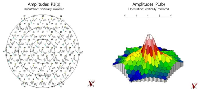
Amplitudes P1(b)
Orientation: vertically mirrored
Amplitudes P1(b)
Orientation: vertically mirrored
Figure 3. Multifocal Electroretinography (Mf-ERG). Neither the trace array nor three-dimensional response density plot demon-
strates any retinal dysfunction corresponding to the region of field defect.시야장애가 지속된 증례를 경험하였기에 이를 보고하는 바 이다.
REFERENCES
1) Rhee DJ, Katz LJ, Spaeth GL, Myers JS. Complementary and alter- native medicine for glaucoma. Surv Ophthalmol 2001;46:43-55.
2) Rhee DJ, Spaeth GL, Myers JS, et al. Prevalence of the use of com- plementary and alternative medicine for glaucoma. Ophthalmology 2002;109:438-43.
3) Kavoussi B, Ross BE. The neuroimmune basis of anti-in- flammatory acupuncture. Integr Cancer Ther 2007;6:251-7.
4) Bäcker M, Grossman P, Schneider J, et al. Acupuncture in mi- graine: investigation of autonomic effects. Clin J Pain 2008;24:106-15.
5) Uchida S, Hotta H. Acupuncture affects regional blood flow in var- ious organs. Evid Based Complement Alternat Med 2008;5:145-51.
6) Calonge M. The treatment of dry eye. Surv Ophthalmol 2001;
45:S227-39.
7) O'Brien K. Complementary and alternative medicine: the move in- to mainstream health care. Clin Exp Optom 2004;87:110-20.
8) Smith JR, Spurrier NJ, Martin JT, Rosenbaum JT. Prevalent use of complementary and alternative medicine by patients with in- flammatory eye disease. Ocul Immunol Inflamm 2004;12:203-14.
9) Patel BC, Clinch TE, Burns TA, et al. Prospective evaluation of topical versus retrobulbar anesthesia: a converting surgeon's experience.
J Cataract Refract Surg 1998;24:853-60.
10) Schrader WF, Schargus M, Schneider E, Josifova T. Risks and se- quelae of scleral perforation during peribulbar or retrobulbar anesthesia. J Cataract Refract Surg 2010;36:885-9.
11) Fielden M, Hall R, Kherani F, et al. Ocular perforation by an acu- puncture needle. Can J Ophthalmol 2011;46:94-5.
12) Oh KT, Boldt HC, Maturi RK, et al. Evaluation of patients with vis- ual field defects following macular hole surgery using multifocal electroretinography. Retina 2000;20:238-43.
13) Cullinane AB, Cleary PE. Prevention of visual field defects after macular hole surgery. Br J Ophthalmol 2000;84:372-7.
=ABSTRACT=
Ocular Perforation and Visual Field Defect Caused by an Acupuncture Needle: a Case Report
Hyunseung Kang, MD, Dong Kyu Lee, MD, Su Jin Lim, MD, Hyoung Eun Kim, MD, Oh Woong Kwon, MD, PhD
Nune Eye Hospital, Seoul, Korea
Purpose: To report a case of globe perforation and linear retinal tear after periocular acupuncture therapy which resulted in persistent temporal field defect with normal retinal function evidenced by multifocal electroretinogram (MERG).
Case summary: A 42-year-old female presented with decreased visual acuity and pain in her right eye after a periocular acupuncture therapy for blepharospasm. At initial presentation, the best corrected visual acuity (BCVA) was 0.08 in the in- jured eye and the intraocular pressure was 15 mmHg. Ultrasonography showed minimal vitreous hemorrhage and fundus examination revealed a linear retinal tear in the posterior pole sparing the macula. Consequently, barrier laser photo- coagulation was performed around the lesion. The patient suffered from metamorphopsia and persistent decreased visual acuity even after 3 months. On fundus examination, epiretinal membrane with macular pucker was observed on the macula. Spectral domain optical coherence tomography (SD-OCT) revealed retinal nerve fiber layer defect with a full-thickness posterior wall tear. Multifocal electroretinogram showed normal retinal function; however, Humphrey visual field test demonstrated field defect corresponding to the injury. A 25-gauge pars plana vitrectomy was performed with membranectomy and ILM peeling. One month postoperatively, improvement in BCVA and metamorphopsia was achieved;
however, the scotomata remained unchanged.
Conclusions: Ocular perforation or retinal tear caused by an acupuncture needle is a rare condition that has not been re- ported previously in Korea. Furthermore, no case of traumatic visual field defect with preserved retinal function has been reported elsewhere. Hence, the authors present a case of isolated visual field defect without retinal dysfunction following full-thickness retinal tear caused by an acupuncture needle.
J Korean Ophthalmol Soc 2013;54(9):1475-1479
Key Words: Acupuncture, Globe perforation, MERG, Ocular perforation, Visual field defect
Address reprint requests to Oh Woong Kwon, MD, PhD Nune Eye Hospital
#404 Seolleung-ro, Gangnam-gu, Seoul 135-841, Korea
Tel: 82-2-2086-7766, Fax: 82-2-2086-7894, E-mail: [email protected]