© 2018 The Korean Ophthalmological Society
This is an Open Access article distributed under the terms of the Creative Commons Attribution Non-Commercial License (http://creativecommons.org/licenses /by-nc/3.0/) which permits unrestricted non-commercial use, distribution, and reproduction in any medium, provided the original work is properly cited.
Original Article
Effectiveness of Intravitreal Ranibizumab for Diabetic Macular Edema with Serous Retinal Detachment
Mahmut Kaya1, Eyyup Karahan2, Taylan Ozturk1, Nilufer Kocak1, Suleyman Kaynak1
1Department of Ophthalmology, Dokuz Eylul University School of Medicine, Izmir, Turkey
2Department of Ophthalmology, Van Training and Research Hospital, Van, Turkey
Purpose: To evaluate the effectiveness of intravitreal injection of ranibizumab (IVR) in treating diabetic macular edema (DME) with serous retinal detachment (SRD) based on spectral domain optical coherence tomography (SD-OCT) patterns.
Methods: One hundred thirty-four eyes of 134 patients with DME who underwent SD-OCT evaluation were includ- ed in this study. We retrospectively analyzed the medical records of patients who received IVR for the treatment of DME. Their eyes were classified into three groups according to the following SD-OCT features: SRD, diffuse retinal thickness and cystoid macular edema. The three groups were compared regarding changes in best-cor- rected visual acuity and central foveal thickness (CFT) after IVR.
Results: The mean age was 61.4 ± 9.2 years (range, 44 to 81 years). The average length of the follow-up period was 9.4 ± 3.4 months (range, 6 to 24 months). The mean CFT value was significantly reduced in all groups (p <
0.001) after treatment. Increases in best-corrected visual acuity were statistically significant for the diffuse retinal thickness and cystoid macular edema groups (p < 0.001 and p < 0.001, respectively). However, there was no significant improvement after IVR injection in the SRD group (p = 0.252). In the SRD group, patients with ellipsoid zone disruption and external limiting membrane disruption demonstrated poorer visual gains at the last follow-up visit (p < 0.005 and p = 0.002, respectively).
Conclusions: A significant reduction in CFT with required IVR injections in DME with SRD was achieved but was accompanied by a worse functional outcome in the SRD group. The presence of subretinal fluid on SD-OCT in study eyes may be a poor prognostic factor for visual acuity.
Key Words: Diabetic macular edema, Ranibizumab, Serous retinal detachment, Spectral domain optical coher- ence tomography, Visual acuity
Macular edema occurs in a wide variety of ocular diseas- es and is a common cause of vision loss in patients with di-
abetic retinopathy [1]. Its complex and multifactorial patho- genesis is not yet fully understood. What is apparent is that the multifactorial disruption of inner and outer blood-reti- nal barriers leads to an abnormal inflow of fluid into the neurosensory retina that exceeds the outflow, producing in- traretinal and subretinal fluid accumulation [2-5]. Using spectral domain optical coherence tomography (SD-OCT), Otani et al. [6] described three patterns of diabetic macular
Received: October 11, 2017 Accepted: December 20, 2017
Corresponding Author: Mahmut Kaya, MD. Department of Ophthal- mology, Dokuz Eylul University School of Medicine, Mithatpasa Cad.
2. Karatas, No:338 D:12, Konak, Izmir, Turkey. Tel: 90-505-525-2216, E-mail: [email protected]
edema (DME): sponge-like swelling, cystoid macular ede- ma (CME), and serous retinal detachment (SRD). SRD as- sociated with CME can only be diagnosed using SD-OCT because it can be hidden beneath CME and therefore missed during fundus fluorescein angiography [7].
Vascular endothelial growth factor (VEGF) is a potent endothelial cell angiogenic factor and a powerful mediator of vascular permeability. It leads to the breakdown of the blood-retinal barrier in diabetic retinopathy, causing leak- age of intravascular fluid from abnormal retinal capillaries and resulting in DME [8]. Therefore, treatment with an- ti-VEGF agents is one of the most promising approaches for the treatment of vision loss due to DME [9,10].
Various studies have established the safety and efficacy of anti-VEGF agents, including ranibizumab and bevaci- zumab, in the treatment of DME [11-13]. However, only a few publications have addressed the issue of why some eyes respond to this treatment more readily than others.
The presence of SRD in retinal vascular diseases, such as diabetes, may affect the treatment results for macular ede- ma associated with retinal vascular leakage and may also limit the ability to perform effective macular laser treat- ment.
There are few reports about the results of bevacuzimab injection on different optical coherence tomographic pat- terns of DME [14,15]. Koytak et al. [14] reported that the CME and SRD subtypes were associated with a greater re- duction in central foveal thickness (CFT) than the diffuse retinal thickness (DRT) subtype. However, changes in vi- sual acuity were not significantly different among the three groups.
The aim of this study was to evaluate the anatomical and functional outcomes based on various patterns of SD-OCT morphology in DME following treatment with intravitreal ranibizumab (IVR) injection.
Materials and Methods
A retrospective chart review of patients with DME who underwent SD-OCT evaluation in the department of oph- thalmology between January 2011 and August 2014 was performed. This study adhered to the tenets of the Decla- ration of Helsinki and was approved by our local ethics committee (2015/01-21). Informed consent was obtained before the investigation began.
In this study, the medical records of patients who had re- ceived an IVR injection for the treatment of DME were retrospectively analyzed. Eyes that had clinically signifi- cant macular edema and a CFT of 300 µm or more deter- mined by SD-OCT (Spectralis HRA+SD-OCT; Heidelberg Engineering, Heidelberg, Germany) were included in the analysis, regardless of diabetic retinopathy stage. If both eyes of the same patient met the inclusion criteria, only one eye was assigned randomly for the study.
All patients underwent macular SD-OCT measurements prior to IVR injection. Eyes that had poor quality SD-OCT scans were excluded from the study. The causes of these poor scans included media opacities, excessive blinking, or persistent eye movement. Other exclusion criteria were oc- ular surgery or trauma, intravitreal or periocular injection of any drug, or laser photocoagulation within six months of the injection; history of any previous vitreoretinal surgi- cal procedure; presence of concomitant retinal pathologies and glaucoma or evidence of vitreomacular traction on SD-OCT. Patients with a follow-up time shorter than six months were also excluded from the study.
During SD-OCT examination, the macula was scanned in six radial sections, including the horizontal, vertical, and oblique planes, through the center of the fovea. The macular thickness was measured automatically by the to- pography software built into the SD-OCT device.
The IVR injections were performed in the operating room under aseptic conditions. Topical anesthesia was achieved by instillation of at least three drops of 0.5%
proparacaine hydrochloride (Alcaine; Alcon Laboratories, Fort Worth, TX, USA). Povidone iodine (5%) was applied to the lids and eyelashes and instilled in the conjunctiva before draping. Next, 0.5 mg/0.05 mL of ranibizumab (Lu- centis; Novartis Pharma AG, Basel, Switzerland) was in- jected using a 30-gauge needle 4 mm posterior to the lim- bus (3.5 mm in pseudophakic eyes). Finally, a drop of 5%
povidone iodine was instilled at the injection site. An eye pad was placed, and 0.5% moxitifloxacin topical drops were prescribed for instillation four times daily.
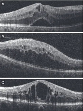
Best-corrected visual acuity (BCVA) with a Snellen chart (BCVA measurements were converted to logarithm of the minimum angle of resolution [logMAR]) and CFT values assessed with SD-OCT prior to the IVR injection and at the last visit were recorded. Eyes were divided into SRD, DRT, and CME groups according to the assessment of macular edema morphology on SD-OCT (Fig. 1A-1C). When more
than one edema pattern was observed, the eye was included in the group of the most obviously predominant pattern. In cases where more than one pattern was present and none was obviously predominant, the eye was not included in the study. The BCVA, macular appearance and SD-OCT find- ings were used to determine whether the patient should re- ceive a repeat injection of IVR. However, it was left at the discretion of the treating physician to follow a specific regi- men or treat patients on an as-needed basis.
All data were analyzed using the SPSS ver. 11.5 (SPSS Inc., Chicago, IL, USA). BCVA measurements were con- verted to logMAR equivalents for statistical analysis. Pear- son chi-square test was used for comparative analyses of categorical variables. The independent sample t-test and paired t-test were employed to analyze changes in BCVA and CFT. One-way analysis of variance (post-hoc Tukey) was used to assess variations among the three groups. For all statistical tests, p < 0.05 was considered significant.
Results
One hundred thirty-four eyes of 134 patients with a min- imum follow-up period of six months were included in the study. The mean follow-up time was 9.4 ± 3.4 months (range, 6 to 24 months). Of the 134 patients, 69 (51.5%) were female, and 65 (48.5%) were male. The mean age was 61.4 ± 9.2 years (range, 44 to 81 years). The baseline demo- graphic and clinical properties of the patients in each group are summarized in Table 1. The three groups did not
Table 1. Demographic and clinical properties of the study groups
SRD (n = 46) DRT (n = 50) CME (n = 38) p-value
Age 59.8 ± 7.1 61.8 ± 10.4 63.0 ± 9.8 0.266
Sex (female / male) 24 / 22 27 / 23 18 / 20 0.822
Type of diabetes (type 1 / type 2) 18 / 28 20 / 30 16 / 22 0.961
Duration of diabetes 16.1 ± 5.7 14.5 ± 6.4 16.8 ± 7.2 0.214
Baseline HbA1c (%) 7.6 ± 1.1 7.5 ± 1.3 7.8 ± 0.9 0.560
PRP (yes / no) 38 / 8 37 / 13 26 / 12 0.311
MGL or focal laser (yes / no) 19 / 27 20 / 30 16 / 22 0.979
Mean number of intravireal injections 4.2 ± 1.5 4.5 ± 1.5 4.4 ± 1.4 0.477
Lens (phakic / pseudophakic) 40 / 6 44 / 6 32 / 6 0.871
Rise in IOP 3 / 46 2 / 50 2 / 38 0.857
SRD = serous retinal detachment; DRT = diffuse retinal thickness; CME = cystoid macular edema; HbA1c = glycated haemoglobin; PRP
= panretinal photocoagulation; MGL = macular grid laser; IOP = intraocular pressure.
Fig. 1. Diabetic macular edema (DME) classification based on spectral domain optical coherence tomography. (A) Patients with predominant serous retinal detachment DME had an associated subretinal collection of fluid under the fovea. (B) Diffuse DME pa- tients demonstrated widespread retinal thickening with a sponge- like appearance of the macula. (C) Patients with focal cystoid DME had a mound-like appearance of the fovea due to focal collection of fluid at the fovea.
A
B
C
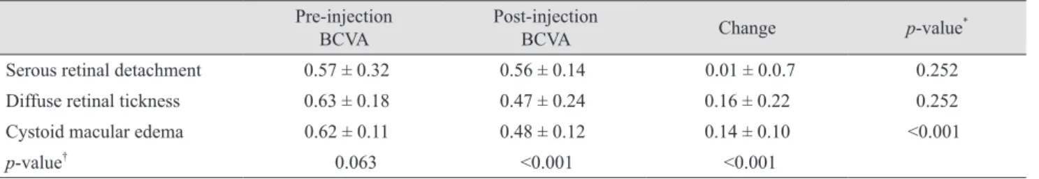
differ significantly in terms of age and gender (p = 0.266 and p = 0.822 respectively). Table 2 and 3 summarize the BCVA (logMAR) and SD-OCT measurement data before and after the injection. Pre-injection mean BCVA did not differ significantly between groups (p = 0.063). The differ- ence between mean pre-injection CFT values of the three groups was also not statistically significant (p = 0.362).
When the pre-injection and post-injection data were com- pared within each group, increases in the BCVA were sta- tistically significant in the DRT and CME groups (p <
0.001 and p < 0.001, respectively), but was not significant in the SRD group (p = 0.252). However, the mean CFT values significantly decreased in all three groups (p <
0.001). There was also a statistically significant difference between groups in terms of postoperative BCVA (p <
0.001). The three groups showed no significant variation in post-injection CFT (p = 0.825). In the SRD group, 34.8%
(16 / 46) had visual improvement; 45.7% (21 / 46) had the same BCVA at the last visit as at preinjection; and 19.6% (9 / 46) of the patients demonstrated deterioration of BCVA at the last visit compared with the pre-injection BCVA. In the DRT group, 80% (40 / 50) experienced visual improve- ment; the BCVA was the same as the pre-injection BCVA at the last visit in 8.0% (4 / 50); and 12.0% (6 / 50) showed deterioration of the BCVA at the last visit compared with
pre-injection BCVA. In the CME group, 78.9% (30 / 38) displayed visual improvement; 13.2% (5 / 38) had no change in BCVA at the last visit; and 7.9% (3 / 38) experi- enced deterioration in BCVA at the last visit compared with pre-injection BCVA.A decrease ˃50 µm in the CFT was accepted as a decreased CFT, while any change
≤50 µm was accepted as no change in CFT. An increase
˃50 µm was defined as an increased CFT. In the SRD group, 71.7% (33 / 46) had a decreased CFT, while 80% (40 / 50) in the DRT group and 81.6% (31 / 38) in the CME group experienced a decreased CFT. Fig. 2 shows the re- sults of all three groups with regard to the change in CFT.
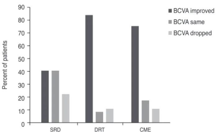
We also evaluated the improvement rates of BCVA in eyes with a decreased CFT. In the SRD group, 39.4% of eyes with a decreased CFT also demonstrated improvement in the BCVA; these rates were 82.9% and 74.2% in the DRT and CME groups, respectively (Fig. 3).
Shifts in the pathomorphology of the macular edema were assessed in each subgroup. In the DRT group, 48.0%
(24 / 50) of the eyes became dry with no or minimal fluid, and 16.0% (8 / 50) retained DRT morphology; 28% became CME (14 / 50), while only 8% (4 / 50) were found to have SRT. In the CME group, 63.2% (24 / 38) of the eyes devel- oped dryness, 36.8% (14 / 38) continued to have CME, and no patient was found to have SRD or DRT. For the SRD Table 2. Pre-injection and post-injection measurements of BCVA (logMAR)
Pre-injection
BCVA Post-injection
BCVA Change p-value*
Serous retinal detachment 0.57 ± 0.32 0.56 ± 0.14 0.01 ± 0.0.7 0.252
Diffuse retinal tickness 0.63 ± 0.18 0.47 ± 0.24 0.16 ± 0.22 0.252
Cystoid macular edema 0.62 ± 0.11 0.48 ± 0.12 0.14 ± 0.10 <0.001
p-value† 0.063 <0.001 <0.001
BCVA = best-corrected visual acuity; logMAR = logarithm of the minimum angle of resolution.
*Paired t-test; †One-way ANOVA (post-hoc Tukey).
Table 3. Pre-injection and post-injection CFT measurements Pre-injection
CFT Post-injection
CFT Change p-value*
Serous retinal detachment 538.2 ± 163.9 382.3 ± 101.5 -155.9 ± 152.8 <0.001
Diffuse retinal thickness 531.2 ± 158.8 356.2 ± 88.7 -174.9 ± 160.2 <0.001
Cystoid macular edema 492.0 ± 145.4 326.5 ± 95.4 -165.5 ± 134.3 <0.001
p-value† 0.362 0.030 0.825
CFT = central foveal thickness.
*Paired t-test; †One-way ANOVA (post-hoc Tukey).
group, 21.7% (10 / 46) of the eyes became dry, 19.6% (9 / 46) and 17.4% (8 / 46) of the eyes converted to having DRT and CME, respectively, while 41.3% (19 / 46) remained with SRD.
An analysis of the outer retinal structures (including the external limiting membrane [ELM] and inner segment el- lipsoidal band layer [previously known as the boundary of the inner segment and outer segment junction]) was car- ried out; ELM was found to be disrupted in 12 eyes (26%) in the SRD group, 6 eyes (12%) in the DRT group, and 6 eyes (15.8%) in the CME group. The ellipsoidal layer was disrupted in 10 eyes (21.7%) in the SRD group, 7 eyes (14%) in the DRT group, and 5 eyes (13.2%) in the CME group. In the DRT and CME groups, patients with an in- tact ELM and integrity of the ellipsoid zone layer showed significant visual improvement post-injection compared with eyes with disruptions of the ELM and the ellipsoid zone layer (p = 0.004 and p = 0.008, respectively). Howev- er, in the SRD group, patients with ellipsoid zone and ELM disruption experienced poorer visual gains at the last follow-up visit (p < 0.005 and p = 0.002, respectively).
No inflammation or severe decreases in vision immedi- ately following the injection were noted, although 7 (5.2%) eyes (3 in the SRD group, 2 in the DRT group, and 2 in the CME group) showed elevated IOP. These cases were man- aged with conservative medical treatment. At the final fol- low-up, no ocular or systemic adverse events, such as thromboembolic events (cerebrovascular accidents, tran- sient ischemic attacks, myocardial infarctions, or peripher- al vascular diseases), were reported.
Discussion
The main cause of vision impairment in diabetic patients is DME. A large epidemiological study indicated that mac- ular edema was present in 15% of patients with diabetic retinopathy [16]. The pathogenesis of DME is complex and multifactorial. Disruption of the inner and outer blood-ret- inal barriers leads to abnormal inflow of fluid into the neu- rosensory retina that exceeds the outflow, producing intra- retinal and subretinal fluid accumulation [2,3]. However, the specific details of the pathogenesis of DME remain un- clear. Classification of DME according to the SD-OCT findings might be helpful to obtain more information about the pathogenesis of DME. The first SD-OCT classi- fication of DME was reported by Otani et al. [6]. Although various patterns of DME have been recognized on SD- OCT, few published studies have reported the visual out- comes for these SD-OCT patterns in DME patients treated with anti-VEGF therapy. Kim et al. [17] concluded that the intravitreal injection of bevacizumab was more effective in the DRT type than in the CME or SRD types of DME.
Wu et al. [18] found that patients with cystoid changes gained greater improvements in visual acuity and macular thickness and volume after intravitreal bevacizumab injec- tion. However, Koytak et al. [14] stated that there was no statistically significant variation between focal, cystoid, and neurosensory detachment groups regarding changes in BCVA after injection of intravitreal bevacizumab.
Shimura et al. [19] reported that the foveal thickness in all patterns was reduced, but the reduction ratios in the DRT and CME groups were significantly greater than that Fig. 2. Central foveal thickness (CFT) changes of the groups. SRD
= serous retinal detachment; DRT = diffuse retinal thickness;
CME = cystoid macular edema.
Fig. 3. Results of best-corrected visual acuity (BCVA) in eyes with decreased diabetic macular edema. SRD = serous retinal detachment;
DRT = diffuse retinal thickness; CME = cystoid macular edema.
90 80 70 60 50 40 30 20 10 0
Percent of patients
Groups
SRD DRT CME
Decreased CFT Same CFT Increased CFT
90 80 70 60 50 40 30 20 10 0
Percent of patients
Groups
SRD DRT CME
BCVA improved BCVA same BCVA dropped
seen in the SRD group following intravitreal bevacuzimab injection. Similarly, improvement in visual acuity in the DRT and CME groups was significantly greater than that in the SRD group; therefore, it was concluded that the ef- fectiveness of intravitreal bevacuzimab injection in reduc- ing macular edema varied depending on the SD-OCT pat- tern and was greatest in the DRT group, intermediate in the CME group, and weakest in the group with the SRD pattern. In our study, the pre-injection BCVA and CFT val- ues were similar in all three groups. The observed decrease in CFT at the last follow-up visit was not statistically sig- nificant between the three groups, whereas the improve- ment in BCVA was statistically worse in the SRD group compared with the DRT and CME groups. The ELM and ellipsoid zone integrities were significantly correlated with post-treatment visual acuity and were significantly lower in the SRD type than in the other types. In eyes with SRD, outer retinal structures were found to be associated with poor visual recovery. In our series, 34 of 41 DRT eyes (82.3%) with a decrease ˃50 µm also showed improvement in BCVA. In the CME group, 23 of 31 (74.2%) eyes with a decrease ˃50 µm demonstrated improvement in BCVA.
However, in the SRD group, only 13 of 33 (39.4%) eyes with a decrease ˃50 µm experienced an improvement in BCVA. In 2016, Seo et al. [20] reported that vision gains and retinal anatomy improvement were maintained in all three types during the first year of IVR administration, which is in agreement with our study findings. Additional- ly, the BCVA of SRD (20 / 60) patients was significantly worse than that of the other types (DRT = 20 / 38; CME = 20 / 43) after 12 months. In contrast, Giocanti-Auregan et al. [21] found in 2017 that similar BCVA gains were ob- served regardless of the presence of SRD. The higher visu- al gain usually observed in DME with SRD could be asso- ciated with a lower baseline BCVA.
The pathogenesis of DRT involves the persistent break- down of the inner blood retinal barrier from the loss of an- chor proteins in the tight junctions of the capillary endo- thelial cells [22]. Vascular endothelial growth factor is a pluripotent growth factor that acts as an endothelial cell-specific mitogen and vasopermeability factor and thus plays a critical role in promoting angiogenesis and vascular leakage [23-25]. Therefore, IVR reduces hypervasoperme- ability, leading to a reduction of DRT. Although the patho- genesis of CME remains unknown, the reduction of CME seen with intravitreal bevacizumab and IVR injections
suggests that CME formation is partly dependent on VEGF. The pathogenesis of SRD associated with DME oc- curs due to transient migration of fluid from the cystoid spaces in the retina to the subretinal space [7]. Another theory is that SRD occurs subsequent to failure of the reti- nal pigment epithelium (RPE) pump mechanism [26]. The breakdown of the RPE pump or disruption of the tight junctions between adjacent RPE cells results in intraretinal edema and SRD [27-29]. IVR may have little efficacy in the regression of diabetes-induced RPE impairment, which suggests that VEGF is not primarily responsible for SRD formation in DME.
In conclusion, the visual improvement noted in DME eyes with SRD was lower than that experienced by eyes with DRT and CME. Disruption of the photoreceptor in- tegrity was correlated with a poorer visual outcome and appeared more frequently in the SRD group. Based on our findings, we believe that different treatment modalities should be considered for DME with SRD. More prospec- tive studies are warranted to better understand the patho- genesis of DME.
Conflict of Interest
No potential conflict of interest relevant to this article was reported.
References
1. Patz A, Schatz H, Berkow JW, et al. Macular edema: an overlooked complication of diabetic retinopathy. Trans Am Acad Ophthalmol Otolaryngol 1973;77:OP34-42.
2. Do Carmo A, Ramos P, Reis A, et al. Breakdown of the inner and outer blood retinal barrier in streptozotocin-in- duced diabetes. Exp Eye Res 1998;67:569-75.
3. Sander B, Larsen M, Moldow B, Lund-Andersen H. Dia- betic macular edema: passive and active transport of fluo- rescein through the blood-retina barrier. Invest Ophthalmol Vis Sci 2001;42:433-8.
4. Tso MO, Cunha-Vaz JG, Shih CY, Jones CW. Clinicopath- ologic study of blood-retinal barrier in experimental diabe- tes mellitus. Arch Ophthalmol 1980;98:2032-40.
5. Bhagat N, Grigorian RA, Tutela A, Zarbin MA. Diabetic macular edema: pathogenesis and treatment. Surv Ophthal-
mol 2009;54:1-32.
6. Otani T, Kishi S, Maruyama Y. Patterns of diabetic macu- lar edema with optical coherence tomography. Am J Oph- thalmol 1999;127:688-93.
7. Kang SW, Park CY, Ham DI. The correlation between flu- orescein angiographic and optical coherence tomographic features in clinically significant diabetic macular edema.
Am J Ophthalmol 2004;137:313-22.
8. Ciulla TA, Harris A, Latkany P, et al. Ocular perfusion abnor- malities in diabetes. Acta Ophthalmol Scand 2002;80:468-77.
9. Thomas BJ, Shienbaum G, Boyer DS, Flynn HW Jr. Evolving strategies in the management of diabetic macular edema:
clinical trials and current management. Can J Ophthalmol 2013;48:22-30.
10. Stewart MW. Anti-vascular endothelial growth factor drug treatment of diabetic macular edema: the evolution contin- ues. Curr Diabetes Rev 2012;8:237-46.
11. Massin P, Bandello F, Garweg JG, et al. Safety and ef- ficacy of ranibizumab in diabetic macular edema (RE- SOLVE Study): a 12-month, randomized, controlled, double-masked, multicenter phase II study. Diabetes Care 2010;33:2399-405.
12. Diabetic Retinopathy Clinical Research Network, Scott IU, Edwards AR, et al. A phase II randomized clinical trial of in- travitreal bevacizumab for diabetic macular edema. Ophthal- mology 2007;114:1860-7.
13. Arevalo JF, Sanchez JG, Wu L, et al. Primary intravitre- al bevacizumab for diffuse diabetic macular edema: the Pan-American Collaborative Retina Study Group at 24 months. Ophthalmology 2009;116:1488-97.
14. Koytak A, Altinisik M, Sogutlu Sari E, et al. Effect of a single intravitreal bevacizumab injection on different op- tical coherence tomographic patterns of diabetic macular oedema. Eye (Lond) 2013;27:716-21.
15. Cheema HR, Al Habash A, Al-Askar E. Improvement of vi- sual acuity based on optical coherence tomography patterns following intravitreal bevacizumab treatment in patients with diabetic macular edema. Int J Ophthalmol 2014;7:251-5.
16. Petrella RJ, Blouin J, Davies B, Barbeau M. Prevalence, demographics, and treatment characteristics of visual im- pairment due to diabetic macular edema in a representative Canadian cohort. J Ophthalmol 2012;2012:159-67.
17. Kim M, Lee P, Kim Y, et al. Effect of intravitreal bevaci- zumab based on optical coherence tomography patterns of diabetic macular edema. Ophthalmologica 2011;226:138-44.
18. Wu PC, Lai CH, Chen CL, Kuo CN. Optical coherence tomographic patterns in diabetic macula edema can predict the effects of intravitreal bevacizumab injection as prima- ry treatment. J Ocul Pharmacol Ther 2012;28:59-64.
19. Shimura M, Yasuda K, Yasuda M, Nakazawa T. Visual outcome after intravitreal bevacizumab depends on the optical coherence tomographic patterns of patients with diffuse diabetic macular edema. Retina 2013;33:740-7.
20. Seo KH, Yu SY, Kim M, Kwak HW. Visual and morpho- logic outcomes of intravitreal ranibizumab for diabetic macular edema based on optical coherence tomography patterns. Retina 2016;36:588-95.
21. Giocanti-Auregan A, Hrarat L, Qu LM, et al. Functional and anatomical outcomes in patients with serous retinal detachment in diabetic macular edema treated with ranibi- zumab. Invest Ophthalmol Vis Sci 2017;58:797-800.
22. Antonetti DA, Barber AJ, Khin S, et al. Vascular permea- bility in experimental diabetes is associated with reduced endothelial occludin content: vascular endothelial growth factor decreases occludin in retinal endothelial cells. Penn State Retina Research Group. Diabetes 1998;47:1953-9.
23. Aiello LP, Avery RL, Arrigg PG, et al. Vascular endothe- lial growth factor in ocular fluid of patients with diabetic retinopathy and other retinal disorders. N Engl J Med 1994;331:1480-7.
24. Neufeld G, Cohen T, Gengrinovitch S, Poltorak Z. Vas- cular endothelial growth factor (VEGF) and its receptors.
FASEB J 1999;13:9-22.
25. Grant MB, Afzal A, Spoerri P, et al. The role of growth factors in the pathogenesis of diabetic retinopathy. Expert Opin Investig Drugs 2004;13:1275-93.
26. Gaucher D, Sebah C, Erginay A, et al. Optical coherence tomography features during the evolution of serous retinal detachment in patients with diabetic macular edema. Am J Ophthalmol 2008;145:289-96.
27. Ozdemir H, Karacorlu M, Karacorlu S. Serous macular detachment in diabetic cystoid macular oedema. Acta Oph- thalmol Scand 2005;83:63-6.
28. Fatt I, Shantinath K. Flow conductivity of retina and its role in retinal adhesion. Exp Eye Res 1971;12:218-26.
29. Nagelhus EA, Veruki ML, Torp R, et al. Aquaporin-4 water channel protein in the rat retina and optic nerve: po- larized expression in Müller cells and fibrous astrocytes. J Neurosci 1998;18:2506-19.