pISSN: 0378-6471 eISSN: 2092-9374
http://dx.doi.org/10.3341/jkos.2011.52.9.1114
= 증례보고 =
황반박리가 동반된 시신경유두소와에서 레이저 없이 시행한 유리체절제술 후 장기관찰 1예
홍정흠⋅김윤영
대구가톨릭대학교 의과대학 안과학교실
목적: 시신경유두소와에 의한 황반박리에서 레이저치료 없이 유리체절제술과 가스 주입술을 행한 후 장기 관찰을 한 증례를 보고하고자 한다.
증례요약: 좌안 시력 저하를 주소로 내원한 13세 남자 환자에서 좌안 시신경 하이측 부위의 유두소와에 동반된 황반박리가 관찰되었다.
빛간섭단층촬영상 유리체견인을 시사하는 유리체가닥이 시신경위에서 발견되었고 망막층간분리가 확인되었다. 치료는 후유리체박리 를 완전히 일으킨 후 유리체절제술을 시행하였으며, 가스를 주입한 후 엎드린 자세를 유지하였으며 레이저치료는 시행하지 않았다.
술 후 6개월째 망막이 완전히 유착되었으나, 15개월 후 황반박리가 재발되었다. 가스 주입술만을 다시 시행한 후 2년 후까지 완전한 망막유착을 보이고 있으며 시력도 향상되었다.
결론: 레이저치료 없이 유리체절제술 후 가스 주입술만으로도 유두소와와 관련된 황반박리의 다른 치료방법이 될 수 있다. 그러나 재발 이 일어날 수 있으며 이 경우 가스 주입술만으로 좋은 효과를 볼 수 있었다. 이러한 결과는 유리체 견인 외 다른 요소가 유듀소와와 관련된 황반박리를 일으킬 수 있음을 시사한다. 좀 더 많은 증례를 통하여 유두소와에 동반된 황반박리의 치료에, 레이저치료 없이 시행한 유리체절제술과 가스 주입술의 효과가 규명되어야 할 것이다.
<대한안과학회지 2011;52(9):1114-1118>
■ 접 수 일: 2010년 9월 28일 ■ 심사통과일: 2011년 1월 24일
■ 게재허가일: 2011년 8월 5일
■ 책 임 저 자: 김 윤 영
대구시 남구 대명 4동 3056-6 대구가톨릭대학교병원 안과
Tel: 053-650-4737, Fax: 053-627-0133 E-mail: [email protected]
* 본 논문의 요지는 2010년 한국망막학회 학술대회에서 구연으로 발표되었음.
시신경유두소와는 드물게 나타나는 선천성 질환으로 약 2/3에서 황반박리가 발생하는 것으로 보고되고 있다.1자연 경과상 황반박리의 회복이 일어날 수 있지만 오래 방치할 경우 황반의 낭포성 변화, 표층 황반원공, 망막색소상피세 포의 탈색소, 시신경세포의 변성 등으로 시력의 예후가 좋 지 않다.2그러나 아직까지 황반박리가 일어나는 정확한 병 태생리와 장액성 망막하액의 기원 등에 대해서는 논란이 있으며 이로 인하여 이 질환의 치료를 선택하는 데 많은 혼 란이 있다. 시신경유두 경계부위의 레이저치료, 유리체강내 가스주입술, 황반돌융술, 술자에 따라 변형된 방법들이 적 용된 유리체절제술 등이 시행되어 왔으며 다양한 결과들이 보고되었다.3-8
최근 빛간섭단층촬영을 이용한 검사에서 황반박리의 형 태가 단순한 황반박리가 아니라 유두소와와 연결된 망막층 간분리와 동반되어 있는 형태학적인 특징들이 보고되었고
많은 예에서 유리체황반견인, 시신경에 부착된 유리체가닥, 부분 후유리체박리와 같은 유리체 이상들이 보고되면서 유 리체 견인이 이 질환의 발생에 중요한 역할을 할 것이라는 견해들이 많다.9,10
저자들은 상기한 빛간섭단층촬영상의 특징을 보인 환자 를 대상으로 유리체 견인이 황반박리를 일으킨 것으로 판 단하여 유리체절제술을 시행하고 레이저 없이 안내가스만 을 주입한 후 경과관찰을 한 증례를 보고하고자 하며 이후 발생한 재발에 대해 안내가스 주입술만으로 비교적 장기간 의 황반유착을 보인 경우를 보고하면서 질환의 병태생리와 재발, 이후 치료와 관련된 사항들에 대한 저자들의 생각을 발표하고자 한다.
증례보고
13세의 남자가 한 달간의 좌안 시력의 저하를 주소로 내 원하였다. 내원 당시 좌안의 교정시력은 0.06이었으며 외 상이나 특이한 전신질환의 병력은 없었다. 안저검사상 좌안 시신경 하이측 부위에 난원형의 결손이 관찰되었으며 황반 박리가 동반되었다. 형광안저촬영상 후기에 시신경 결손부 위의 염색에 의한 과형광과 장액성황반박리 부위의 누출로 인한 과형광을 관찰할 수 있었다. 빛간섭단층촬영상 시신경
A B
C D
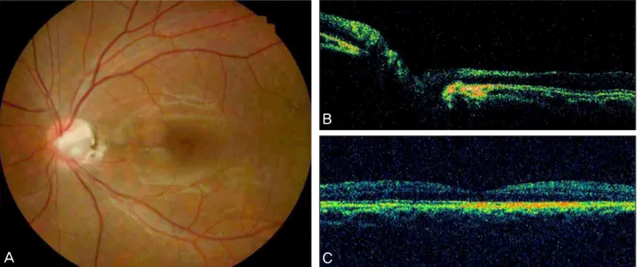
Figure 1. (A) Preoperative fundus photograph of the left eye showing inferotemporal optic disc pit associated serous macular
detachment. (B) Late phase of the fluorescein angiography showing hypofluorescence of the optic disc pit and dye pooling into sub- retinal fluid. (C, D) Optical coherence tomography image showing serous macular detachment, schisis-like separation of the retinal layers and dense vitreous strands (white arrow) attached to the optic disc pit.A B
C Figure 2. (A) Optical coherence tomography (OCT) image
showing macular attachment but residual subretinal fluid inferior to the macula. (B) OCT image showing complete resorption of subretinal fluid at 6 months after first operation and (C) re- accumulation of subretinal fluid in the papillomacular area at 15 months after first operation.
유두소와에 인접한 망막의 층간분리와 황반박리를 관찰할 수 있었으며 시신경위에 비정상적인 유리체가닥이 관찰되 었다(Fig. 1).
수술은 표준적인 20 G 유리체절제술을 시행하였으며 술 중 트리암시놀론을 이용하여 후유리체박리를 유도하였다.
이 때 황반박리의 가장자리부터 후유리체막이 상당히 단단 히 붙어 있음을 확인하였으나 별다른 합병증 없이 후유리
체박리를 만들었으며 유리체절제술 후 액체 공기치환술을 시행하면서 유두소와의 함몰부위를 통하여 배액을 시도하 였으나 충분하게 이루어지지 않았다. 14% C3F8 가스를 주 입 후 엎드린 자세를 일주일간 유지시켰다. 환자는 술 후 1 개월째 황반은 유착되었으나 황반 하측부위에 약간의 망막 하액이 빛간섭단층촬영상 관찰되었으며 이는 약 6개월째까 지 지속되다가 완전히 소실되었으며 이 때 시력은 0.25로
A
B
C
Figure 3. Fundus photograph (A) and optical coherence tomography image (B, C) of the left eye reveals complete attached macula
at two years after only gas tamponade procedure.향상되었다(Fig. 2). 술 후 15개월째 환자가 다시 시력저하 를 호소하여 빛간섭단층촬영을 시행한 뒤 황반박리가 재발 된 것을 확인하고 14% C3F8 가스만을 주입하고 술 후 22 일째 완전히 망막이 유착됨을 확인할 수 있었으며 가스 주 입술 후 2년째까지 망막은 잘 유착된 상태를 유지하면서 시 력은 0.4로 향상되었다(Fig. 3).
고 찰
본 증례가 기존의 국내보고와 다른 점은 시신경유두소와 에 동반된 황반박리의 치료를 위해 확실한 후유리체박리를 유도한 후 유리체절제술을 시행하면서 레이저 치료를 하지 않았다는 점과 비록 한 증례이지만 비교적 긴 장기간의 추 적관찰을 했다는 점이다. 이와 더불어 빛간섭단층촬영상 시 신경에 유착된 비정상적인 유리체가닥과 망막층간분리와 동반된 황반박리 등의 소견을 확인할 수 있었다는 점이다.
이 질환에서 발생하는 황반박리의 치료를 위해 레이저를 시행하는 것은 시신경유두의 경계부위에 망막맥락막의 유 착을 통해 시신경유두소와와 망막사이의 통로를 영구적으 로 막기 위한 이론적인 배경을 갖고 있으나3단독으로 사용 할 경우 실패의 빈도가 높아 최근에는 유리체절제술과 함 께 시도되는 경향이 있다.6 그러나 본 증례같이 레이저 시 술 부위에 망막층간 분리가 있는 경우 레이저치료로 이 통 로를 막는다는 것은 이론적으로 맞지 않고 큰 장액성망막 박리가 있는 경우에는 레이저치료가 불가능하다. Ryu et al5은 이러한 점을 고려하여 유리체절제술과 가스 주입술 후 레이저치료 할 부위의 망막박리가 유착되고 나서 레이 저치료를 함으로써 치료의 효과를 높일 수 있었다는 보고
를 한 바 있다. 그러나 아무리 약한 레이저 치료를 하더라 도 시신경 주위 혹은 황반유두다발 내 레이저의 치료는 진 한 암점과 같은 부작용이 발생할 수 있으며 이 레이저의 목 적이 장벽을 만들어 준다는 것을 고려한다면 레이저의 세 기를 높일 수 밖에 없기 때문에 레이저로 인한 부작용이 커 질 수 있다는 점이다.11,12저자들은 이 질환의 발생에 유리 체 견인이 중요한 역할을 한다고 판단하였으며 수술 중 확 실한 후유리체박리를 만들어주면 망막내층으로 유입되는 새로운 체액의 유입을 막아 결국 황반박리가 호전될 것으 로 생각하여 레이저 치료를 추가하지 않았다. 유리체견인과 같은 이상이 이 질환의 발생에 영향을 미칠 수 있다는 견해 는 최근의 빛간섭단층촬영을 이용한 연구결과를 통해 확인 될 수 있으며 또한 황반돌융술을 통해 성공적으로 황반을 유착시킬 수 있다는 보고에서 그 근거를 찾을 수 있는데 Theodossiadis et al10은 황반돌융술이 유리체견인의 방향 을 바꾸어 줌으로써 이 질환과 관련된 황반박리를 치료할 수 있다고 발표하였다.
본 증례의 경우 수술 후 1개월 내로 황반은 유착이 되었 지만 황반의 아래쪽으로 망막하액이 남아있다가 술 후 6개 월째 망막이 완전히 유착됨을 확인할 수 있었다. Hirakata et al7의 연구에 의하면 술 후 완전한 망막의 유착은 2개월 에서 12개월 사이에 일어난다고 발표한 바 있어 일치되는 소견으로 볼 수 있다.
황반견인을 제거하는 것이 수술 성공의 가장 중요한 요 인으로 생각될 수 있지만 본 증례처럼 술 후 15개월째 다시 망막하액이 발생했다는 사실을 통해 저자들은 아마도 황반 견인 외 다른 요인이 황반박리를 일으킬 수 있을 것으로 생 각하였다. 물론 레이저 치료를 하지 않았다는 점이 큰 요인
일 수 있지만 레이저 치료 후에도 재발이 될 수 있다는 보 고가 있으며6새로운 망막의 견인이 그 원인이 될 수 있겠 지만 본 증례처럼 유리체강내 가스 주입술만으로 황반이 오랫동안 다시 유착되고 있다는 사실을 볼 때 저자들은 다 른 요인이 다시 황반박리를 일으킨 것으로 생각하였다.
Doyle et al13은 고해상도의 빛간섭단층촬영을 이용한 연구 에서 유두소와를 가진 환자에서 시신경 유두함몰부위를 가 로지르는 막을 관찰할 수 있었으며 황반박리가 있는 환자 에서는 이를 확인할 수 없었다고 하였다. 그들은 이러한 막 들이 비정상적인 신경 섬유와 색소성 조직을 포함한 원시 망막조직으로 이루어져 있으며 망막하액의 유입을 막아주는 장벽이 될 수 있다고 발표하였으며 Johnson and Johnson14 은 이러한 막에 작은 구멍이나 열공을 관찰할 수 있었다고 발표한 바 있다.
유리체절제술을 시행한 경우에도 재발이 많이 일어날 수 있다는 보고는 있지만 그 원인에 대한 고찰들은 드물다. 비 록 한 증례이지만 최근의 보고들에 근거하여 재발성 황반 박리의 이유에 대한 저자들의 의견은 비록 본원의 빛간섭 단층촬영으로는 확인할 수 없었지만 유두소와를 덮고 있는 막이 다시 손상을 입어 망막하액의 유입을 가능하게 할 수 있다는 것이다. 본 증례처럼 가스주입술만으로 다시 황반이 유착되는 것은 가스가 막의 손상된 구조의 치유에 도움을 주면서 체액의 유입을 막기 때문인 것으로 생각하지만 향후 좀 더 많은 증례를 통해 이 부분이 규명되어야 될 것이다.
결론적으로 저자들은 유두소와와 관련된 황반박리환자 에서 레이저 치료 없이 유리체절제술과 가스주입술을 이용 하여 오랫동안 황반을 유착시켰으나 재발이 되어 가스주입 술만으로 다시 황반을 유착시킬 수 있었다. 좀 더 많은 증 례와 해상도가 높은 진단 장비를 통하여 질환의 병태생리 와 치료에 관한 연구가 필요할 것으로 생각한다.
참고문헌
1) Sobol WM, Blodi CF, Folk JC, Weingeist TA. Long term visual outcome in patients with optic nerve pit and serous retinal detach- ment of the macula. Ophthalmology 1990;97:1539-42.
2) Brown GC, Brown MM. Repair of retinal detachment associated with congenital excavated defects of the optic disc. Ophthalmic Surg 1995;26:11-5.
3) Gass JD. Serous detachment of the macula: secondary to congenital pit of the optic nervehead. Am J Ophthalmol 1969;67:821-41.
4) Lincoff H, Yannuzzi L, Singerman L, et al. Improvement in visual function after displacement of the retinal elevation emanating from optic pits. Arch Ophthalmol 1993;111:1071-9.
5) Ryu JW, Ra H, Lee WK. A case of surgically treated serous macular detachment associated with optic disc pit. J Korean Ophthalmol Soc 2010;51:155-8.
6) Ghosh YK, Banerjee S, Konstantinidis A, et al. Surgical manage- ment of optic pit associated maculopathy. Eur J Ophthalmol 2008;18:142-6.
7) Hirakata A, Okada AA, Hida T. Long term results of vitrectomy without laser treatment for macular detachment associated with an optic disc pit. Ophthalmology 2005;112:1430-5.
8) Theodossiadis GP. Treatment of maculopahty associated with optic disc pit by sponge explant. Am J Ophthalmol 1996;121:630-7.
9) Karacorlu SA, Karacorlu M, Ozdemir H, et al. Optical coherence tomography in optic pit maculopahty. Int Ophthalmol 2007;27:293-7.
10) Theodossiadis PG, Grigoropoulos VG, Emfietzoglou J, Theodossiadis GP. Vitreous findings in optic disc pit maculopahty based on opti- cal coherence tomography. Grafe’s Arch Clin Exp Ophthalmol 2007;245:1311-8.
11) Blair CJ, Gass JD. Photocoaulation of the macula and papil- lomacular bundle in the human. Arch Ophthalmol 1972;88:167-71.
12) Roider J. Laser treatment of retinal disease by subthreshold laser effect. Semin Ophthalmol 1999;14:19-26.
13) Doyle E, Trivedi D, Good P, et al. High resolution optical coher- ence tomography demonstration of membranes spanning optic disc pits and colobomas. Br J Ophthalmol 2009;93:360-5.
14) Johnson TM, Johnson MW. Pathogenesis implications of sub- retinal gas migration through pits and atypical colobomas of the optic nerve. Arch Ophthalmol 2004;122:1793-800.
=ABSTRACT=
A Case of Vitrectomy without Laser for Serous Macular Detachment Associated with Optic Disc Pit
Jung Heum Hong, MD, Yun Young Kim, MD, PhD
Department of Ophthalmology, Catholic University of Daegu College of Medicine, Daegu, Korea
Purpose: To report a long term result of vitrectomy, gas tamponade without laser retinopexy for serous macular detach- ment associated with an optic disc pit (ODP).
Case summary: A 13 year old boy with visual disturbance in the left eye showed serous macular detachment associated with an inferior temporal ODP. The abnormal vitreous strand over the optic disc implying vitreous traction and retinoschisis were revealed by the optical coherence tomography (OCT) examination. Pars plana vitrectomy after complete induction of posterior vitreous detachment without laser retinopexy, and gas tamponade with postoperative facedown positioning were performed. Complete retinal attachment occurred at 6 months after first operation but recurrent macula detachment occurred at 15 months after first operation. Additional gas tamponade resulted in successful retinal attachment for more than 2 years and visual improvement.
Conclusions: Vitrectomy and gas tamponade without additional laser photocoagulation could be another option for the treatment of ODP maculopathy. But recurrent macular detachment might occur and simple gas tamponade was effective in this case. This result supports another factor in addition to vitreous traction may play a role in the development of the macular detachment associated ODP. Further studies are required to evaluate the effect of vitrectomy, gas tamponade without laser retinopexy for the treatment of ODP maculopathy.
J Korean Ophthalmol Soc 2011;52(9):1114-1118
Key Words: Maculopathy, Optic disc pit, Vitectomy without laser retinopexy
Address reprint requests to Yun Young Kim, MD, PhD
Department of Ophthalmology, Daegu Catholic University Medical Center
#3056-6 Daemyeong 4-dong, Nam-gu, Daegu 705-718, Korea Tel: 82-53-650-4737, Fax: 82-53-627-0133, E-mail: [email protected]