Received April 20, 2018, Revised August 1, 2018, Accepted for publication August 2, 2018
Corresponding author: Weon Ju Lee, Department of Dermatology, Kyungpook National University Hospital, 130 Dongdeok-ro, Jung-gu, Daegu 41944, Korea. Tel: 82-53-420-5838, Fax: 82-53-426-0770, E-mail:
weonju@knu.ac.kr
ORCID: https://orcid.org/0000-0001-5708-1305
This is an Open Access article distributed under the terms of the Creative Commons Attribution Non-Commercial License (http://creativecommons.
org/licenses/by-nc/4.0) which permits unrestricted non-commercial use, distribution, and reproduction in any medium, provided the original work is properly cited.
Copyright © The Korean Dermatological Association and The Korean Society for Investigative Dermatology
Ann Dermatol Vol. 31, No. 1, 2019 https://doi.org/10.5021/ad.2019.31.1.22
ORIGINAL ARTICLE
Efficacy of Bacteriophages in Propionibacterium acnes-Induced Inflammation in Mice
Min Ji Kim, Dong Hyuk Eun, Seok Min Kim, Jungmin Kim1, Weon Ju Lee
Departments of Dermatology and 1Microbiology, School of Medicine, Kyungpook National University, Kyungpook National University Hospital, Daegu, Korea
Background: Bacteriophages have been introduced as living drugs for infectious diseases; thus, they may provide an alter- native to conventional acne therapeutics in patients with non-responsive acne. Objective: We investigated the effect of bacteriophages using an acne mouse model with Propionibacterium acnes-induced inflammatory nodules by clinical examination, pathology, and immunohistochemical analysis. Methods: A human-isolated P. acnes suspension (109 colony forming units/μl) was injected into the backs of HR-1 mice. Group A was used as a control, Group B was in- jected on the back with P. acnes 4 weeks following the initial P. acnes suspension injection, and group C was injected on the back with P. acnes and bacteriophages 4 weeks following the initial P. acnes suspension injection. Clinical and histo- pathological evaluations were performed. Results:
Inflammatory nodule size decreased with time in all groups.
Group C showed the greatest decrease in size, followed by group B and group A. The histopathological findings showed a decrease in epidermal thickness and the number and size of microcomedone-like cysts in groups B and C compared to group A. Immunohistochemistry revealed similar expression of integrin α6, the epidermal proliferation marker, infiltra- tion of CD4/CD8 T cells and neutrophils, and expression of
myeloperoxidase, interleukin-1β, toll-like receptor-2, LL-37, and matrix metalloproteinase-2/3/9 in all three groups.
Conclusion: Using an acne mouse model with P. acnes-in- duced inflammatory nodules, we demonstrate that bacter- iophages may constitute an alternative to conventional acne therapies. However, additional studies are needed for hu- man applications. (Ann Dermatol 31(1) 22∼28, 2019) -Keywords-
Acne vulgaris, Bacteriophage, Mice, Propionibacterium acnes
INTRODUCTION
Acne vulgaris is a very common skin disease of the pilose- baceous follicles characterized by comedones, papules, pustules, nodules, and cysts1. One of the main causative factors of acne is colonization of the follicular infundibulum and sebaceous duct by Propionibacterium acnes. P. acnes plays an important role in inducing an inflammatory event2. In addition, P. acnes can lead to the development of abnormal differentiation and keratinization in epi- dermal keratinocytes1. Therefore, P. acnes may act as a major causative factor in early inflammatory papules of acne.
The establishment of animal models for various diseases is helpful for expanding research fields and developing the production of new therapeutic modalities. Although there are various acne animal models, such as the Mexican hair- less dog, the Rhino mouse, and the rabbit ear assay, eluci- dative models are still needed3. Previously, we reported the establishment of an acne mouse model; HR-1 mice de- velop acne-like inflammatory nodules on their backs fol- lowing injection of P. acnes, in contrast to BALB/c, VDR
k/o, and SCID mice4.
Bacteriophages are bacterial viruses consisting of proteins that encapsulate a DNA or RNA genome5; they are widely distributed in sites populated by bacteria. Bacteriophages replicate within bacteria after injecting their genome into the bacterial cytoplasm. They tend to kill only specific pathogens without damaging the normal flora; thus, they can be used as living drugs for bacterial infections, includ- ing acne6. Bacteriophages targeting P. acnes have been isolated from acne patients7.
In this study, we investigated the efficacy of bacter- iophages in a P. acnes-induced acne mouse model by clinical examination, histopathology, and immunohisto- chemical studies.
MATERIALS AND METHODS
P. acnes culturing
P. acnes (ATCC 11828) were isolated from pustular le- sions of Korean patients with moderate inflammatory acne. P. acnes from post log phase cultures were grown on brain heart infusion agar under anaerobic conditions at 37oC. P. acnes cells were harvested by centrifugation at 5,000×g for 10 minutes, washed three times with phos- phate buffered saline (PBS), and resuspended to a concen- tration of 109 colony forming units/ml in PBS for the experiments.
Acne mouse model using HR-1 mice and injection of P. acnes and/or bacteriophages into HR-1 mice
Six-week-old female Hos:HR-1 mice (HR-1; SLC Inc., Hamamatsu, Japan) were maintained under conventional laboratory conditions following 1 week of acclimation. A live P. acnes suspension was injected intradermally (20 μl aliquots), using a 30-gauge needle, on both sides of the backs of three mice per group (group A, group B, and group C). Group A was used as the control; Group B was re-injected with P. acnes on the back 4 weeks following the first P. acnes injection; and Group C was injected with P. acnes and bacteriophages on the back 4 weeks follow- ing the first P. acnes injection. Bacteriophages were in- jected intradermally in 20 μl aliquots at the P. acnes in- jection site. This procedure was performed in accordance with the institutional guidelines for the care and use of laboratory animals (KNU-2016-0112).
Bacteriophage preparation
Bacteriophages (KNU, Daegu, Korea) against P. acnes were cultured and diluted using tryptone agar plates, tryp- tone soft agar tubes, and tryptone broth tubes. The bacter- iophages were prepared at a concentration of 2.5×10⁶
plaque forming units (PFU)/ml. The concentration was de- termined using the following formula: PFU/ml=number of plaques/d×v (d: dilution, v: volume of diluted virus added to the plate).
Evaluation of clinical changes
The degree of clinical inflammatory changes was eval- uated using digital photography at baseline and at week 1, 2, 3 and 4 post the first injection in group A and after the second injection in groups B and C.
Histological examination
Mouse tissue samples were collected at week 2 and 4 af- ter the first injection in group A and after the second in- jection in groups B and C. Eight tissue samples were col- lected from group A, six from group B, and six from group C. Paraffin-embedded tissue sections (3-μm thick) were processed for hematoxylin and eosin staining. The histo- logical changes were compared with an emphasis on changes in inflammation, epidermal/follicular wall thick- ness, and formation of cystic structures containing kerati- nized plugs (microcomedone-like cystic structures) in the dermis of groups A, B, and C.
Immunohistochemistry
Immunohistochemical staining was performed at week 2 after the first injection in group A and after the second in- jection in groups B and C using standard techniques. The primary antibodies included: integrin α6 (used at 1:150 dilution; Santa Cruz Biotechnology, Inc., Santa Cruz, CA, USA), CD4+ T cells (1:300 dilution; Abcam, Cambridge, UK), CD8+ T cells (1:100 dilution; Abcam), neutrophil (1:80 dilution; Abcam), myeloperoxidase (MPO, 1:200 di- lution; Abcam), interleukin (IL)-1β (1:150 dilution;
Abcam), matrix metalloproteinase (MMP)-2 (1:300 dilu- tion; Abcam), MMP-3 (1:100 dilution; Abcam), MMP-9 (1:250 dilution; Abcam,), toll-like receptor (TLR)-2 (1:500 dilution; Abcam), and LL-37 (1:300 dilution; Abcam). The expression of inflammatory biomarkers per unit area in groups A, B, and C was measured using Image J (NIH Image, Bethesda, MD, USA).
Western blot analysis
Western blot analysis was performed at week 2 after the first injection in group A and after the second injection in groups B and C to detect the anti-immunoglobulin G (IgG) antibody against P. acnes. P. acnes was lysed in a lysis buffer (25 mM Tris-HCl [pH 7.2], 150 mM KCl, 0.1% so- dium dodecyl sulfate, 1% Triton X-100, 5 mM EDTA [pH 8.0], and 2 mM phenylmethylsulfonyl fluoride) supple- mented with a protease inhibitor cocktail. Equal protein
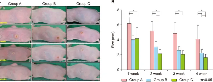
Fig. 1. (A) Changes in inflammatory nodule size in groups A, B, and C. The size of the inflammatory nodules in all groups decreased with time. (B) Changes in inflammatory nodule size in groups A, B, and C. The size of the inflammatory nodules in all groups decreased with time. The changes in size between groups B/C and group A were significantly different at weeks 1, 2, 3, and 4. The changes in size between group B and group C were significantly different at week 2 and 4.
amounts were resolved by sodium dodecyl sulfate-poly- acrylamide gel electrophoresis, transferred to a poly- vinylidene difluoride membrane, and blotted with the mouse serum obtained from each group, followed by en- hanced chemiluminescence and autoradiography.
Statistical analysis
The data were statistically analyzed using ANOVA. A p-value <0.05 was considered significant. All statistical analyses were performed using PASW software (Version 18.0; IBM Co., Armonk, NY, USA).
RESULTS
Changes in inflammatory nodules in acne mouse model groups A, B, and C
The size of the inflammatory nodules in all three groups decreased with time (Fig. 1A, B). There was a statistically significant difference in the size of the inflammatory nod- ules in groups B and C compared to group A at week 1, 2, 3, and 4. Group C showed a statistically significant de- crease in inflammatory nodule size compared to group B at week 2 and 4.
Characteristics of histopathological findings in groups A, B, and C
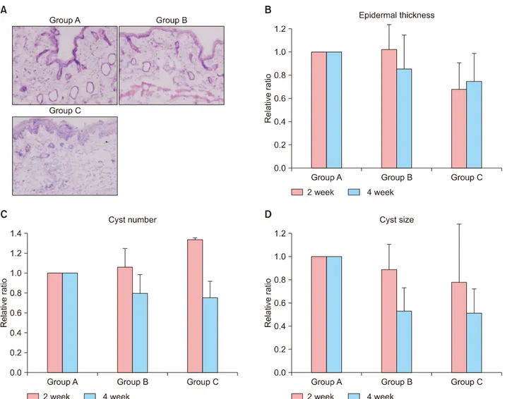
Epidermal thickness and the number and size of the mi- crocomedone-like cysts were decreased in groups B and C compared to group A at week 4 (Fig. 2A∼D). However, there were no statistically significant differences.
Expression of inflammatory biomarkers in groups A, B, and C
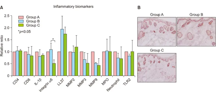
The immunohistochemistry results showed that infiltration of CD8 T cells and neutrophils and the expression of IL-1β, MMP-3 and MMP-9 in group B and group C were de- creased when compared with group A (Fig. 3A). On the contrary, infiltration of CD4 T cells and the expression of LL-37 in group B and group C were increased when com- pared with group A (Fig. 3A). However, there were no statistically significant differences in all three groups (Fig.
3A). Integrin α6 expression was increased in group B and decreased in group C when compared with group A (Fig.
3A, B). Integrin α6 expression showed a statistically sig- nificant decrease in group B when compared with group B (p<0.05) (Fig. 3A).
Western blot analysis using an anti-IgG antibody No immunoreactivity for an anti-IgG antibody against P.
acnes was observed at week 2 after the first injection in the group A or at week 2 after the second injection in the group C. However, western blot analysis could detect an IgG antibody component against P. acnes (approximately 47 kDa) at week 2 after the second injection in the group B (Fig. 4).
DISCUSSION
Topical and systemic medications have been used for the treatment of acne8. However, bacterial resistance to anti- biotics and adverse retinoid events have led to the devel-
Fig. 2. (A) Histopathological findings of group A, group B and group C at week 4 (H&E, ×200). (B) Changes in epidermal thickness.
The epidermal thickness of the inflammatory nodules decreased with time, especially in group C, followed by group B and group A. However, there were no statistically significant differences. (C) Changes in the number of microcomedone-like cysts. The number of microcomedone-like cysts decreased with time, especially in group C, followed by group B and group A. However, there were no statistically significant differences. (D) Changes in the size of microcomedone-like cysts. The size of the microcomedone-like cysts decreased with time, especially in group C, followed by group B and group A. However, there were no statistically significant differences.
opment of new therapeutic modalities for acne9,10; vacci- nation has been introduced as one of the promising ther- apeutics for acne11.
Killed-pathogen-based vaccines have been used as a vac- cine for the treatment of acne12 and an intranasal killed whole P. acnes preparation has been tested12. P. acnes has been established as a potent immunomodulator13, which can produce antibodies against secretary enzymes or cell wall molecules14. However, antibodies against P. acnes do not completely protect against the recurrence of acne, most likely because of insufficient production of pro- tective antibodies against P. acnes. This study also showed the production of antibody in mice after treatment with P.
acnes. Bacteriophages inhibited the production of the anti-
body in mice after treatment with P. acnes.
Vaccination against cell wall-anchored sialidase has also been introduced15. Sialidase, a P. acnes virulence factor, is involved in degrading host tissue and inducing in- flammation16. Nakatsuji et al.15 have reported that a siali- dase-based acne vaccine showed good effects on P.
acnes-induced ear inflammation. However, this vaccine may not be able to neutralize virulence factors secreted by P. acnes17. In addition, this vaccine may lack therapeutic effects for acne.
Monoclonal antibodies targeting Christie-Atkins-Munch- Peterson (CAMP) factor have demonstrated improvement of acne17. P. acnes possesses a CAMP that acts as a poten- tial secretory virulence factor to enhance hemolysis and
Fig. 3. (A) Immunohistochemistry analysis. Inflammatory biomarker expression was similar in all groups. There were no statistically significant differences except for integrin α6 between group B and group C. (B) Immunohistochemistry with integrin α6 in group A, group B and group C at week 2 (×200). IL: interleukin, MMP: matrix metalloproteinase, MPO: myeloperoxidase, TLR: toll-like receptor.
Fig. 4. Western blot analysis showed immunoreactivity to the immunoglobulin G antibody against Propionibacterium acnes (P.
acnes) following treatment with P. acnes.
cytolysis16,18. Nakatsuji et al.17 also investigated the treat- ment of acne using vaccines. They showed that passive immunization targeting secretory CAMP factors proteins can neutralize P. acnes virulence without disturbing other bacteria17. Active immunization using a CAMP factor-tar- geted vaccine and passive neutralization of the CAMP fac- tor showed roughly equal potency in terms of improving P. acnes-induced inflammation.
TLR vaccines and antimicrobial peptides (AMPs) constitute other optional vaccines for the treatment of acne. Recent studies have shown that TLRs and AMPs play an important role in the pathogenesis of inflammatory acne. Therefore,
regulation of TLRs and AMPs could provide different ther- apeutic options for acne19.
Bacteriophages targeting P. acnes are also promising acne vaccine candidates20. Bacteriophages have several advan- tages over antibiotics against P. acnes. They are environ- mentally friendly, rapidly processed, less expensive, and well tolerated5. In this study, we confirmed the potential of bacteriophages for the treatment of acne. The in- flammatory nodules of HR-I mice decreased in size and microcomedone-like cysts was reduced in number and in size following bacteriophage treatment. In addition, epi- dermal thickness was decreased in bacteriophage treat- ment group. Epidermal hyperplasia can be induced by skin barrier disruption, calcium channel activation, metab- otrophic receptor activation or unsaturated fatty acids21. Epidermal hyperplasia may be related to the induction of abnormal keratinization, which can be one of the causa- tive factors of comedone formation. Furthermore, the de- creased expression of integrin α6 after bacteriophage ap- plication shows its beneficial effect for the treatment of acne. Integrin α6 expression can be a response to in- flammation and related to epidermal hyperplasia, leading to the abnormal differentiation2. Inflammatory biomarkers and cells, such as CD8 T cells, neutrophils, IL-1β, MMP-3 and MMP-9, were also decreased after bacteriophage treatment. Antimicrobial peptides can play a protective role against P. acnes or induce inflammatory signal in acne vulgaris22. In this study, the increased expression of LL-37 by P. acnes was not inhibited by bacteriophages.
Several parameters for bacteriophages need to be resolved
for a comprehensive acne vaccine: delivery route, time point of delivery, dose, phage stability, infection rate, availability of highly efficient antibiotics, poor under- standing of general aspects of phage life cycles, bacterial resistance to phage infection, and the possibility of tem- perate phages that integrate into the human genome23. Understanding the integration of temperate phages into the human genome is critical for the safety of phage therapy.
Recently, bacteriophages have been applied in a semi-sol- id preparation for the treatment of acne24. In addition, bac- teriophages have also been formulated in a water-oil nanoemulsion25. Bacteriophages against Acinetobacter baumannii have been applied in humans in the form of an antiseptic gel or a paraffin-oil-based product26. In light of these reports, a variety of formulations for the treatment of acne using bacteriophages that target P. acnes should be examined.
In conclusion, further investigation of bacteriophages tar- geting P. acnes should be conducted to gain a better un- derstanding of bacteriophage characteristics and to devel- op more beneficial vaccines for the treatment of acne.
ACKNOWLEDGMENT
This research was supported by the Basic Science Research Program through the National Research Foundation of Korea (NRF) funded by the Ministry of Education, Science and Technology (2012R1A1A2007017).
CONFLICTS OF INTEREST
The authors have nothing to disclose.
ORCID
Min Ji Kim, https://orcid.org/0000-0001-6574-6485 Dong Hyuk Eun, https://orcid.org/0000-0003-4044-0679 Seok Min Kim, https://orcid.org/0000-0001-6470-7986 Jungmin Kim, https://orcid.org/0000-0002-1795-1844 Weon Ju Lee, https://orcid.org/0000-0001-5708-1305
REFERENCES
1. Shaheen B, Gonzalez M. Acne sans P. acnes. J Eur Acad Dermatol Venereol 2013;27:1-10.
2. Jeremy AH, Holland DB, Roberts SG, Thomson KF, Cunliffe WJ. Inflammatory events are involved in acne lesion initiation.
J invest dermatol 2003;121:20-27.
3. Mirshahpanah P, Maibach HI. Models in acnegenesis.
Cutan Ocul Toxicol 2007;26:195-202.
4. Jang YH, Lee KC, Lee SJ, Kim DW, Lee WJ. HR-1 mice: a new inflammatory acne mouse model. Ann Dermatol 2015;
27:257-264.
5. Jończyk-Matysiak E, Weber-Dąbrowska B, Żaczek M, Międzybrodzki R, Letkiewicz S, Łusiak-Szelchowska M, et al. Prospects of phage application in the treatment of acne caused by Propionibacterium acnes. Front Microbiol 2017;
8:164.
6. Jassim SA, Limoges RG. Natural solution to antibiotic resistance: bacteriophages ‘The Living Drugs’. World J Microbiol Biotechnol 2014;30:2153-2170.
7. Marples RR. The microflora of the face and acne lesions. J Invest Dermatol 1974;62:326-331.
8. Sandoval LF, Hartel JK, Feldman SR. Current and future evidence-based acne treatment: a review. Expert Opin Pharmacother 2014;15:173-192.
9. Bienenfeld A, Nagler AR, Orlow SJ. Oral antibacterial therapy for acne vulgaris: an evidence-based review. Am J Clin Dermatol 2017;18:469-490.
10. Vallerand IA, Lewinson RT, Farris MS, Sibley CD, Ramien ML, Bulloch AGM, et al. Efficacy and adverse events of oral isotretinoin for acne: a systematic review. Br J Dermatol 2018;178:76-85.
11. Nakatsuji T, Rasochova L, Huang CM. Vaccine therapy for P. acnes-associated diseases. Infect Disord Drug Targets 2008;8:160-165.
12. Nakatsuji T, Liu YT, Huang CP, Zoubouis CC, Gallo RL, Huang CM. Antibodies elicited by inactivated Propionibac- terium acnes-based vaccines exert protective immunity and attenuate the IL-8 production in human sebocytes: relevance to therapy for acne vulgaris. J Invest Dermatol 2008;128:
2451-2457.
13. Mussalem JS, Vasconcelos JR, Squaiella CC, Ananias RZ, Braga EG, Rodrigues MM, et al. Adjuvant effect of the Propionibacterium acnes and its purified soluble polysaccharide on the immunization with plasmidial DNA containing a Trypanosoma cruzi gene. Microbiol Immunol 2006;50:253-263.
14. Lodes MJ, Secrist H, Benson DR, Jen S, Shanebeck KD, Guderian J, et al. Variable expression of immunoreactive surface proteins of Propionibacterium acnes. Microbiology 2006;152:3667-3681.
15. Nakatsuji T, Liu YT, Huang CP, Zouboulis CC, Gallo RL, Huang CM. Vaccination targeting a surface sialidase of P.
acnes: implication for new treatment of acne vulgaris. PLoS One 2008;3:e1551.
16. Brüggemann H. Insights in the pathogenic potential of Propionibacterium acnes from its complete genome. Semin Cutan Med Surg 2005;24:67-72.
17. Nakatsuji T, Tang DC, Zhang L, Gallo RL, Huang CM.
Propionibacterium acnes CAMP factor and host acid sphingomyelinase contribute to bacterial virulence: potential targets for inflammatory acne treatment. PLoS One 2011;6:
e14797.
18. Lo CW, Lai YK, Liu YT, Gallo RL, Huang CM. Staphylococcus aureus hijacks a skin commensal to intensify its virulence:
immunization targeting beta-hemolysin and CAMP factor. J
Invest Dermatol 2011;131:401-409.
19. Ozlu E, Karadag AS, Ozkanli S, Oguztuzun S, Kilic M, Zemheri E, et al. Comparison of TLR-2, TLR-4, and antimicrobial peptide levels in different lesions of acne vulgaris. Cutan Ocul Toxicol 2016;35:300-309.
20. Górski A, Dąbrowska K, Hodyra-Stefaniak K, Borysowski J, Międzybrodzki R, Weber-Dąbrowska B. Phages targeting infected tissues: novel approach to phage therapy. Future Microbiol 2015;10:199-204.
21. Katsuta Y, Iida T, Inomata S, Denda M. Unsaturated fatty acids induce calcium influx into keratinocytes and cause abnormal differentiation of epidermis. J Invest Dermatol 2005;124:1008-1013.
22. Borovaya A, Dombrowski Y, Zwicker S, Olisova O, Ruzicka T, Wolf R, et al. Isotretinoin therapy changes the expression of antimicrobial peptides in acne vulgaris. Arch Dermatol
Res 2014;306:689-700.
23. Brüggemann H, Lood R. Bacteriophages infecting propioni- bacterium acnes. Biomed Res Int 2013;2013:705741.
24. Brown TL, Petrovski S, Dyson ZA, Seviour R, Tucci J. The formulation of bacteriophage in a semi solid preparation for control of Propionibacterium acnes growth. PLoS One 2016;10:e0151184.
25. Esteban PP, Alves DR, Enright MC, Bean JE, Gaudion A, Jenkins AT, et al. Enhancement of the antimicrobial properties of bacteriophage-K via stabilization using oil-in-water nano-emulsions. Biotechnol Prog 2014;30:932- 944.
26. Chen LK, Liu LY, Hu A, Chang KC, Lin NT, Lai MJ, et al.
Potential of bacteriophage ΦAB2 as an environmental biocontrol agent for the control of multidrug-resistant Acinetobacter baumannii. BMC Microbiol 2013;13:154.