pISSN: 0378-6471 eISSN: 2092-9374 DOI : 10.3341/jkos.2011.52.1.112
= 증례보고 =
양안에 발생한 코우츠병 1예
마대중1 최 진1 장지웅1 김정훈1,2 김성준1,2 유영석1,2
서울대학교 의과대학 안과학교실1, 서울대학교병원 임상의학연구소 인공안구센터2
목적: 양안에 발생한 코우츠병 증례를 경험하였기에 문헌 고찰과 함께 보고자 한다.
증례요약: 19개월 남아가 5개월 전부터 양안이 안으로 몰림을 주소로 내원하였다. 안저검사에서 우안 전체 망막의 심한 삼출망막박리 및 망막표면의 실핏줄확장증이 관찰되었고, 좌안 망막 하측의 삼출망막박리가 관찰되었다. 형광안저혈관조영술에서 좌안 망막 하이측 의 적도부위에서 형광누출을 동반한 망막실핏줄확장증 소견과 무혈관성 망막이 발견되었다. 이에 우안은 망막하액 외배액술 및 유리 체강 내 공기주입술을 시행하였으며, 좌안은 아르곤 레이저광응고술 및 냉동치료를 시행하였다. 우안 망막하액에 대한 세포검사에서 악성세포는 발견되지 않았다. 수술 후 44개월째 추적관찰에서 최대교정시력 우안 광각무, 좌안 0.4이며 양안 망막은 편평하게 유지되 었다. 녹내장 및 망막박리의 재발, 통증 등의 합병증은 발생하지 않았다.
결론: 코우츠병은 양안에 매우 드물게 발생하는 질환으로 양안의 진행이 비대칭일 수 있고, 진행 정도가 단안에 발생한 경우보다 더 심한 경향이 있으며, 장기간 경과 후에도 질병이 진행할 수 있어 양안에 대한 적절한 치료 및 장기간의 세밀한 검사가 필요하다.
<대한안과학회지 2011;52(1):112-116>
접 수 일: 2010년 7월 11일 심사통과일: 2010년 7월 28일 게재허가일: 2010년 11월 12일
책 임 저 자: 유 영 석
서울특별시 종로구 연건동 28 서울대학교병원 안과
Tel: 02-2072-2438, Fax: 02-741-3187 E-mail: [email protected]
* 본 논문의 요지는 2009년 대한안과학회 제102회 학술대회에서 e-poster로 발표된 바 있음.
코우츠병은 특발성 망막실핏줄확장증, 혈관삼출, 이로 인 한 삼출망막박리를 보이는 질환으로 유리체망막견인을 포 함한 다른 원인질환이 동반되지 않는 경우이다.1-3
코우츠병은 주로 십대 이전의 남아에서 단안에 발생하며, 삼출망막박리 발생으로 인한 시력저하가 발생할 수 있고, 수정체폐쇄각녹내장, 신생혈관녹내장의 합병될 수 있어, 동 반된 통증으로 안구적출이 필요한 경우도 있다.
국외에서 시행된 다수의 코우츠병의 증례보고 및 임상양 상 보고에서 양안에 발생한 코우츠병의 비율이 전체 코우 츠병 증례의 0%에서 26%까지 보고된 바 있으나, 국내에서 는 아직 보고된 바가 없다.6-19
본 증례는 양안에 발생한 코우츠병 환아로 이에 대해 보 고를 수행 하고자 한다.
증례보고
19개월 남아가 내원 약 5개월 전부터 시작된 양안이 안
으로 몰림을 주소로 타병원 안과에 내원하여 양안 망막박 리로 진단받은 후 전원되었다.
환아는 만삭에 2.9 kg로 출생하였으며, 과거력 및 가족력 에서 특이 사항은 없었다.
초진 당시 주시 및 추종은 우안은 불량하였고 좌안은 양 호하였다. 사시각 검사에서 근거리 주시시에 30프리즘 디 옵터의 내사시가 관찰되었으며, 외안부 및 전안부 검사에서 는 특이소견이 관찰되지 않았다.
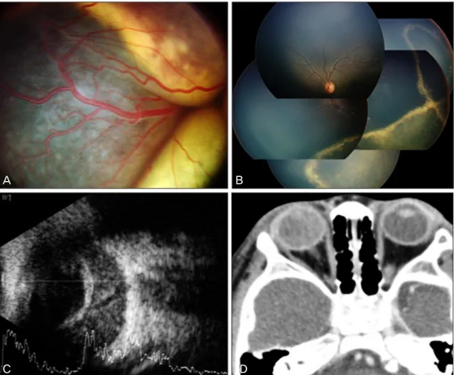
안저검사에서 우안 전체 망막의 심한 삼출망막박리 및 망막표면의 실핏줄확장증이 관찰되었고(Fig. 1A), 좌안 망 막하측의 삼출망막박리가 관찰되었다(Fig. 1B).
악성 종양의 감별을 위해 시행한 안구초음파검사 및 안 와단층촬영에서는 양안 안구 내 종괴 소견은 관찰되지 않 았다(Fig. 1C, D).
전신 마취하여 시행한 형광안저혈관조영술에서 좌안 망 막 하이측의 적도부위에서 형광누출을 동반한 망막실핏줄 확장증 소견과 무혈관성 망막이 관찰되었다(Fig. 2).
이에 우안은 공막 천자를 통한 망막하액 외배액술 및 섬 모체평면부 천자를 통한 유리체강 내 공기주입술을 시행하 였으며, 좌안은 망막실핏줄확장증 및 무혈관 망막부위에 대 하여 아르곤 레이저광응고술 및 냉동치료를 시행하였다.
악성종양 감별 위하여 시행한 우안 망막하액에 대한 세 포검사에서 악성세포는 발견되지 않았으며 다수의 염증세 포와 지질을 포함한 대식세포가 관찰되었다.
수술 후 2개월째 추적관찰에서 우안 망막은 편평하였으며,
A B
C D
Figure 1. Initial examination findings. (A) Total bullous exudative retinal detatchment with retinal vascular te-
langiectasia in the right eye. (B) Fundus photograph showing characteristic features of Coats’ disease with localized exudative retinal detachment with vascular telangiectasia at the inferior periphery in the left eye. (C) Ocular ultra- sonography showing no mass-like lesion. (D) Orbit CT showing no mass-like lesion.A B
Figure 2. Fluorescein angiogram of the left eye before the laser photocoagulation. (A) Retinal telangiectatic vessels and
avascular area. (B) Fluorescein leakeage from telangiectatic vessels.좌안 망막은 망막하액 감소한 소견이 관찰되었다(Fig. 3).
이후 지속적으로 우안 내사시 소견 보여 수술 34개월 후 우안 내직근후전술 및 외직근절제술 시행하였다.
수술 후 44개월 째 추적관찰에서 최대교정시력 우안 광 각무, 좌안 0.4이었으며 양안 망막은 편평하였다(Fig. 4).
녹내장 및 망막박리의 재발, 통증 등의 합병증은 발생하지
A B
Figure 3. Fundus photograph taken 2 months after operation. (A) Right eye, flat. (B) Left eye, subretinal fluid de-
creased a great deal.A B
Figure 4. Fundus photograph taken 33 months after operation. (A) Right eye. (B) Left eye. Both fundi are flat and
stable.않았다.
고 찰
코우츠병은 특발성 망막실핏줄확장증, 혈관삼출, 이로 인 한 삼출망막박리를 보이는 질환으로 유리체망막견인을 포 함한 다른 원인질환이 동반되지 않는 경우이며, 대부분 단 안에 발생한다고 알려져 있다.1-3
1908년 Coats1가 단안의 삼출망막박리 6예를 보고한 이 후로 코우츠병에 대한 다수의 증례보고 및 임상양상 분석 이 수행되었다. 1967년 Green,4 1987년 McGettrick and Loeffler,52002년 Alexandridou and Stavrou6가 각각 양안 에 발생한 코우츠병 증례를 1예씩 보고한 바 있으며, 다수 의 코우츠병의 임상양상 보고에서 양안에 발생한 코우츠병
의 비율이 전체 코우츠병 증례의 0%에서 26%까지 보고된 바 있으나, 2001년 Shields et al12은 이전에 보고된 양안에 발생한 코우츠병 증례 중 다수에서 코우츠병의 진단기준에 부합하지 않은 것을 보고한 바 있다.7-17 실제로 1967년 Green4이 보고한 증례는 좌안은 하이측 변연부에 망막실핏 줄확장과 동반된 삼출망막박리가 관찰된 반면, 우안은 망막 종괴 소견이 동반되어 양안에 발생한 코우츠병의 진단기준 에 부합하지 않는다고 할 수 있다.
코우츠병에서 인종간에 발생 빈도 및 임상양상의 차이는 없는 것으로 알려져 있으나, 한국인을 포함한 동양인에 대 한 보고는 많지 않다.15 2007년 Lai et al18은 대만인 30명 32안에 대하여 코우츠병의 임상양상을 보고하였고, 이 중 2예에서 양안에 발생하였다.
국내에서는 1969년에 Kim et al19이 최초로 코우츠병 증
례를 보고한 이후, 수 차례 증례보고가 있었다. 1999년에 Choi and Yu20는 코우츠병으로 진단받은 30명 30안, 2006 년에 Han et al21은 67명 67안의 임상양상 및 치료 결과에 대하여 보고한 바 있으나, 국내에는 아직 양안에 발생한 코 우츠병 증례가 보고된 바 없다.
2001년 Shields et al7이 보고한 150명, 158안 중 양안 에 발생한 8명 16안, 1987년 McGettrick and Loeffler5및 2002년 Alexandridou and Stavrou6의 증례 보고, 그리고 본 증례 모두에서 양안에 발생한 코우츠병에서 양안 질병 의 진행 정도가 차이가 있었다. 2001년 Shields et al7은 질병의 진행 정도가 경한 편안의 경우 대부분 증상 없이 망막 변연부에 경미한 망막실핏줄확장증 소견만 관찰됨을 보고한 반면, 2002년 Alexandridou and Stavrou6의 증례 보 고에서는 망막실핏줄확장증과 동반된 중심와 바깥쪽의 삼출 물, 본 증례에서는 망막실핏줄확장증과 동반된 부분 삼출망 막박리 소견이 관찰되어 이전 보고와는 차이가 있었다.
2001년 Shields et al7의 보고에 따르면 코우츠병에서 초 진 시의 주소는 시력저하(43%), 사시(23%), 백색동공 (20%) 순이었으며 초진 시에 전망막박리가 관찰되는 빈도 는 48%였다. 반면, 1987년 McGettrick and Loeffler5 및 2002년 Alexandridou and Stavrou6의 증례 보고의 주소는 백색동공이었으며, 본 증례는 내사시였다. 또한 진행 정도 가 심한 편안에서 모두 전망막박리가 관찰되어 양안에서 발생한 코우츠병의 경우 단안에서 발생한 경우보다 질병의 진행 정도가 더 심한 경향이 있음을 유추할 수 있다.
또한 2002년 Alexandridou and Stavrou6의 증례에서는 진행 정도가 경한 편안의 망막실핏줄확장증과 동반된 중심 와 바깥쪽의 삼출물에 대하여 치료하지 않고 경과관찰하였 을 때, 17년 경과 후 병변이 진행함을 보고한 바 있어 양안에 발생한 코우츠병에서는 진행 정도가 경한 편안에 대해서도 적절한 치료 및 장기적인 경과관찰이 필요함을 알 수 있다.
따라서 코우츠병은 양안에 발생이 가능하고, 양안 질병 의 진행이 비대칭일 수 있으며, 진행 정도가 단안에 발생한 경우보다 더 심한 경향이 있어 초기에 정확한 진단 및 치료 가 필수적이다. 또한 진행 정도가 심한 편안은 단안에서 발 생한 코우츠병보다 예후가 좋지 않은 경향이 있어 시력 및 안구 보존을 위해서는 진행 정도가 경한 편안에 대해서도 장기간의 세밀한 경과관찰이 필요하다.
결론적으로, 본 증례는 양안에 발생한 코우츠병에 대하 여 치료 및 장기 경과 관찰한 국내 최초의 증례보고로, 적 절한 치료를 통하여 편안의 시력보존이 가능하였고, 합병증 의 병발을 예방하여 양안을 보존할 수 있었다.
참고문헌
1) Coats G. Forms of retinal diseases with massive exudation. Roy Lond Ophthalmol Hosp Rep 1908;17:440-525.
2) Leber T. Ueber eine durch Vorkommen multipler Miliaraneurysmen charakterisierte Form von Retinaldegeneration. Albrecht von Graefes Arch Ophthalmol 1912;81:1-14.
3) Reese AB. Telangiectasis of the retina and Coats' disease. Am J Ophthalmol 1956;42:1-8.
4) Green WR. Bilateral Coats' disease. Massive gliosis of the retina.
Arch Ophthalmol 1967;77:378-83.
5) McGettrick PM, Loeffler KU. Bilateral Coats' disease in an infant (a clinical, angiographic, light and electron microscopic study).
Eye 1987;1:136-45.
6) Alexandridou A, Stavrou P. Bilateral Coats' disease: long-term fol- low up. Acta Ophthalmol Scand 2002;80:98-100.
7) Shields JA, Shields CL, Honavar S, Demirci H. Coats disease.
Clinical variations and complications of Coats disease in 150 cases.
The 2000 Sanford Gifford Memorial Lecture. Am J Ophthalmol 2001;131:561-71.
8) Woods AC, Duke JR. Coats's disease. I. Review of the literature di- agnostic criteria, clinical findings, and plasma lipid studies. Br J Ophthalmol 1963;47:385-412.
9) Gomez Morales A. Coats' disease. Natural history and results of treatment. Am J Ophthalmol 1965;60:855-65.
10) Egerer I, Tasman W, Tomer TT. Coats disease. Arch Ophthalmol 1974;92:109-12.
11) Tarkkanen A, Laatikainen L. Coat's disease: clinical, angiographic, histopathological findings and clinical management. Br J Ophth- almol 1983;67:766-76.
12) Chang MM, McLean IW, Merritt JC. Coats' disease: a study of 62 histologically confirmed cases. J Pediatr Ophthalmol Strabismus 1984;21:163-8.
13) Silodor SW, Augsburger JJ, Shields JA, Tasman W. Natural history and management of advanced Coats' disease. Ophthalmic Surg 1988;19:89-93.
14) Shields JA, Shields CL, Honavar SG, et al. Classification and man- agement of Coats disease. The 2000 Proctor Lecture. Am J Ophthalmol 2001;131:572-83.
15) Shields JA, Shields CL. Review: Coats disease: the 2001 LuEsther T. Mertz lecture. Retina 2002;22:80-91.
16) Shienbaum G, Tasman WS. Coats disease: a lifetime disease.
Retina 2006;26:422-4.
17) Rishi P, Rishi E, Uparkar M, et al. Coats' disease: an Indian perspective. Indian J Ophthalmol 2010;58:119-24.
18) Lai CH, Kuo HK, Wu PC, et al. Manifestation of Coats' disease by age in Taiwan. Clin Experiment Ophthalmol 2007;35:361-5.
19) Kim WS, Jung BJ, Kim JW, et al. A Case of Coats' disease. J Korean Ophthalmol Soc 1969;10:37-9.
20) Choi SY, Yu YS. Treatment and clinical results of coats' disease. J Korean Ophthalmol Soc 1999;40:2190-7.
21) Han ES, Choung HK, Heo JW, et al. The Clinical analysis and treatment results of Coats' disease in children. J Korean Ophthalmol Soc 2006;47:423-30.
=ABSTRACT=
Bilateral Coats' Disease: A Case Report
Dae Joong Ma, MD1, Jin Choi, MD1, Ji Woong Jang, MD1, Jeong Hun Kim, MD1,2, Seong Joon Kim, MD1,2, Young Suk Yu, MD, PhD1,2
Department of Ophthalmology, Seoul National University College of Medicine1, Seoul, Korea Seoul Artificial Eye Center, Seoul National University Hospital Clinical Research Institute2, Seoul, Korea
Purpose: To report a case of bilateral Coats' disease.
Case summary: A 19-month-old boy presented with esodeviation of his eyes, which started 5 months prior. A fundus exam showed total bullous exudative retinal detachment with retinal vascular telangiectasia in the right eye and localized exuda- tive retinal detachment with vascular telangiectasia at the inferior periphery in the right eye. Fluorescein angiogram of the left eye showed retinal telangiectatic vessels, avascular area and fluorescein leakeage from telangiectatic vessels. The patient received external drainage of subretinal fluid and intravitreal air injection of the right eye and Argon LASER photo- coagulation and cryotheraphy of the left eye. A cytologic exam of the subretinal fluid drained from the right eye showed no malignant cells. Forty-four months after the operation, his best corrected visual acuity was no light perception in the right eye and 0.4 in the left eye. Both fundi were flat and stable. No complications, such as glaucoma, recurred retinal detach- ment, or pain, occurred.
Conclusions: Coats' disease rarely occurs bilaterally and can be involved asymmetrically. The disease presents more se- verely when bilateral and can progress after long-term observation. Proper treatment and long-term follow-up of both eyes are necessary to prevent visual loss and preserve eyes.
J Korean Ophthalmol Soc 2011;52(1):112-116
Key Words: Coats' disease, Exudative retinal detachement, Retinal telangiectasia
Address reprint requests to Young Suk Yu, MD, PhD
Department of Ophthalmology, Seoul National University Hospital
#28 Yongon-dong, Chongno-gu, Seoul 110-744, Korea
Tel: 82-2-2072-2438, Fax: 82-2-741-3187, E-mail: [email protected]