463 대한안과학회지 2015년 제 56 권 제 3 호
J Korean Ophthalmol Soc 2015;56(3):463-465 ISSN 0378-6471 (Print)⋅ISSN 2092-9374 (Online)
http://dx.doi.org/10.3341/jkos.2015.56.3.463
Case Report
출산 후 젊은 여성에서 발생한 황반원공 1예
A Case of Postpartum Macular Hole in a Young Woman
김병선1⋅김병재1⋅한용섭1,2⋅박종문1,2⋅정인영1,2
Byoung Seon Kim, MD1, Byung Jae Kim, MD1, Yong Seop Han, MD, PhD1,2, Jong Moon Park, MD, PhD1,2, In Young Chung, MD, PhD1,2
경상대학교 의학전문대학원 안과학교실1, 경상대학교 건강과학연구원2
Department of Ophthalmology, Gyeongsang National University School of Medicine1, Jinju, Korea Gyeongsang Institute of Health Science, Gyeongsang National University2, Jinju, Korea
Purpose: To describe a case of macular hole (MH) in a 29-year-old non-myopic woman after uncomplicated delivery.
Case summary: A 29-year-old woman visited our clinic complaining of decreased visual acuity in her left eye after uncomplicated delivery. Fundoscopy and optical coherence tomography showed a full thickness macular hole in the left eye. However, we found not posterior vitreous detachment or vitreomacular traction in the posterior pole. The patient underwent pars plana vi- trectomy, internal limiting membrane peeling, and SF6 gas tamponade. Three months after vitrectomy, the patient’s visual acuity was improved and the macular hole was closed successfully.
Conclusions: We experienced and treated a case of postpartum MH developed in a young woman without posterior vitreous de- tachment or vitreomacular traction in the posterior pole. This suggests another mechanism of MH formation. Postpartum MH was successfully treated by existing idiopathic macular hole surgery.
J Korean Ophthalmol Soc 2015;56(3):463-465 Key Words: Postpartum macular hole, Vitrectomy
■Received: 2014. 9. 19. ■ Revised: 2014. 12. 1.
■Accepted: 2015. 2. 4.
■Address reprint requests to In Young Chung, MD, PhD Department of Ophthalmology, Gyeongsang National University Hospital, #79 Gangnam-ro, Jinju 660-702, Korea Tel: 82-55-750-8171, Fax: 82-55-750-4158
E-mail: [email protected]
ⓒ2015 The Korean Ophthalmological Society
This is an Open Access article distributed under the terms of the Creative Commons Attribution Non-Commercial License (http://creativecommons.org/licenses/by-nc/3.0/) which permits unrestricted non-commercial use, distribution, and reproduction in any medium, provided the original work is properly cited.
특발성 황반원공은 후유리체 박리에 의한 견인력으로 인 하여 발생하거나 유리체와 황반 사이의 접선 견인력에 의 하여 발생한다고 알려졌으며 대부분 60-70대의 고령에서 발생하며 여성에서 호발한다.1 젊은 성인에서 황반 원공이 발생하는 경우는 대부분 외상이며, 특발성 황반원공과 달 리 후유리체가 완전히 박리되지 않아도 발생할 수 있으며 자연 회복되는 경우도 있다.2 특발성 황반원공 여성에서 뚜 렷한 호발의 경향(70%)이 있기에 에스트로젠과의 관련성
에 관한 의문이 있었고, 폐경기 전후 또는 호르몬 대체 요 법 등의 호르몬 변화가 가능한 위험 인자로서 제시되기도 하였다.3,4 기저질환이나 외상의 병력이 없는 젊은 여자 환 자에서 출산 후 황반원공이 발생하는 경우는 최근 국외에 서 30세 여자환자에서 출산 후 발생한 양안 황반원공을 보 고한 바 있으나 아직 그 예가 국내에 보고된 적이 없는 매 우 드문 경우이다. 이에 본 저자들은 기저 질환이 없는 젊 은 여자환자에서 출생 후 발생한 황반원공을 치험하였기에 이를 보고하고자 한다.
증례보고
기저 질환 없는 29세 여자 환자가 내원 1달 전 출산 직후 부터 좌안 시력저하 및 변형시를 주소로 본원으로 내원하 였다. 내원 당시 우안의 최대교정시력은 20/20으로 측정되
464
- 대한안과학회지 2015년 제 56 권 제 3 호 -
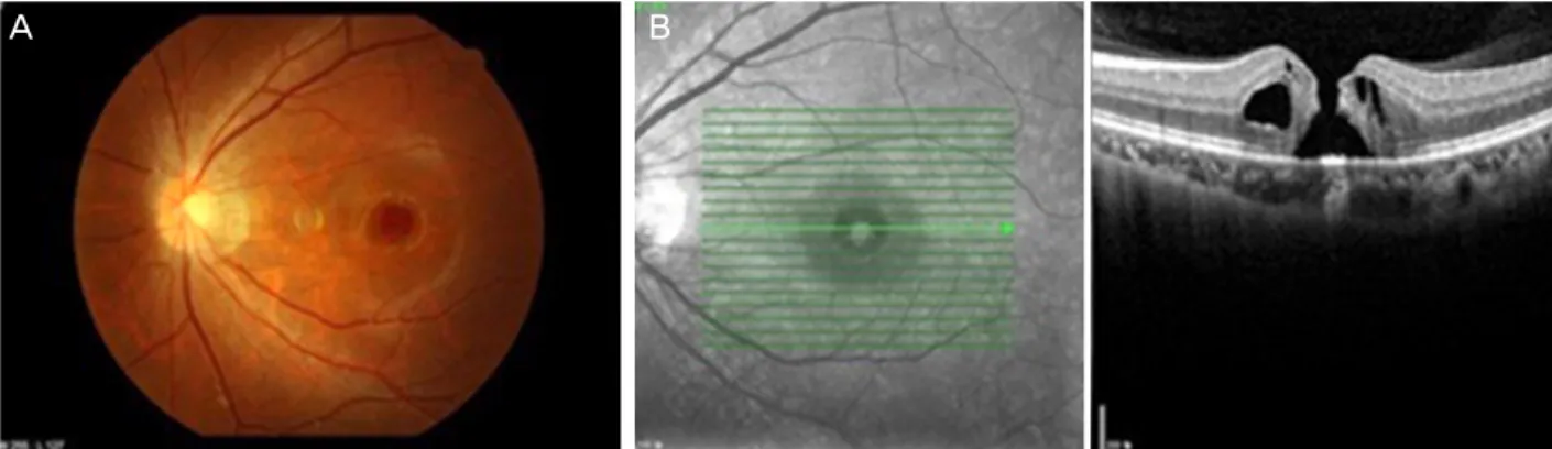
Figure 1. Preoperative fundus photography (A) and optical coherence tomography (B) of the left eye.
Figure 2. Fundus photography (A) and optical coherence tomography (B) 3 months after pars plana vitrectomy and posterior vitre-
ous detachment formation, internal limiting membrane peeling, 20% SF6 gas tamponade shows closed macular hole.었으며 좌안의 최대교정시력은 20/63으로 측정되었으며 양 안 안압은 정상 소견을 보였다. 안저검사 및 빛간섭단층촬 영 검사에서 좌안의 전층황반원공 소견을 보였으나 후유리 체 박리 및 견인력을 일으킬 만한 소견은 보이지 않았다 (Fig. 1). 이에 좌안 유리체 절제술, 후유리체 박리, 내경계 막 제거술 및 20% SF6 가스 주입술을 시행하였다. 수술 후 3개월째 좌안의 최대교정시력은 20/32으로 측정되었고 빛 간섭단층촬영 검사상 전층황반원공은 폐쇄된 소견을 보였 다(Fig. 2).
고 찰
황반원공의 발생기전은 아직 명확히 밝혀지지는 않았지 만 연령이 증가함에 따라 발생하는 경우, 외상, 고도 근시, 염증이 동반된 낭포황반부종, 망막혈관질환, 망막전막 등과 관련하여 생길 수 있다.5-9
특발성 황반원공의 발생기전은 빛간섭단층촬영 검사가 나온 후로부터 유리체와 망막 사이의 견인력에 의할 것이 라고 추정되고 있으며 후유리체 박리가 불안정하게 발생하 면서 생기는 견인력이라고 보고하고 있고 이는 대부분 연 령의 증가에 따라 발생하게 된다.10
젊은 성인에서는 후유리체 박리가 일어나면서 발생하는 경우는 드물며 대부분 유리체와 망막 사이의 견인력이 아 닌 이차적인 외상에 의해 황반원공이 발생하며 갑작스러운 압박과 팽창이 안구에 전해져 유리체 부착 부위 중 중심와 에 전달되어 파열되면서 발생한다고 가정되고 있다.11,12 또 한 Yamada et al13은 외상성 황반원공은 항상 외상 직후에 생기지 않고 유리체 견인력에 의하여 외상성 황반 원공이 발생할 수 있다고 보고하였다.
본 증례에서는 출산 후 갑자기 발생한 시력저하를 주소 로 본원에 내원한 젊은 여성의 빛간섭단층촬영 검사에서 전층 황반원공의 소견을 보였으며 특발성 황반 원공의 발 생 원인으로 생각되는 후유리체 박리나 망막의 견인력 소 견은 보이지 않았으며 수술 소견에서도 후유리체 박리가 보이지 않았다. 그리고 이 환자는 출산 전후 외상의 병력은 없었으며, 출산 중에도 심한 머리 흔들림이나 특이한 외상 없이 정상 분만하였다. 즉, 본 증례는 이전에 알려져 있는 기전을 보이지 않았다. 국외에서 2013년 Cloché et al14이 보고한 30세 여자 환자에서 출산 후 양안에서 발생한 황반 원공의 증례에도 출산에 특이 소견이 없었으며 3기 황반원 공을 보인 우안의 경우 수술적 치료로 호전되었으며, 1기 황반원공이었던 좌안은 자연 호전되었음을 보고하였다. 국
A B
A B
465
= 국문초록 =
출산 후 젊은 여성에서 발생한 황반원공 1예
목적: 특이질환이 없는 젊은 여자 환자에서 정상분만 출산 후 발생한 황반원공을 치험하였기에 이를 보고자 한다.
증례요약: 29세 여자 환자가 출산 후 좌안 시력 저하를 주소로 본원 내원하였다. 안저검사 및 빛간섭단층촬영 검사에서 좌안의 전층황 반원공 소견을 보였으나 후유리체 박리 및 견인력 소견은 없었다. 이에 좌안 유리체 절제술, 후유리체 박리, 내경계막 제거술 및 20%
SF6 가스 주입술을 시행하였다. 술 후 3개월 뒤 시력은 호전되었으며 황반원공도 폐쇄되었다.
결론: 본 저자들은 젊은 여자 환자에서 출산 후 후유리체박리 및 견인력이 없이 발생한 황반원공을 경험하였다. 이 증례는 황반원공이 발생하는 다른 기전을 시사하나, 기존 특발성 황반원공과 동일한 수술로 성공적으로 폐쇄되었음을 보고하고자 한다.
<대한안과학회지 2015;56(3):463-465>
- 김병선 외 : 출산 후 발생한 황반원공 -
내에서는 Chung et al15이 36세 여자 환자에서 유방암의 보 조 치료약으로 항 에스트로젠 약물인 Tamoxifen을 경구복 용하고 있는 중에 발생한 양안의 황반원공으로 양안 유리 체 절제술을 시행받고 호전되었음을 보고한 바 있다.이 증 례에서 폐경기 혹은 에스트로젠 약물을 복용 중인 여성에 서 황반원공이 발생할 수 있으며 이는 혈중 에스트로젠 농 도가 영향을 줄 수 있다고 보고하였다.
본 증례에서는 이전 연구들에서 제시한 황반원공의 발생 기전과 달리 유리체 액화나 후유리체 박리와 관계없이 황 반원공이 발생하였다. 따라서 이 증례는 드물지만 출산 후 황반원공이 발생할 수 있음을 시사하며 이는 기존에 제시 된 여성 호르몬의 변화와의 관련성을 가장 크게 의심해 볼 수 있으나 그 빈도가 너무 희박하여 이 가설을 뒷받침하기 어려운 제한점이 있다. 그러나 황반원공의 발생기전을 명 확히 알 수 없으나 기존의 특발성 황반원공과 동일하게 유 리체 절제술, 후유리체 박리, 내경계막 제거술 및 가스 주 입술로 성공적으로 황반원공의 폐쇄를 유도할 수 있었다.
외상 등의 병력이 없는 젊은 여성에서 출산 후에 후유리체 박리 혹은 견인력 없이 황반원공이 발생된 희귀한 증례를 경험하고 이를 기존 황반원공과 같은 수술적 치료로 성공 적으로 치료하였기에 이를 보고하고자 한다.
REFERENCES
1) Gass JD. Idiopathic senile macular hole. Its early stages and pathogenesis. Arch Ophthalmol 1988;106:629-39.
2) Mitamura Y, Saito W, Ishida M, et al. Spontaneous closure of trau- matic macular hole. Retina 2001;21:385-9.
3) McDonnell PJ, Fine SL, Hillis AI. Clinical features of idiopathic macular cysts and holes. Am J Ophthalmol 1982;93:777-86.
4) James M, Feman SS. Macular holes. Albrecht Von Arch Klin Clin Exp Ophthalmol 1980;215:59-63.
5) Johnson MW, Van Newkirk MR, Meyer KA. Perifoveal vitreous detachment is the primary pathogenic event in idiopathic macular hole formation. Arch Ophthalmol 2001;119:215-22.
6) Lewis ML, Cohen SM, Smiddy WE, Gass JD. Bilaterality of idio- pathic macular holes. Graefes Arch Clin Exp Ophthalmol 1996;
234:241-5.
7) Aaberg ThM. Macular holes: a review. Surv Ophthalmol 1970;
15:62.
8) Ho AC, Guyer DR, Fine SL. Macular hole. Surv Ophthalmol 1998;
42:393-416.
9) Sjaarda RN. Macular hole. Int Ophthalmol Clin 1995;35:105-22.
10) Gaudric A, Haouchine B, Massin P, et al. Macular hole formation:
new data provided by optical coherence tomography. Arch Ophthalmol 1999;117:744-51.
11) Yanagiya N, Akiba J, Takahashi M, et al. Clinical characteristics of traumatic macular holes. Jpn J Ophthalmol 1996;40:544-7.
12) Johnson RN, McDonald HR, Lewis H, et al. Traumatic macular hole: observations, pathogenesis, and results of vitrectomy surgery.
Ophthalmology 2001;108:853-7.
13) Yamada H, Sakai A, Yamada E, et al. Spontaneous closure of trau- matic macular hole. Am J Ophthalmol 2002;134:340-7.
14) Cloché V, Sablon JC, Berrod JP. [Bilateral postpartum macular hole]. J Fr Ophtalmol 2013;36:e11-4.
15) Chung SE, Kim SW, Chung HW, Kang SW. Estrogen antagonist and development of macular hole. Korean J Ophthalmol 2010;
24:306-9.