목적: 후공막염은 드문 질환으로 알려져 있다. 갑상선 기능항진증을 동반한 환자의 단안에서 발생하여 1년 만에 반대편 눈에 재발한 후공막염 1예를 경험하고 이에 대한 보고를 하고자 한다.
증례요약: 39세 여자환자가 좌안의 안구동통과 두통을 주소로 내원하였다. 안저 검사, 초음파검사, CT 및 MRI 검사 등을 이용해 후공막 염으로 진단하고 Methylprednisolone을 경구투여 조치를 하여 효과적인 치료결과를 얻었다. 퇴원 후 4개월 뒤에 갑상선유두암으로 좌측 갑상선부분절제술을 시행받은 뒤 그로부터 7개월 뒤 1주일간의 우안의 안구동통과 두통으로 다시 내원한 환자를 안저검사, 초음 파검사, MRI 검사의 임상소견 결과로 후공막염으로 진단하였다. Methylprednisolone을 경구투여 조치 후 효과적인 치료결과를 얻었고 환자는 퇴원 후 5개월까지 경과관찰하였으나 재발의 소견은 보이지 않았다.
결론: 갑상선 기능항진증 환자에서 안구동통이나 두통이 있을 때 후공막염의 동반 가능성을 생각해 볼 수 있고, 후공막염의 진단 후에 도 양안에 재발이 가능하다는 점을 염두에 두어야 할 것으로 판단된다.
<대한안과학회지 2010;51(12):1659-1664>
접 수 일: 2010년 4월 7일 심사통과일: 2010년 10월 28일
책 임 저 자: 권 정 도
부산광역시 금정구 남산동 374-75 왈레스 기념 침례병원 안과
Tel: 051-580-1359, Fax: 051-512-1354 E-mail: [email protected]
* 본 논문의 요지는 2010년 대한안과학회 제103회 학술대회에서 포스터로 발표되었음.
Figure 1. Fundus Photograghy shows exudative retinal detach- ment, retinal hemorrhage, and whitish subretinal whitish le- sion at 3 o’clock.
후공막염은 전체 공막염 환자의 약 2∼20% 정도에서만 나타나는 희귀한 질환이다.1 염증반응이 적도부 앞쪽까지 확산되기도 하나 후안부에만 독립적으로 발생할 수도 있 어 초기에는 병변 위치상 안과질환 중 진단이 그리 쉽지 않은 질환 중의 하나이다. 하지만 이 질환은 부신피질호르 몬을 이용한 면역억제요법으로 치료될 수 있는 안과질환 으로 알려져 있다.1-4 병변의 위치상 그 동안 진단이 쉽지 않았지만 전산화 단층촬영, 자기공명영상 등으로 현재는 비교적 진단이 용이해졌다. 저자들은 갑상선 기능항진증 환자의 단안에서 발생하여 1년 만에 반대편 눈에 재발한 후공막염의 사례를 초음파검사와 전산화 단층촬영, 자기공 명영상 등으로 조기 진단하였다. 그리고 이 질환에 대하여 부신피질호르몬 제제의 조기투여로 효과적인 치료결과를 얻었기에 구체적 경과를 보고하는 바이다.
증례보고
39세 여성 환자가 좌안의 안구동통과 두통을 주소로 내
원하였다. 내원 1주 전부터 좌안의 충혈 및 안구동통으로 개인병원에서 유행성각결막염으로 진단받고 1주일간의 치 료를 받았으나 통증이 호전되지 않은 상태로 본원에 내원 하게 되었다.
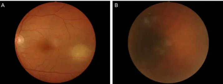
초진 시 안 소견은 나안시력 우안 0.8, 좌안 0.25이었고, 교정시력 우안 1.0, 좌안 0.63이었다. 골드만 압평안압계 로 측정한 안압은 우안 14 mmHg, 좌안 14 mmHg이었으 며 세극등검사상 좌안에서 결막충혈과 결막부종이 관찰되 었고, 안저 검사상 3시 방향 적도부에서 장액성망막박리 등의 소견을 확인할 수 있었다(Fig. 1). 초음파 검사상 공
A B C
Figure 2. (A) B-scan ultrasonogram shows scleral thickening and “T-sign” in the left eye. The squaring off of the normally rounded optic nerve shadow with extension of the edema along the back of the eye is called the “T-sign.” (B) Orbit CT shows lining pattern contrast enhancement of the sclera in the left eye. (C) Orbit MRI shows soft tissue density of the bulbar area and lining pattern con- trast enhancement of the sclera in the left eye.
A B
Figure 3. (A) Exudative retinal detachment, retinal hemorrhage, and whitish subretinal lesion are decreased in the fundus photo- graph 7 days later. (B) Exudative retinal detachment, retinal hemorrhage, and whitish subretinal lesion are resolved in the fundus photograph 2 weeks later.
막벽의 확장과 망막박리 소견이 뚜렷하게 관찰되었고, 전 산화 단층촬영 검사와 자기공명영상에서 공막벽의 확장소 견을 볼 수 있었다(Fig. 2). 안구운동장애검사 및 흉부방사 선검사에서는 특이소견 없이 정상소견을 보였으나 혈액검 사상 TSH는 0.00으로 감소되어 있었으며, Free T4가 2.25, Thyroglobulin Ab가 82.3, TSH receptor Ab 8.15로 증가되어 갑상선 기능항진증의 소견을 보였다.
이상의 소견으로 후공막염으로 진단한 후 부신피질호르 몬 제제인 Methylprednisolone 60 mg을 1일 1회 일주일 간 경구투여 조치를 하였던 결과 입원 2일째 좌안의 안구 동통 및 두통이 완화되었다. 입원 7일째에는 안저 검사상 3시 방향 적도부에서 장액성망막박리가 감소되었고(Fig. 3), 결막부종과 결막충혈 증상도 거의 회복되었으며 양안 나 안 시력은 0.63으로 증가하였다. Methylprednisolone 60 mg을 1일 1회 일주일간 경구투여 후 40 mg, 30 mg, 20 mg으로 일주일 단위로 서서히 감량하여 효과적인 치료결
과를 얻을 수 있었다.
환자는 퇴원 후 4개월 뒤에 갑상선유두암으로 좌측 갑상 선부분절제술을 시행받은 뒤 그로부터 7개월 뒤 1주일간 의 우안의 안구동통과 두통으로 다시 내원하였다. 초진 시 안소견은 나안시력 우안 0.63, 좌안 0.5였고 골드만 압평 안압계로 측정한 안압은 우안 8 mmHg, 좌안 8 mmHg이 었다. 안저 검사와 빛간섭단층촬영상 우안의 시신경 부근 의 장액성 망막박리와 시신경유두부종소견이 관찰되었다 (Fig. 4). 초음파검사와 자기공명영상 소견상 우안의 공막 비후 소견이 관찰되었다(Fig. 5). 좌안에서는 안저 검사상 3시 방향 적도부에서 장액성망막박리가 있었던 자리의 변 성이 남아 있었다(Fig. 6).
혈액검사상 total protein은 6.2, albumin 3.6, potassium 3.6으로 약간 감소되어 있었고 류마티스인자(RF), 항핵항 체(ANA), 인체백혈구항원(HLA) B27, 매독면상반응(RPR), B형 간염항원(HBsAg)은 음성으로 나왔으며 다른 검사결
Figure 4. Fundus photogragh shows serous retinal detachment and blurred margin at the optic disc. OCT images show edema of the optic disc.
A
Figure 5. (A) B-scan ultrasonogram showed scleral thickening and “T-sign” in the right eye. (B) Orbit MRI shows soft tissue density of the bulbar area and lining pattern contrast enhance- ment of the sclera in the right eye.
B
Figure 6. Fundus Photogragh shows degeneration that exuda- tive retinal detachment resolved 11 months later.
고 찰
공막염은 안구외벽을 침범하는 염증반응 중 가장 심각 한 질환 중의 하나이다. 공막염은 국소적, 또는 전신적 감
A B
Figure 7. (A) Serous retinal detachment and blurr the margin at the optic disc is decreased in the fundus photograph 3 days later.
OCT images shows that edema of the optic disc is decreased. (B) Serous retinal detachment and blurred margin at the optic disc the s resolved in the fundus photograph 2 months later. OCT images show that edema of the optic disc is resolved.
염, 종양에 의한 침윤, 화학적 손상, 수술, 외상, 이물질 등 에 의해 일어날 수도 있다. 하지만 면역학적 전신질환에 동 반되거나 특발성으로 발생하는 사례가 많다.4-7 류마티스 성 관절염은 공막염 환자의 20-30%를 차지하는 가장 흔 한 원인이며, 류마티스성 관절염 환자에서 평균적으로 14.5년 후에 공막염이 생긴다.8 그리고 다른 교원성 혈관 질환은 삼출성 망막박리가 동반된 후공막염을 일으킬 수 있다.9 전신성 홍반성 루프스는 결절성 공막염을 일으킬 수 있고 양성 맥락막주름과 관련이 있다.10Watson et al11 은 그가 조사한 공막염 환자의 46%에서 장미여드름, 베체 트병, 크론병, 피부근염, 통풍, 대상포진, IgA 신증, 세균 감염, 곰팡이, 결절성동맥주위염, 포르피린증, 괴저성 농피 증, 라이터병, 재발성 다발 연골염, 류마티스 관절염, 유육 종증, 스틸병, 매독, 전신성 홍반성 루푸스, 타카야수병, 측 두동맥염, 외상, 결핵, 궤양성 대장염, 베게너육아종증 등 의 전신질환을 가지고 있다고 보고하였다. 본 증례에서도 위와 같은 면역학적 전신질환을 배제하기 위한 다양한 면 역학적 검사를 시행하였다. 그 결과 RF, ANA, HLA B27 등은 음성으로 나왔으며 특히 류마티스성 관절염과 전신 성 홍반성 루푸스의 진단 기준을 만족하는 소견은 보이지 않았다. 그 외 RPR, HBsAg 등의 다른 검사결과에서도 정 상소견을 보였다.
본 증례의 환자에서는 면역학적 전신질환인 갑상선 기
능항진증을 동반하고 있었으며 갑상선 조직검사상 갑상선 유두암으로 진단받고 좌측 갑상선부분절제술을 시행받았 다. 그리고 갑상선부분절제술을 시행 후에도 1년만에 반대 편 눈에 재발하였다. 이에 전신적 면역질환인 갑상선기능 항진증과 후공막염의 연관성에 대해 고려할 필요가 있으 며, 후공막염의 재발에 있어서 갑상선 기능항진증이 원인 일 가능성에 대해서도 염두에 두어야 할 것으로 판단된다.
후공막염은 주로 여성 환자에게서 편측성 경향으로 나 타나며 보통 전공막염과 동반되는 것이 일반적이다. 이와 더 불어 선행하는 전부염증반응과 후부염증반응 소견이 있다.
후공막염 시 동반되는 증상 및 소견으로는 안구동통, 안 구돌출, 장액성 망막박리, 시신경 유두부종, 망막색소상피 박리, 안구내출혈, 급성협우각형 녹내장, 포도막염 및 속발 성 녹내장, 각막주변부 궤양 등이 있다.12후공막염은 매우 드문 질환이고 임상증상이 다양하기 때문에 맥락막 주름, 시신경 유두부종, 삼출성 망막박리 등을 유발하는 다른 안 과적 질환과의 감별진단이 반드시 고려되어야 한다. 맥락 막 주름은 안구 종양, 유두부종, 저장성, 그레이브스병 등 에 의해 나타난다.13,14 안구 종양은 보통 전공막염을 유발 하지 않으며, 초음파 검사나 컴퓨터 단층촬영으로 감별할 수 있다.13뇌압 상승은 유두 부종을 유발할 수 있지만 공 막염의 특징적 소견인 안와주위 통증을 유발하지 않는다.
저장성에 의한 맥락막 주름은 불규칙하며, 가지를 치는 형
망막색소상피 박리를 동반한 후공막염의 환자에서 부신 피질호르몬 제제를 투여하여 증상의 호전을 보여 투여량 을 줄이자 급격히 재발하는 양상을 보인 증례에서와는 다 르게,3 본 증례의 경우 우안에서는 안저 검사상 시신경 부 근의 장액성 망막박리와 시신경유두부종소견이 관찰되었 으며, 좌안에서는 3시 방향 적도부에서 장액성망막박리의 소견이 관찰되었던 바, 후공막염이 각 눈에 다른 임상 양상 으로 나타날 수도 있다는 점도 이 환자를 통하여 확인할 수 있었다.
최근에는 후공막염에 대한 인식의 증가와 함께 초음파 검사 및 전산화 단층촬영, 자기공명영상 등으로 안구 후벽 의 확장을 관찰함으로써 비교적 진단이 용이해졌다고 할 수 있다. Cappaert et al17은 후공막염 환자에서 초음파검 사를 이용하여 안구 후면부의 편평화와 안후막의 비후를 관찰할 수 있다고 하였다. 전산화 단층촬영에서는 두꺼워 진 공막이 보이며, 이는 조영제에 의해서 증강되고 이러한 양상은 자기공명영상에서도 관찰할 수 있다.
본 증례의 경우에서도 초음파검사와 전산화 단층촬영, 자기공명영상에서 공막의 비후의 소견을 보였고 조영제에 의한 증강효과로 후공막염을 진단할 수 있었다.
후공막염의 치료로는 면역억제요법으로 다량의 부신피 질호르몬 투여가 주로 이용되고 있다. McCluskey et al7 와 Wakefield et al18은 효율적인 치료 및 장기치료를 위 해 고용량의Pulse methylprednisolone 정주를 제안한 바 있다. 본 증례에서도 후공막염으로 조기진단함과 동시에 Methylprednisolone 60 mg을 1일 1회 일주일간 경구투여 후 40 mg, 30 mg, 20 mg으로 일주일 단위로 서서히 감량하 여효과적인 치료결과를 얻을 수 있었다.
Watson et al19과 Rosenbaum et al2은 공막염의 치료에 있어서 비스테로이드성 항염 제재의 사용에 대한 보고를 하였다. 스테로이드를 사용할 수 없는 경우, 혹은 고용량의 스테로이드를 사용함에도 불구하고 지속적인 증상이 있는 경우에는 다른 방법의 면역억제 치료가 필요하다고 판단 된다.
Cyclophosphamide는 진전된 공막염의 치료에 있어서 가 장 널리 사용되는 약제이다. Wakefield et al20와 Nussenblatt
른 면역억제제 치료도 하나의 방안이 될 것이다.
저자들은 갑상선 기능항진증을 동반한 환자의 단안에서 발생하여 1년만에 반대편 눈에 재발한 후공막염을안저검 사, 초음파 검사 및 전산화 단층촬영, 자기공명영상 등을 이용하여 조기진단한 후 부신피질호르몬의 조기 경구투여 로 효과적인 치료결과를 얻었기에 그 경과를 보고하는 바 이다.
참고문헌
1) Kim SW, Lee KH, Lee EK. A case of posterior scleritis associated with ciliochoroidal detachment and anterior uveitis in background diabetic retinopathy patient. J Korean Ophthalmol Soc 1995;36:
1234-8.
2) Rosenbaum JT, Robertson JE. Recognition of posterior scleritis and its treatment with indomethacin. Retina 1993;13:17-21.
3) Kim MW, Chung YT. Retinal pigment epithelial detachment in posterior scleritis. J Korean Ophthalmol Soc 1989;30:823-7.
4) Benson WE. Posterior scleritis. Surv Ophthalmol 1988;32:297-316.
5) Joo SH, Choi JK. A case of posterior scleritis associated with re- lapsing polychondritis. J Korean Ophthalmol Soc 1989;30:665-70.
6) Benson WE, Shields IA, Tasman W, et al. Posterior scleritis: a cause of diagnostic confusion. Arch Ophthalmol 1979;97:1482-6.
7) McCluskey P, Wakefield D. Intrascleritis. Arch Ophthalmol 1987;
105:793-7.
8) McGavin DD, Williamson J, Forrester JV, et al. Episcleritis and scleritis. A study of their clinical manifestations and association with rheumatoid arthritis. Br J Ophthalmol 1976;60:192-226.
9) Anderson B Sr. Ocular lesions in relapsing polychondritis and oth- er rheumatoid syndromes. Am J Ophthalmol 1967;64:35-50.
10) Foster GS. Immunosuppressive therapy for external ocular in- flammatory disease. Ophthalmology 1980;87:140-50.
11) Watson PG, Hazleman BL. The sclera and systemic disorders.
London: Saunders, 1976.
12) Vitale AT, Maza MS. Scleral inflammatory disease. In: Stephen J Ryan, eds. Retina, 4th revised ed. Los Angeles: Elsevier Mosby, 2006; v. 2. chap. 98.
13) Hedges TR Jr, Leopold IH. Parallel retinal folds; Their significance in orbital space-taking lesions. Arch Ophthalmol 1959;62:353-5.
14) Nettleship E. Peculiar lines in the choroid in a case of postpapillitic atrophy. Trans Ophthalmol 1981;88:565-74.
15) Jellinek EH. The orbital pseudotumor syndrome and its differ- entiation from endocrine exophthalmos. Brain 1969;92:35-8.
16) Berger B, Reeser F. Retinal pigment epithelial detachment in poste-
=ABSTRACT=
A Case of Recurrent Posterior Scleritis With Hyperthyroidism in Both Eyes
Eung Lee, MD, Sang Moon Jeoung, MD, Jeong Do Kwon, MD
Department of Ophthalmology, Wallace Memorial Baptist Hospital, Busan, Korea
Purpose: Posterior scleritis is known to be a rare disease. The authors of the present study herein report a case of posteri- or scleritis, which occurred in a patient’s eye, accompanied by hyperthyroidism and recurring in the other eye one year later.
Case summary: A 39-year-old female patient visited the hospital for ocular pain in the left eye and a headache. The patient was diagnosed with posterior scleritis through fundus examination, ultrasonography, CT and MRI, and an effective out- come of treatment was obtained by oral administration of methylprednisolone. Four months after discharge, the patient re- ceived left subtotal thyroidectomy for thyroid papillary cancer. Seven months after surgery she visited again, due to ocular pain that started 1 week earlier in the left eye, as well as a headache, and was diagnosed with posterior scleritis upon fun- dus examination, ultrasonography and MRI. Methylprednisolone was administered orally and an effective treatment result was obtained. After discharge, the patient was followed up for 5 months and did not show any signs of recurrence.
Conclusions: When a hyperthyroidism patient has ocular pain or a headache, the possibility of posterior scleritis accom- paniment should be considered, as well as the possibility that posterior scleritis, which already occurred in one eye, may recur in the other eye.
J Korean Ophthalmol Soc 2010;51(12):1659-1664
Key Words: Hyperthyroidism, Methylprednisolone, Posterior scleritis, Recurrent posterior scleritis, Thyroid papillary cancer
Address reprint requests to Jeong Do Kwon, MD
Department of Ophthalmology, Wallace Memorial Baptist Hospital
#374-75 Namsan-dong, Kumjung-gu, Busan 609-340, Korea Tel: 82-51-580-1359, Fax: 82-51-512-1354, E-mail: [email protected]
rior scleritis. Am J Ophthalmol 1980;90:604-7.
17) Cappaert WE, Purnell EW, Frank KE. Use of B-sector scan ultrasound in the diagnosis of benign choroidal folds. Am J Ophthalmol 1977;84:375-9.
18) McCluskey P, Wakefield D. Current concept in the management of scleritis: a study of their clinical manifestations and association with rheumatoid arthritis. Br J Ophthalmol 1988;16:169-76.
19) Watson PG, Hayreh SS. Scleritis and episcleritis. Br J Ophthalmol 1976;60:163-91.
20) Wakefield D, McCluskey P. Cyclosporin therapy for severe scleritis.
Br J Ophthalmol 1989;73:743-6.
21) Nussenblatt RB, Palestine AG. Cyclosporine: immunology, phar- macology and therapeutic uses. Surv Ophthalmol 1986;31:159-69.