ISSN 0378-6471 (Print)⋅ISSN 2092-9374 (Online)
https://doi.org/10.3341/jkos.2018.59.4.379
Case Report
열공망막박리 환자에서 유리체절제술 이후에 발생한 망막주름의 2예
Two Cases of Outer Retinal Folds Developing after Vitrectomy in Patients Exhibiting Rhegmatogenous Retinal Detachment
이정후1⋅윤창기1⋅김현웅2
Jung Hoo Lee, MD1, Chang Ki Yoon, MD, PhD1, Hyun Woong Kim, MD, PhD2
인제대학교 의과대학 부산백병원 안과학교실1, 인제대학교 의과대학 해운대백병원 안과학교실2 Department of Ophthalmology, Busan Paik Hospital, Inje University College of Medicine1, Busan, Korea Department of Ophthalmology, Haeundae Paik Hospital, Inje University College of Medicine2, Busan, Korea
Purpose: We report two cases of retinal folds developing after pars plana vitrectomy in patients exhibiting rhegmatogenous reti- nal detachment.
Case summary: (Case 1) A healthy 52-year-old male visited our clinic complaining of blurred vision in his right eye. His visual acuity was 0.8 in that eye. Fundal examinations revealed upper retinal detachment and retinal tears at the 12 and 1 o’clock positions. He underwent pars plana vitrectomy with gas injection, and 1 week later, the retina was reattached. A retinal fold was detected at the 4 o’clock position; the fold extended for two disc diameters from the optic disc to the equator. The fold resolved spontaneously after 3 months. (Case 2) A 59-year-old male visited our clinic complaining of blurred vision in his right eye. His vis- ual acuity was “counting fingers” in that eye. Fundal examination revealed a retinal tear at the 11 o’clock position and upper reti- nal detachment involving the macula. He underwent pars plana vitrectomy with gas injection. A retinal fold was detected in the temporal region of the disc running from the 7 o’clock position to the equator. Over 11 months of observation without treatment, optical coherence tomography (OCT) revealed that the retinal fold resolved.
Conclusions: We report the first two Korean cases of spontaneous relief of retinal folds developing after vitrectomy, and the OCT patterns of the folds.
J Korean Ophthalmol Soc 2018;59(4):379-383
Keywords: Optical coherence tomography, Pars plana vitrectomy, Retinal fold
■Received: 2017. 8. 17. ■ Revised: 2017. 12. 26.
■Accepted: 2018. 3. 29.
■Address reprint requests to Hyun Woong Kim, MD, PhD Department of Ophthalmology, Inje University Haeundae Paik Hospital, #875 Haeun-daero, Haeundae-gu, Busan 48108, Korea Tel: 82-51-890-6114, Fax: 82-51-890-8722
E-mail: [email protected]
*Conflicts of Interest: The authors have no conflicts to disclose.
ⓒ2018 The Korean Ophthalmological Society
This is an Open Access article distributed under the terms of the Creative Commons Attribution Non-Commercial License (http://creativecommons.org/licenses/by-nc/3.0/) which permits unrestricted non-commercial use, distribution, and reproduction in any medium, provided the original work is properly cited.
망막주름은 열공망막박리 환자에서 유리체절제술 이후 발생하는 드문 합병증이다.1,2 망막주름과 같은 해부학적 변화가 나타나면 변시증, 복시 등의 증상을 호소할 수 있 다.1 이와 같은 시력장애가 나타날 수 있는 망막주름에 관
하여 수술적 치료를 포함한 적극적인 치료가 필요하며, 주 변부 망막주름의 경우 보존적 치료가 적절하다는 보고가 있다.2-6 국내에서는 황반부 주름에 대해 황반하 평형염액 주입과 부분 액체-가스 교환술을 이용하여 치료한 증례는 있으나 보존적 치료를 시행한 증례와 망막주름을 스펙트 럼영역 빛간섭단층촬영을 사용하여 관찰한 보고는 없었 다.7 저자들은 빛간섭단층촬영을 사용하여 망막주름이 자 발적으로 호전된 사례를 경험하였기에 이를 보고하는 바 이다.
A
B
C
D
Figure 1. Fundus photography of case 1 before and after
vitrectomy. (A) Upper retinal detachment was detected with retinal tear (at 12 and 1 o’clock) before vitrectomy. (B) Retinal fold was detected from the optic disc to the equator at 3 weeks after vitrectomy. (C) Retinal fold remained at 2 months after vitrectomy.(D) Retinal fold was relieved spontaneously after 3 months.
증례보고
증례1
건강한 52세 남성이 내원 3개월 전부터 시작된 우안의 하이측 시야 가림을 주소로 내원하였다. 타 병원에서 2년 전 양안에 백내장수술을 시행받은 병력이 있었다. 첫 방문 에서 우안 교정시력은 0.8, 안압은 골드만편평안압계로 14 mmHg였으며 안저검사에서 12시(2 유두직경크기)와 1시 (1.5 유두직경크기) 방향의 망막열공과 10시부터 4시 방 향의 황반부를 포함하지 않는 상부 망막박리가 관찰되었 다(Fig. 1A). 국소 마취하에 유리체절제술, 안내레이저광 응고술 및 육불화황 가스(28% SF6) 주입술을 시행하였다.
술 후 엎드린 자세를 유지하였고 망막은 잘 유착되었으나 수술 1주일째 시행한 안저검사에서 시신경유두의 4시 방 향으로 2시신경유두 직경 떨어진 부위로부터 경선방향으 로 적도부까지 이어지는 망막주름이 관찰되었다(Fig. 1B), 수술 2달째 이측 주름의 크기가 다소 작아졌으나 여전히 남아있었다(Fig. 1C).
환자는 망막주름에 대한 특별한 치료 없이 경과관찰 중 에 수술 3달째 안저검사에서 자발적으로 망막주름의 호전 이 관찰되었고(Fig. 1D), 빛간섭단층촬영에서도 동일부위 에 망막주름의 자발적인 호전이 확인되었다(Fig. 2). 이후 수술 8달째 망막주름의 재발은 관찰되지 않았고 환자의 우안 교정 시력은 1.0으로 측정되었으며 시야이상과 시각 의 왜곡증상 등은 없었다.
증례2
59세 남성이 내원 2일 전부터 시작된 우안의 시력 저하 를 주소로 내원하였다. 1년 전 양안에 백내장수술을 받은 병력이 있으며 다른 전신 질환 및 안과적 질환의 병력은 없었다. 내원 당시 우안의 교정시력은 안전수지였고 안압 은 골드만 편평안압계로 11 mmHg였다. 안저검사에서 좌 안은 특이소견이 관찰되지 않았고, 우안은 11시 방향의 망막 열공(2 유두직경크기)이 있었고 열공의 주변부로 격 자변성이 있었으며 10시부터 3시까지 황반부를 포함한 상 부 망막박리가 관찰되었다. 유리체절제술 및 육불화황 가 스(28% SF6) 주입술을 시행하였다. 술 후 엎드린 자세를 유지하였고 술 후 10일째 망막박리의 폐쇄를 확인하였으 나 황반부를 포함하여 시신경유두의 이측에서부터 7시 방 향의 적도부까지 이어지는 망막주름이 형성되었다. 경과 관찰 중 빛간섭단층촬영에서 2주째에 동일한 위치에 망막 의 전층을 침범한 주름을 확인할 수 있었다.
11개월 동안 특별한 치료 없이 경과를 관찰하였고 우안 의 교정시력은 0.6으로 호전되었으며 망막의 주름은 혈관
Figure 2. Optical coherence tomography findings of case 1 at 3 months after vitrectomy. Retinal fold was not detected, but hyper-
reflective lesions were detected at inner retinal layer.궁 안쪽에서는 크게 감소하였으며 주변부에서도 주름의 크기가 감소하였다. 빛간섭단층촬영에서도 주름의 범위와 두께의 감소를 보여 망막주름의 호전을 확인할 수 있었다 (Fig. 3, 4).
고 찰
물리적인 힘에 의해서 신경망막층과 브루크막, 망막색 소상피가 물결모양으로 접힌 것을 망막주름이라고 한다.8 이러한 망막주름이 발병하는 기전에 대해 여러 가지 가설 들이 제시되었으나 정확히 밝혀진 바는 없다. 그중에 유력 한 가설은 망막박리 수술 과정에서 주입되는 유리체강 내 가스 방울과 중력과 관련 있는 자세에 의해 떨어진 망막 과 붙어있는 망막 사이 공간에 망막하액이 축적되어 발병 한다는 주장이 있다. 유리체절제술 중에 열공으로부터 멀 리 떨어진 일부의 망막하액은 빠져나오지 못하게 되고, 공 기의 유입이 증가함에 따라 남은 망막 하액이 후극부로 이동하여 망막주름을 형성하게 된다.4-6,9 본 증례의 두 번 째 환자의 경우 11시 방향에 열공이 있는 상부 망막박리 로 열공으로부터 멀리 떨어진 박리 영역에 망막하액이 빠 져나가지 못하고 남게 되고, 이 부위가 엎드린 자세와 가 스로 인해 압박을 받게 되어 이측 하부로 주름이 형성된 것으로 생각할 수 있다. 이 기전의 관점에서 망막주름의 예방을 위해서 망막하액의 완벽한 제거, 적절한 양의 가스 주입, 술 후 즉시 엎드린 자세 유지가 중요하다고 주장되 고 있다.4,5,9
망막주름을 그대로 둘 경우 망막 광수용체 등에 손상을 줄 수 있기 때문에 적극적인 치료가 필요하다는 의견도 있다.10 국내에서는 평형염액 주입과 부분 액체-가스 치환 술을 사용하여 황반 주름을 제거하였고, 해부학적 및 기능 적으로 개선을 보였다는 보고가 있었다.7 반면에 적극적
치료가 망막에 더 많은 손상을 줄 수 있다는 반대되는 의 견도 있다.4,11 El-Amir et al4의 경우, 황반 주름을 치료하 기 위해 유리체절제술을 시행 후 황반하 평형염액 주입, 액체 가스 주입술, 그리고 얼굴을 아래로 엎드린 자세를 사용하여 치료하였는데, 그 결과 영구적인 황반 주름의 해 소는 있었지만 광수용체층에 손상을 주었다고 보고하였다. 이처럼 적극적인 치료를 하지 않고 보존적 치료를 시행 하였던 연구도 있었는데 Larrison et al12은 32명의 망막주 름환자를 관찰하였다. 일부 환자는 수개월에 걸쳐 망막주 름의 자연호전이 되기도 했으나 대부분의 환자는 망막주 름이 지속되어 자연호전의 어려움이 있음을 보고하였다.12 상반된 결과를 보였던 연구로 Gotzaridis et al6은 공막돌 륭술 이후 변시증 및 시력저하(20/80)를 동반한 황반부 망 막주름이 생겼고 특별한 치료 없이 경과관찰하였는데 수 술 후 1년째 망막주름이 완전히 호전되었고 시력호전 (20/20)이 있었다고 보고하였다. 이러한 망막주름의 자발 적 호전이 발생한 기전에는 망막의 기억력과 탄력성이 관 여한다고 제시되고 있다.5,6
Wong13은 망막주름 환자를 분류하여 그에 따라 적극적 치료가 필요한 환자군과 보존적 치료가 필요한 환자군으 로 구분지었다. 그는 10명의 망막주름환자를 빛간섭단층 촬영 소견을 기준으로 ‘ripple, taco, displacement’ 3가지 분류로 나누었다. ‘ripple’과 ‘taco’는 내측 망막을 거의 침 범하지 않는 외측 망막의 병변이고 특별한 치료 없이 자 발적 호전이 되었다. 내층을 침범하는 ‘displacement fold’
의 경우에는 망막이 회복을 위해 스스로 재형성되고 움직 이지만 12개월의 추적 관찰 후에도 원래 위치로 돌아가지 않았다고 한다. 이러한 경우 망막주름이 펴지는 해부학적 성공 이후에도 망막전위가 남을 수 있어 수술적 중재가 필요하다고 보고하였다.
본 증례의 첫 번째 환자의 빛간섭단층촬영에서 내층망
A B C
Figure 3. Serial fundus photography of case 2 before and after vitrectomy. (A) Upper retinal detachment involving the macular was
detected with retinal tear (at 11 o’clock) before vitrectomy. (B) Retinal fold was detected from mid periphery to the equator at 10 days after vitrectomy. (C) Retinal fold was partial relieved at 11 months after vitrectomy.A
C
B D
F
E
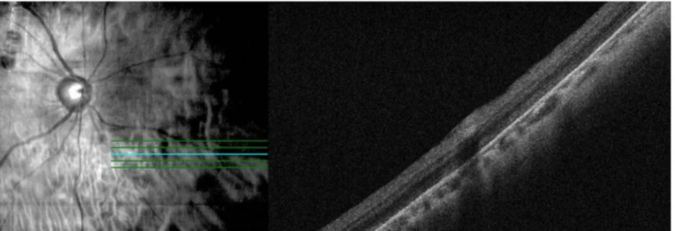
Figure 4. Consecutive spectral-domain optical coherence tomography (OCT) findings of case 2 at 10 days after vitrectomy (A-C) and
at 11 months after vitrectomy (D-F). (A) Infrared fundus image. Blue line: level of OCT cut. (B) Fundus image with cube scan over- lay showed the range of retinal fold. (C) Raster scan. Retinal fold was detected through full thickness retinal layer with nerve fiber layer undulation and inner segment ellipsoid zone disruption at 10 days after vitrectomy. (D) Infrared fundus image. Blue line: level of OCT image. (E) Fundus image with cube scan overlay. The range of retinal folds was reduced in macular cube scan. (F) Retinal fold was relieved at 11 months after vitrectomy. Retinal fold’s thickness was decreased and inner segment ellipsoid zone disruption was ameliorated in Raster scan image. The range of retinal folds was reduced in macular cube scan image.막의 국소적인 고반사 병변만 남기고 망막주름이 호전된 소견을 관찰할 수 있었다(Fig. 2). 두 번째 환자의 빛간섭 단층촬영에서 망막의 전층을 포함하는 황반의 주름과 그 주변으로 신경섬유층의 주름도 관찰되었다. 처음과 비교하 여 술 후 11개월째 빛간섭단층촬영의 래스터 스캔(raster scan)에서 망막주름이 호전되었으며 광수용체 타원구역 (inner segment ellipsoid zone) 단절면이 감소한 것을 관찰 할 수 있었다. 황반 큐브스캔(macular cube scan)에서도 망막주름의 범위가 줄어들었고 두께가 감소하였음을 알 수 있었다(Fig. 3, 4).
망막주름은 임상적 심각성과 관련된 보고는 있으나, 드 문 합병증이기에 적절한 치료 방법이 제시되지 않았고, 현 재까지도 뚜렷한 해결방법이 정립되지 않았다. 본 증례는 국내에서 이전 보고된 증례들과 달리 망막주름의 수술적 치료 없이도 기능적, 해부학적 회복이 가능함을 제시하였
으며 이를 빛간섭단층촬영으로 확인한 의의를 가진다. 만 약 주변부 망막주름의 경우, 정기적인 안저검사를 하여, 합병증의 발생이 없을 경우, 보존적 치료만으로도 호전을 기대할 수 있겠다. 추후 다양한 영상촬영장비로 장기간 관 찰하여 망막주름이 해소되는 기전에 대한 많은 추가적인 연구가 필요하다고 생각된다.
REFERENCES
1) dell'Omo R, Costagliola C. Longitudinal study of macular folds by spectral-domain optical coherence tomography. Am J Ophthalmol 2012;154:757-8; author reply 8-9.
2) Iafe NA, Law S, Sarraf D, Tsui I. Outer retinal folds following pars plana vitrectomy with membrane peel. Retin Cases Brief Rep 2017;11 Suppl 1:S31-3.
3) Pavan PR. Retinal fold in macula following intraocular gas. An avoidable complication of retinal detachment surgery. Arch
= 국문초록 =
열공망막박리 환자에서 유리체절제술 이후에 발생한 망막주름의 2예
목적: 열공망막박리 환자에서 유리체절제술 이후에 발생한 망막주름의 2예를 보고하고자 한다.
증례요약: (증례 1) 52세 남성이 우안의 시력저하를 주소로 내원하였다. 우안의 교정시력은 0.8이었고 12시와 1시 방향의 망막 열공과 황반부를 침범하지 않은 상부 망막박리가 관찰되었다. 치료를 위해 유리체절제술 및 가스 주입술을 시행하였으며 망막은 재유착되었 다. 유리체절제술 시행 1주일째 망막은 유착되었으나 시신경유두의 4시 방향으로 2시신경유두 직경 떨어진 부위로부터 경선방향으로 적도부까지 이어지는 망막주름이 형성되었다. 특별한 치료 없이 경과관찰 중에 안저소견에서 3달째에 주름이 자발적으로 호전된 소견 이 관찰되었다. 이후 재발 없이 안정적인 경과를 보였다. (증례 2) 59세 남성이 우안의 시력저하를 주소로 내원하였다. 우안의 교정시 력은 안전수지였고 11시 방향의 망막 열공과 황반부를 침범한 상부 망막박리가 관찰되었다. 유리체절제술 및 가스 주입술을 시행하였 으며 망막은 재유착되었다. 유리체절제술 시행 10일째 망막박리의 폐쇄를 확인하였으나 시신경유두의 이측에서 7시 방향으로 적도부 까지 이어지는 망막주름이 형성되었다. 경과관찰 중 빛간섭단층촬영에서 2주째에 동일한 위치에 망막주름이 관찰되었고 11달째 망막 주름의 호전이 관찰되었다.
결론: 유리체절제술 이후에 발생한 망막주름의 자연호전과 빛간섭단층촬영 소견은 국내에 보고된 바가 없어 이를 보고하고자 한다.
<대한안과학회지 2018;59(4):379-383>
Ophthalmol 1984;102:83-4.
4) El-Amir AN, Every S, Patel CK. Repair of macular fold following retinal reattachment surgery. Clin Exp Ophthalmol 2007;35:791-2.
5) Ruiz-Moreno JM, Montero JA. Sliding macular fold following ret- inal detachment surgery. Graefes Arch Clin Exp Ophthalmol 2011;249:301-3.
6) Gotzaridis E, Georgalas I, Ladas I. Spontaneous regression of a ret- inal fold a year after scleral buckling and intravitreal injection of gas. Eye (Lond) 2010;24:394-6.
7) Song YJ, Kim YB, Jo YJ. Management of macular folds using sub- macular BSS injection and partial fluid-gas exchange. J Korean Ophthalmol Soc 2003;44:2022-7.
8) Albert DM, Jakobiec FA. Principles and Practice of Ophthalmology, 2nd ed. Philadelphia: W.B. Saunders Co., 2000; 889-98.
9) Heinrich H, Silvia B. Retinal folds following retinal detachment surery. Ophthalmologia 2011;226 Suppl 1:18-26.
10) Hayashi A, Usui S, Kawaguchi K, et al. Retinal changes after reti- nal translocation surgery with scleral imbrication in dog eyes.
Invest Ophthalmol Vis Sci 2000;41:4288-92.
11) Trinh L, Glacet-Bernard A, Colasse-Marthelot V, et al. Macular fold following retinal detachment surgery. J Fr Ophtalmol 2006;
29:995-9.
12) Larrison WI, Frederick AR Jr, Peterson TJ, Topping TM. Posterior retinal folds following vitreoretinal surgery. Arch Ophthalmol 1993;111:621-5.
13) Wong R. Longitudinal study of macular folds by spectral-domain optical coherence tomography. Am J Ophthalmol 2012;153: 88-92.e1.