ISSN 0378-6471 (Print)⋅ISSN 2092-9374 (Online)
https://doi.org/10.3341/jkos.2020.61.1.111
Case Report
실리콘기름 충전 급성망막괴사 눈에서 Ganciclovir 주입 후 발생한 망막독성
Retinal Toxicity Following the Injection Ganciclovir into Silicone Oil-filled Eye to Treat Acute Retinal Necrosis
조연지⋅최승권⋅박선호⋅이재정⋅이지은⋅박성후
Yeon Ji Jo, MD, Seung Kwon Choi, MD, Sun Ho Park, MD, Jae Jung Lee, MD, Ji Eun Lee, MD, PhD, Sung Who Park, MD, PhD
부산대학교 의과전문대학원 안과학교실
Department of Ophthalmology, Pusan National University School of Medicine, Yangsan, Korea
Purpose: To report a case of retinal toxicity after an intravitreal ganciclovir injection to treat acute retinal necrosis in an eye filled with silicone oil.
Case summary: A 56-year-old male presented with ocular pain and visual loss in his right eye. His best-corrected visual acuity was 20/25, inflammatory cells in the anterior chamber, multiple retinitis lesions and retinal vessel occlusions in the peripheral ret- ina and vitreous opacity were showed. Acute retinal necrosis was suspected, anterior chamber polymerase chain reaction (PCR) test was done. Aciclovir 2,400 mg/day intravenously and ganciclovir 2.0 mg were administered by intravitreal injection. After 4 days, retinitis was worsened and PCR test was positive for varicella zoster virus. Ganciclovir intravitreal injections were in- creased twice a week. After 16 days, retinal detachment occurred, so scleral encircling, vitrectomy, laser photocoagulation, and silicone oil tamponade were conducted. Ganciclovir 1.0 mg was injected at the end of surgery. The patient’s visual acuity de- creased to hand motion, and multiple crystal deposits with multiple retinal hemorrhages were observed in the right eye the next day. Visual acuity did not recover and optical coherent tomography showed that the macula was thinned.
Conclusions: Visual loss seemed to be related with the retinal toxicity of ganciclovir. The increased local concentration due to the silicone oil tamponade is thought to have caused the toxicity.
J Korean Ophthalmol Soc 2020;61(1):111-115
Keywords: Ganciclovir, Retinal necrosis syndrome and acute, Retinal toxicity
■Received: 2019. 7. 26. ■ Revised: 2019. 9. 4.
■Accepted: 2019. 12. 30.
■Address reprint requests to Sung Who Park, MD, PhD Department of Ophthalmology, Pusan National University Hospital, #179 Gudeok-ro, Seo-gu, Busan 49241, Korea Tel: 82-51-240-7326, Fax: 82-51-242-7341
E-mail: [email protected]
*Conflicts of Interest: The authors have no conflicts to disclose.
ⓒ2020 The Korean Ophthalmological Society
This is an Open Access article distributed under the terms of the Creative Commons Attribution Non-Commercial License (http://creativecommons.org/licenses/by-nc/3.0/) which permits unrestricted non-commercial use, distribution, and reproduction in any medium, provided the original work is properly cited.
급성망막괴사는 1971년에 Urayama1에 의해 처음 보고된 괴사성망막염이다. 1994년 American Uveitis Society에서
제시한 진단 기준은 주변부 망막에 하나 이상의 경계가 불 분명한 망막괴사, 급속히 진행하는 병변, 측면으로의 확산, 폐쇄성 소동맥 혈관병증, 심한 유리체 또는 전방의 염증반 응이 보이는 경우이다.2 이후 급성망막괴사의 원인이 바이 러스로 밝혀졌고, 전방수를 이용한 중합효소 연쇄반응(po- lymerase chain reaction, PCR)검사에서 바이러스가 검출된 경우보다 명확한 진단이 가능하고, 바이러스가 특정되면 치료 방향결정에 도움을 준다.3
급성망막괴사는 항바이러스제제의 전신치료 혹은 국소 적 유리체강내 주입으로 치료하며, 부신피질호르몬, 항응고
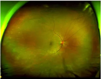
Figure 1. Ultra-widefield fundus image at the initial visit.
Multiple retinal opaque was presented in the periphery. Mild vitreous haziness and retinal vasculitis were combined.
A
B
Figure 2. Ultra-widefield fundus image and optical coherent
tomography at the sixteen day after (A) retinal detachment with giant retinal tear were noticed in the fundus examination.(B) Optical coherence tomography showed fovea center was not involved.
제 등을 보조적으로 사용할 수 있다. 전신 항바이러스제제 는 Aciclovir가 일차약제로 추천되고, 유리체강내 주입에는 Ganciclovir, Forscarnet, Brivudine 등이 사용된다. Flaxel et al4은 항바이러스제제의 전신치료와 유리체강내 주입을 병 용하였을 때 예후가 좋았다고 보고하였다.
1994년 ganciclovir 40 mg를 유리체강 내에 주입 후 망막 독성이 발생하였다는 보고가 있지만,5 2.0 mg 이하의 주입 량은 비교적 안전한 것으로 알려져 있다.6 급성망막괴사는 경과 관찰 중 50-70%의 높은 빈도로 망막박리가 발생할 수 있으며,7 망막박리가 발생한 경우 예후는 매우 나쁘며, 유 리체절제술과 실리콘기름 충전 등이 필요하다.8,9
급성망막괴사로 진단 후 전신 및 국소적 항바이러스제로 치료한 환자에서 망막박리가 발생하였다. 유리체절제술 및 실리콘기름주입술을 시행하고, 안전한 용량이라고 알려진 ganciclovir 1.0 mg를 유리체강내 주입하였으나 망막독성을 경험하였기에 이를 보고하고자 한다.
증례보고
56세 남자가 3일 전부터 발생한 우안의 시력저하와 통증 을 주소로 본원에 내원하였다. 고혈압 및 고지혈증으로 치 료 중이었고 다른 특이병력은 없었다. 최대교정시력은 우안 0.8, 좌안 1.0이었으며 안압은 우안 16 mmHg, 좌안 14 mmHg 였다. 세극등현미경검사에서 우안 결막충혈, 전방 내 염증 세포 1+ 및 유리체의 염증 소견이 관찰되었다. 안저검사에 서는 우안 망막 주변부에 다수의 황백색 침윤이 관찰되었 고 하측에 혈관염 소견이 관찰되었다(Fig. 1). 전방수를 채
취하여 Herpes simplex virus (HSV) type 1, 2와 Varicella zoster virus (VZV)에 대한 PCR을 시행하였고, 검사 결과 VZV 가 검출되었다.
급성망막괴사로 진단하고 aciclovir 2,400 mg/일을 정맥 주사하고, 2.0 mg ganciclovir를 유리체강 내에 주입하였다.
치료 2일째 유리체혼탁이 증가하여 prednisolone 20 mg 하 루 1회 경구투여를 시작하였고, 치료 4일째 망막괴사 범위 가 증가하여 ganciclovir 2.0 mg를 1주일에 2회씩 반복적으 로 주사하였다.
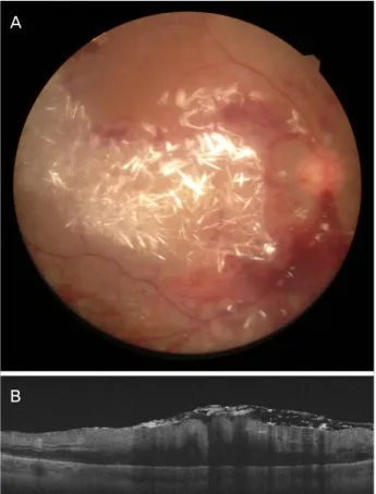
치료 16일째 우안 최대교정시력이 0.5로 감소하였고, 안 저검사에서 이측 망막박리가 관찰되었다(Fig. 2). 우안 두르 기공막돌융술, 유리체절제술, 눈속레이저, 실리콘기름 충전 술을 시행하였다. 1.0 mg Ganciclovir를 유리체강 내에 주 입하였다. 수술 다음날 우안 최대교정시력이 안전수동으로 감소하였고, 안저검사에서 황반부 크리스탈 결정 형태 침 착물과 다발성출혈이 관찰되었고, 빛간섭단층촬영검사에서 심한 망막부종이 관찰되었다(Fig. 3).
수술 6개월 후 교정시력은 안전수동으로 회복되지 않았 다. 안저검사에서 망막은 잘 유착되어 있었고, 급성망막괴 사로 인한 염증반응은 소실되었으나 빛간섭단층촬영검사 에서 중심 황반의 얇아짐이 관찰되었다(Fig. 4).
A
B
Figure 3. Fundus photo and optical coherent tomography at
the next day of the surgery (A) multiple crystal deposit with multiple retinal hemorrhages was noticed on the fundus examination. (B) Optical coherence tomography showed se- vere macular edema.A
B
Figure 4. Ultra-widefield fundus image and optical coherent
tomography at the sixth months after (A) there were no ne- crotic lesion, inflammation and retinal detachment on the fun- dus examination. (B) Optical coherence tomography image showed thinned macula.고 찰
Ganciclovir는 HSV, VZV, Cytomegalovirus (CMV)에서 aciclovir보다 강한 항바이러스 효과를 보이는 것으로 알려 져 있어 aciclovir 치료에 반응이 없는 경우나 면역 저하 환 자에서 사용을 고려할 수 있는 항바이러스제제이다.10 국내 에서도 급성망막괴사환자에서 유리체강내 주입술을 시행 하여 효과를 본 증례들이 보고되었다.11 Ganciclovir는 대부 분의 환자에서 치료 용량으로 유리체강내 주입술을 시행할 경우 안전하다고 알려져 있다. Saran and Maguire5은 40 mg 의 ganciclovir를 유리체강 내에 주입하였던 환자에서 크리 스탈 결정 형태 침착물과 다수의 망막출혈이 관찰되었고, 시기능이 소실되었다고 보고하였다. 높은 알칼리성을 띄는 고농도의 ganciclovir가 직접 망막에 접촉하여 삼투적 손상 을 유발하여 망막독성이 발생한 것으로 추정하였다. 임상 양상이 본 연구의 증례와 매우 유사하여 본 증례도 고농도 의 ganciclovir의 직접 접촉에 의한 손상으로 추정할 수 있 다.
Teoh et al6은 34명의 CMV 망막염 환자에서 ganciclovir 2.0 mg를 2회/주 주입한 후 1.0 mg 1회/주 주입하는 것으로 치료한 결과를 보고하였고, 이는 효과적이면서 안전한 농 도라고 보고하였다.6 급성망막괴사 환자에서 ganciclovir의 적절한 주입량에 대한 보고가 없어, 저자들은 Teoh et al6의 논문을 참조하여 ganciclovir 1회 2.0 mg 주입하고 조절이 잘 되지 않을 경우 주 2회 간격으로 추가하는 것을 원칙으 로 하였다.
토끼를 이용한 동물실험에서 Hegazy et al12는 실리콘기 름이 주입된 경우 ganciclovir 유리체강내 주입술이 망막독 성 발생 가능성을 높일 수 있다고 보고하였다. 반면 Meshi et al13은 실리콘기름이 주입된 1명의 환자에게 2.0 mg의 ganciclovir를 반복적으로 주입하였으나 독성이 발생하지 않고 바이러스 망막염이 효과적으로 치료되었다고 보고하 였다.
Ganciclovir는 수용성이므로 실리콘기름에는 용해되지 않는다. 유리체강내 용적이 5 mL이고, 충전물이 없는 경우 ganciclovir 2.0 mg가 유리체강 내에 주입되면 유리체액내 ganciclovir의 농도는 2.0 mg/5 mL이 된다. 그렇지만 유리 체강내 용적 중 4 mL가 실리콘기름으로 채워진다면 유리 체강내 주입된 2 mg ganciclovir는 1 mL의 유리체액에 용 해되므로 농도는 2.0 mg/1 mL가 되어 충전물이 없는 경우
에 비해 농도가 약 5배 높아진다. 만약 실리콘기름이 4.8 mL 채워져 0.2 mL의 유리체액만 존재하는 눈에 ganciclovir 2.0 mg을 주입할 경우 유리체액 내 ganciclovir의 농도가 2.0 mg/
0.24 mL가 되어, 충전물이 없는 경우에 비해 농도가 약 25배 높아지며, Saran and Maguire5의 증례보고에서의 40 mg을 5 mL 유리체액에 주입한 경우와 유사한 농도가 된다. 따라 서 충전물이 채워진 눈에서 약제를 유리체강 내에 주사하 는 경우 약제 독성 가능성이 높아질 것을 예상할 수 있고, 망막독성 발생 여부는 실리콘기름 충전 비율에 따라 달라 질 수 있음을 추정할 수 있다. 본 증례는 실리콘기름이 95%
정도 채워진 것으로 추정되며, 실리콘기름 충전 비율이 높 아 상대적으로 적은 ganciclovir 주입량(1.0 mg)에도 불구하 고 망막독성이 발생한 것으로 추정된다.
본 증례는 실리콘기름이 충전된 눈에서 일반적인 치료용 량의 절반인 1.0 mg ganciclovir를 유리체강 내에 주입하였 다. 주입 후 다음날 시력은 소실되었고, 망막표면에 크리스 탈 결정 형태의 침착과 다수의 망막출혈을 확인하였고, 이 는 고농도 ganciclovir에 의한 망막독성으로 추정된다. 실리 콘기름 충전으로 유리체액내 ganciclovir의 국소적 농도가 높아져 망막독성을 유발한 것으로 생각된다. 실리콘기름이 주입된 눈에서 유리체내 약제를 주입하는 경우 적절한 주 입량을 예측하기 어려워 주의가 필요하다. 유리체내 주입 술이 꼭 필요한 경우라면 실리콘기름 충전 비율을 고려하 여, 약제 주입 용량을 결정하는 것이 합리적일 것으로 추정 된다.
REFERENCES
1) Urayama A. Unilateral acute uveitis with relinal periarteritis and
detachment. Jpn J Clin Ophthalmol 1971;25:607-19.
2) Holland GN. Standard diagnostic criteria for the acute retinal ne- crosis syndrome. Executive Committee of the American Uveitis Society. Am J Ophthalmol 1994;117:663-7.
3) Bernheim D, Germi R, Labetoulle M, et al. Time profile of viral DNA in aqueous humor samples of patients treated for vari- cella-zoster virus acute retinal necrosis by use of quantitative re- al-time PCR. J Clin Microbiol 2013;51:2160-6.
4) Flaxel CJ, Yeh S, Lauer AK. Combination systemic and intravitreal antiviral therapy in the management of acute retinal necrosis syn- drome (an American Ophthalmological Society thesis). Trans Am Ophthalmol Soc 2013:111:133-44.
5) Saran BR, Maguire AM. Retinal toxicity of high dose intravitreal ganciclovir. Retina 1994;14:248-52.
6) Teoh SC, Ou X, Lim TH. Intravitreal ganciclovir maintenance in- jection for cytomegalovirus retinitis: efficacy of a low-volume, in- termediate-dose regimen. Ophthalmology 2012;119:588-95.
7) Clarkson JG, Blumenkranz MS, Culbertson WW, et al. Retinal de- tachment following the acute retinal necrosis syndrome. Ophthalmology 1984;91:1665-8.
8) Park SW, Shin MK, Byon IS, et al. Risk factors of retinal detach- ment after acute retinal necrosis. J Korean Ophthalmol Soc 2013;54:1694-9.
9) Matsuo T. Vitrectomy and silicone oil tamponade as an initial sur- gery for retinal detachment after acute retinal necrosis syndrome.
Ocul Immunol Inflamm 2005;13:91-4.
10) Duker JS, Blumenkranz MS. Diagnosis and management of the acute retinal necrosis (ARN) syndrome. Surv Ophthalmol 1991;
35:327-43.
11) Yang JW, Kim WJ, Park YH, Two cases of acute retinal necrosis treated with systemic antiviral drugs and intravitreal antiviral injections.
J Korean Ophthalmol Soc 2009;50:794-9.
12) Hegazy HM, Kivilcim M, Peyman GA, et al. Evaluation of toxicity of intravitreal ceftazidime, vancomycin, and ganciclovir in a sili- cone oil-filled eye. Retina 1999;19:553-7.
13) Meshi A, Friehmann A, Sella S, et al. Intravitreal administration of antiviral agents in silicone oil-filled human eyes. Ophthalmol Retina 2017;1:288-93.
= 국문초록 =
실리콘기름 충전 급성망막괴사 눈에서 Ganciclovir 주입 후 발생한 망막독성
목적: 급성망막괴사 환자에서 Ganciclovir 유리체강내 주입 후 발생한 망막독성 1예를 보고하고자 한다.
증례요약: 56세 남자가 우안 시력저하 및 통증으로 내원하였다. 최대교정시력 0.8, 전방 염증과 다수의 주변부 망막염, 혈관폐쇄, 유리 체 혼탁이 관찰되었다. 급성망막괴사로 추정하여 전방수 중합효소 연쇄반응 검사와 aciclovir 2,400 mg/일 정맥주사, ganciclovir 2.0 mg 유리체강내 주입술을 시행하였다. 치료 4일째 병변 악화와 전방수에서 Varicella zoster virus가 확인되었다. Ganciclovir 유리체강내 주입을 주 2회로 증량하였다. 치료 16일째 우안 망막박리 소견으로 두르기공막돌융술, 유리체절제술, 눈속레이저, 실리콘기름 충전술 과 ganciclovir 1.0 mg을 유리체강 내에 주입하였다. 수술 다음날 우안 최대교정시력 안전수동이었고, 황반부의 크리스탈 결정 형태 침착물과 다발성 출혈이 관찰되었다. 이후 시력은 회복되지 않았고 황반의 얇아짐이 관찰되었다.
결론: 시력소실은 ganciclovir 망막독성에 의한 것으로 추정되며, 실리콘기름 충전으로 ganciclovir의 국소적 농도가 높아진 것이 원인 으로 생각된다.
<대한안과학회지 2020;61(1):111-115>
조연지 / Yeon Ji Jo
부산대학교 의과전문대학원 안과학교실 Department of Ophthalmology, Pusan National University Hospital