www.ophthalmology.org 1626
대한안과학회지 2013년 제 54 권 제 10 호 J Korean Ophthalmol Soc 2013;54(10):1626-1629 pISSN: 0378-6471
eISSN: 2092-9374
http://dx.doi.org/10.3341/jkos.2013.54.10.1626
= 증례보고 =
유리체절제술을 받은 눈에서 발생한 황반원공의 자연폐쇄 1예
이승희⋅이근우⋅윤숙현⋅김윤영 대구가톨릭의과대학 안과학교실
목적: 유리체절제술을 시행 받았던 눈에 발생한 황반원공이 자연적으로 폐쇄된 증례를 보고하고자 한다.
증례요약: 좌안의 혈관염에 의한 유리체 혼탁과 2차성 망막전막으로 유리체절제술을 시행 받은 71세 여자 환자가 술 후 전방과 유리체 내 염증 소견이 재발하여 치료를 받던 중 안저 검사상 황반원공이 발생되었다. 당시 좌안의 시력은 0.125였고 빛간섭단층촬영 결과 황반 주위의 낭포성 부종을 동반한 전층의 황반원공이 관찰되었다. 환자에게 유리체절제술, 내경계막제거술 및 가스 주입술을 권유하 였으나 전신적인 쇠약으로 인해 더 이상의 수술적 치료를 원하지 않아 경과 관찰하였다. 황반 원공 발생 2주 후 낭포성 부종 및 원공의 감소를 보였고 13주 후 황반원공의 완전한 폐쇄와 정상적인 황반의 모양을 빛간섭단층촬영상 확인할 수 있었으며 좌안의 시력은 0.4로 회복되었다.
결론: 유리체절제술을 시행 받았던 눈에서 황반원공이 발생한 후 자연적인 폐쇄가 일어나는 경우는 극히 드물지만 안내 염증을 동반한 작은 크기의 황반원공의 경우 자연적인 폐쇄를 보일 수 있어 즉각적인 수술보다는 경과 관찰을 고려할 수 있으며 이러한 현상은 황반 부종의 발생이나 회복 같은 퇴행성 과정이 작용한 것으로 생각한다.
<대한안과학회지 2013;54(10):1626-1629>
■Received: 2013. 3. 4. ■ Revised: 2013. 5. 8.
■Accepted: 2013. 7. 24.
■Address reprint requests to Yoon Young Kim, MD, PhD Department of Ophthalmology, Daegu Catholic University Medical Center, #33 Duryugongwon-ro 17-gil, Nam-gu, Daegu 705-718, Korea
Tel: 82-53-650-4728, Fax: 82-53-627-0133 E-mail: [email protected]
* This study was presented as a poster at the 109th Annual Meeting of the Korean Ophthalmology Society 2013.
특발성 황반원공은 대개 수술적인 치료가 필요하나 자연 폐쇄를 보이는 증례들도 보고된바 있다. 외상성 황반원공을 제외한 특발성 황반원공의 자연폐쇄에 대한 연구에 의하면 0-6% 정도에서 수술적인 치료없이 황반원공의 폐쇄를 보 인다는 보고들이 있었으나 이들의 연구는 빛간섭단층촬영 없이 조사된 연구들이 많으며 최근 스펙트럼 영역 빛간섭 단층촬영을 이용한 연구에서는 약 2% 정도에서 자연폐쇄 가 일어나는 것으로 알려졌다.1
유리체절제술을 받은 눈에서 황반원공의 발생은 그 빈도 가 낮으며 발생된 황반원공이 자연적으로 폐쇄되는 경우는 극히 드문 현상으로 아주 제한적으로 보고된바 있다.2-5그 러나 이러한 황반원공의 발생과 폐쇄를 스펙트럼 영역의 빛간섭단층촬영계로 보고한 경우는 더욱 드물며 국내보고 는 없다.
저자들은 유리체혼탁과 망막전막으로 유리체절제술을 받았던 환자에서 발생된 황반원공의 발생과 자연폐쇄 과정 을 스펙트럼 영역의 빛간섭단층촬영계로 촬영한 영상과 함 께 보고하고자 한다.
증례보고
71세 여자환자가 좌안의 반복되는 시력저하를 호소하여 의뢰되었다. 당시 좌안의 시력은 안전수지 50 cm였으며 전 안부 검사상 특이 소견은 보이지 않았으며 안저 검사상 유 리체 혼탁이 심하여 자세한 안저 관찰이 힘든 상태였다. 환 자와 상의 후 좌안의 백내장 초음파유화술과 함께 유리체 절제술을 시행하였으며 술 중 망막하측주변부의 염증성 침 착물이 관찰되었고, 이미 완전한 후유리체박리는 이루어진 상태였다. 황반부의 전막이 관찰되어 망막전막제거술을 함 께 시행하였고 의도적인 내경계막제거술은 시행하지 않았 다. 술 후 1달째 시력은 0.1 정도로 호전되었으나 술 후 6주 째 1주간의 미열과 근육통으로 내과에 입원 후 안과에 협진 의뢰되어 시행한 안과 검사상 좌안의 전방염증 3+, 각막부 종이 보였으며 유리체에서 또한 2+ 정도의 염증소견을 관 찰할 수 있었다. 당시 시력은 0.02로 저하되어 있었고, 시행 한 빛간섭단층촬영(Cirrus HD OCT, Carl Zeiss Meditec, Inc., Dublin, CA, USA)상 다른 특이소견은 보이지 않았다
www.ophthalmology.org 1627 - 이승희 외 : 유리체절제술안의 황반원공 자연폐쇄 -
Figure 1. Optical coherence tomography (OCT) showing no abnormal finding 6 weeks after vitrectomy.
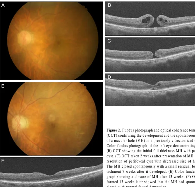
Figure 2. Fundus photograph and optical coherence tomography (OCT) confirming the development and the spontaneous closure of a macular hole (MH) in a previously vitrectomized eye. (A) Color fundus photograph of the left eye demonstrating a MH.
(B) OCT showing the initial full thickness MH with perifoveal cyst. (C) OCT taken 2 weeks after presentation of MH showing resolution of perifoveal cyst with decreased size of hole. (D) The MH closed spontaneously with a small residual foveal de- tachment 7 weeks after it developed. (E) Color fundus photo- graph showing a closure of MH after 13 weeks. (F) OCT per- formed 13 weeks later showed that the MH had spontaneously closed with normal foveal depression.
A B
C
D E
F
(Fig. 1). 내과에서 시행한 혈액 및 소변배양 검사에서는 특 이사항은 없었으나 구강분비물 검사상 구강칸디다증 소견 이 보여 칸디다에 의한 내인성안내염을 배제하기 위하여
전방수 천자술을시행하여 배양검사를 시행하였다.검사상 특 이사항은 보이지 않아 Prednisolone acetate 1% (Pred forte, Allegan)와 Moxifloxacin 0.5% 5 ml (Vigamox, Alcon)로 점안치료 하고 내과적으로 fluconazole 100 mg 복용하며 경과 관찰하였다.
치료 후 좌안의 염증소견이 좋아지고 시력은 0.32로 호 전되었고 안저 검사상 특이 소견은 보이지 않았다. 유리체 절제술 시행 11주 후 다시 환자의 시력이 0.125로 감소하 였으며 안저검사상 황반원공 소견이 관찰되었다(Fig. 2A).
당시 시행한 빛간섭단층촬영상 좌안 황반주위의 낭포성 부 종을 동반한 전층의 황반원공을 확인할 수 있었다(Fig.
2B). 망막내경계막제거술과 함께 유리체강내 가스 주입술 을 권유하였으나 환자의 전신상태가 좋지 않아 더 이상의 수술적인 치료를 원하지 않아 경과 관찰하였다. 황반원공
www.ophthalmology.org 1628
- 대한안과학회지 2013년 제 54 권 제 10 호 -
발생 2주 후 낭포성 부종 및 원공의 크기 감소를 보였고, 7 주 후 황반원공의 폐쇄 및 작은 범위의 황반박리만 남아있 는 것을 확인할 수 있었다(Fig. 2C, D). 황반원공 발생 13 주 후 완전한 황반원공의 폐쇄 및 정상적인 중심와 모양을 확인할 수 있었으며 시력은 0.4로 호전되었다(Fig. 2E, F).
고 찰
특발성 황반원공의 발생 후 자연 폐쇄가 일어나는 경우 는 자연적으로 완전한 후유리체박리가 일어나거나 후유리 체에 의한 견인이 완전히 해소될 때 발생할 수 있으며 이러 한 경우 병기상 1기 혹은 2기의 경우에서 간혹 발생될 수 있다.6
유리체절제술을 받은 눈에서 황반원공의 발생은 기존의 특발성 황반원공의 발생에 관여하는 후유리체견인과의 관 련성은 극히 낮으며 낭포성 황반부종과 같은 망막내층의 퇴행성 과정이 관여하는 것으로 알려져 있고 망막전막의 발생과 내경계막의 수축 등과 같은 망막표면의 견인력도 일부 관여할 수 있는 것으로 알려졌다.2,7그러나 이러한 경 우 황반원공이 자연적으로 폐쇄되는 경우는 보고가 드물며 특히 유리체절제술의 일차 원인이 황반원공이 아닌 경우 유리체절제안에서 황반원공의 발생과 자연폐쇄를 보고한 경우는 현재 5예가 보고되고 있으며 대동맥혈관류로 인한 유리체출혈로 수술한 경우가 1예, 망막전막 1예, 그 외 망 막박리로 유리체절제술을 시행 받은 경우가 3예로 보고되 고 있다.3-5본 증례의 경우처럼 유리체 혼탁과 2차성 망막 전막이 유리체절제술의 원인이었고 황반원공의 발생과 폐 쇄를 스펙트럼 영역의 빛간섭단층촬영으로 보고한 경우는 거의 없다.
본 증례의 경우 황반원공의 발생 기전은 명확하지는 않 으나 유리체절제술 후 염증 소견이 재발되었고 황반주위의 낭포성 황반부종이 빛간섭단층촬영상 관찰된 바 있어, 상기 기술한대로 낭포성 황반부종의 파열로 인한 망막내층의 손 상이 황반원공 발생의 초기 인자로 작용한 것으로 생각한 다. 이전의 유리체절제술 당시 유리체 피질은 충분히 제거
되었고 안저 검사와 빛간섭단층촬영상 황반에 견인력을 제 공할 만한 망막전막이 발견되지 않은 점과 황반원공의 크 기가 작고 균등한 모양으로 보이는 점 또한 이러한 설명에 부합될 수 있다고 생각한다.
또한 본 증례처럼 황반원공의 자연폐쇄는 이전의 보고에 서와 같이 낭포성 황반부종의 호전과 유리체절제술 후 증 가된 망막의 탄성으로 황반원공의 자연 폐쇄가 가능했던 것으로 판단된다.2,3
본 증례는 스펙트럼 영역 빛간섭단층촬영계로 유리체절 제술안에서 황반원공의 발생과 자연폐쇄를 확인한 예로서 유리체절제술안에서 발생한 황반원공의 경우, 수술적 치료 가 어려운 경우나 염증을 동반한 작은 크기의 황반원공은 바로 수술적인 방법을 선택하지 말고 일정 기간 경과 관찰 을 고려할 수 있음을 시사해주는 증례로 생각하나 향후 더 많은 증례를 통한 연구가 필요할 것이다.
REFERENCES
1) Privat E, Tadayoni R, Gaucher D, et al. Residual defect in the fo- veal photoreceptor layer detected by optical coherence tomog- raphy in eyes with spontaneously closed macular holes. Am J Ophthalmol 2007;143:814-9.
2) Yonekawa Y, Hirakata A, Inoue M, Okada AA. Spontaneous clo- sure of a recurrent myopic macular hole previously repaired by pars plana vitrectomy. Acta Ophthalmol 2011;89:e536-7.
3) Tsilimbaris MK, Gotzaridis S, Charisis SK, et al. Spontaneous clo- sure of macular holes developed after pars plana vitrectomy. Semin Ophthalmol 2007;22:39-42.
4) Lo WR, Hubbard GB. Macular hole formation, spontaneous clo- sure, and recurrence in a previously vitrectomized eye. Am J Ophthalmol 2006;141:962-4.
5) Ogawa M, Ohji M. Spontaneous closure of a macular hole after vi- trectomy for an epiretinal membrane. Jpn J Ophthalmol 2010;
54:357-70.
6) Hanano R, Shimoda Y, Kishi S. Tomographic features of sponta- neous closure of full-thickness macular holes. Jpn J Ophthalmol 2007;51:76-7.
7) Lipham WJ, Smiddy WE. Idiopathic macular hole following vi- trectomy: implications for pathogenesis. Ophthalmic Surg Lasers 1997;28:633-9.
www.ophthalmology.org 1629
=ABSTRACT=
A Case of Spontaneous Closure of Macular Hole in a Previously Vitrectomized Eye
Seung Hee Lee, MD, Geun Woo Lee, MD, Sook Hyun Yoon, MD, Yoon Young Kim, MD, PhD
Department of Ophthalmology, Catholic University of Daegu School of Medicine, Daegu, Korea
Purpose: To report a case of spontaneous closure of a macular hole in a previously vitrectomized eye.
Case summary: A 71-year-old female had undergone vitrectomy on the left eye due to a secondary epiretinal membrane with vitreous opacity caused by vasculitis. After the procedure, while the patient was still on medication for the recurrent in- flammation of the anterior and posterior segment of the vitreous, a macular hole was found after fundus examinations.
Visual acuity of her left eye was 0.125 and ocular coherence tomography (OCT) confirmed a full thickness macular hole with a perifoveal cyst. We recommended vitrectomy, internal limiting membrane peeling, and intravitreal gas injection, but the patient refused further intervention due to her poor general condition. After 2 weeks, resolution of the perifoveal cyst with the macular hole was observed. After 13 weeks, OCT revealed the complete closure of the macular hole with normal foveal depression and the patient regained 0.4 visual acuity.
Conclusions: Spontaneous closure of macular hole is a rare phenomenon in vitrectomized eyes, but a small macular hole with inflammation may close spontaneously without additional intervention. Therefore, observation should be considered rather than hasty surgical intervention. Apparently, the spontaneous closure of a macular hole is due to degenerative proc- esses such as development of macular edema and natural recovery.
J Korean Ophthalmol Soc 2013;54(10):1626-1629
Key Words: Macular hole, Spontaneous closure, Vitrectomy
Address reprint requests to Yoon Young Kim, MD, PhD
Department of Ophthalmology, Daegu Catholic University Medical Center
#33 Duryugongwon-ro 17-gil, Nam-gu, Daegu 705-718, Korea Tel: 82-53-650-4728, Fax: 82-53-627-0133, E-mail: [email protected]
- 이승희 외 : 유리체절제술안의 황반원공 자연폐쇄 -