대한안과학회지 제 49 권 제 1 호 2008 J Korean Ophthalmol Soc 49(1):169-176, 2008 DOI : 10.3341/jkos.2008.49.1.169
맥락막골종은 맥락막에 발생하는 양성 골화 종양으 로 10∼20대의 건강한 여성에서 주로 발생하고 75%
정도가 단안에서 발생하지만 드물게 양안에서 발생하기 도 한다. 증상으로는 무증상에서부터 시력감소, 변시 증, 시야결손 등 다양하게 나타나며 망막색소상피 및 신경망막의 변성, 망막하 신생혈관, 망막하출혈 등 합 병증의 발생상태에 따라 시력예후는 다양하다.1,2 종양 은 시신경주위에 황백색에서 황적색의 경계가 명확한 부채꼴 모양 혹은 지도 모양을 보이며 난원형이나 둥근 모양을 하고 있다. 1978년 Gass et al3에 의해 처음 보고되었고 국내에서는 5례가 보고되었으며, 아직 원인 에 대해서는 정확히 밝혀지지 않았다. 최근 몇 년간 황 반부 병변이나 맥락막망막 질환에서 빛간섭단층촬영과 다초점 망막전위도 검사가 활발히 시행되고 있지만 맥 락막골종에 대한 소견은 아직 국내에 보고된 바 없었 다. 저자들은 맥락막골종 환자를 경험하고 빛간섭단층 촬영과 다초점 망막전위도 검사를 시행하였기에 그 소 견을 문헌고찰과 함께 보고하고자 한다.
증례보고
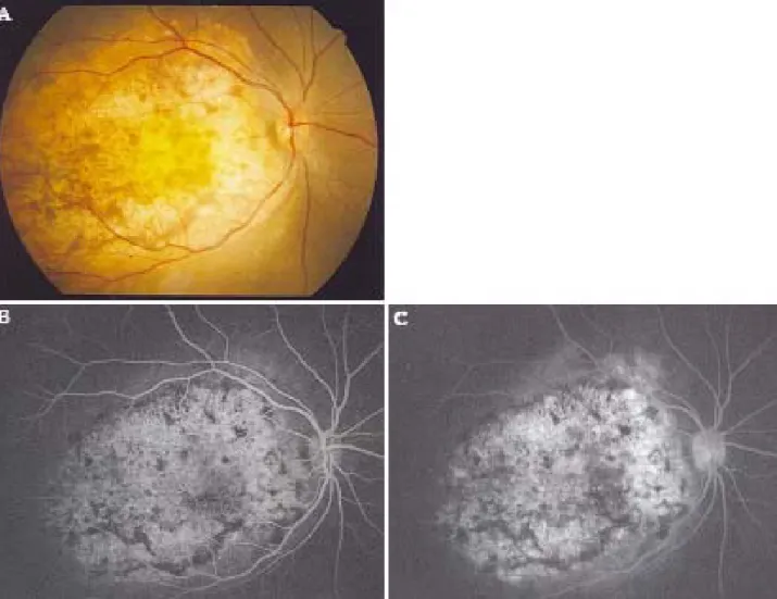
16세 여자가 4년간의 우안 시력감소를 주소로 내원 하였다. 과거력 및 가족력상 특이사항은 없었고 초진시 교정시력은 우안 0.1, 좌안 1.0이었으며 골드만 압평안 압계로 측정한 안압은 우안 14 mmHg, 좌안 15 mmHg이었고, 세극등 현미경검사상 양안 모두 특이소 견이 관찰되지 않았다. 안저검사상 우안의 시신경연접 부위와 황반부를 포함한 후극부에 가로 10 mm, 세로 9 mm 정도 크기의 황백색의 망막하병변이 관찰되었으 며 전반적인 망막색소상피의 변성과 병변부위에 장액성 망막박리가 합병되어 있었다(Fig. 1A). 좌안의 안저소 견과 우안의 주변부망막, 망막혈관, 시신경유두는 정 상이었으며, 형광안저촬영검사상 초기부터 병변과 일 치하는 과형광 소견이 나타나기 시작하여 형광물질 의 누출 없이 말기까지 지속되었고 황반부에 망막하 신생혈관의 소견은 보이지 않았다(Fig. 1B, C).
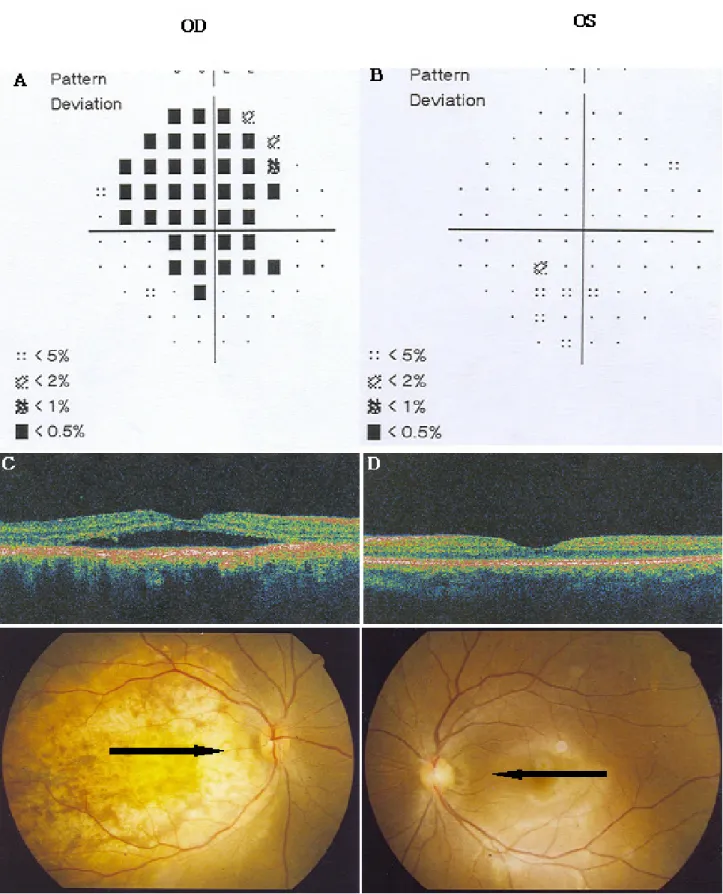
A-scan에서 맥락막골종의 전면에 해당하는 부위에 고도의 spike가 나타났고, B-scan에서 약간 융기 된 high reflectivity의 맥락막종괴와 posterior acoustic shadowing이 관찰되었으며, 전산화단층촬 영상 우안 후극부 맥락막에 골음영과 일치하는 원판 모 양의 병변을 보여 맥락막골종으로 확진할 수 있었다 (Fig. 2A-D). 자기공명촬영을 시행하였으나 특이소견 을 관찰할 수 없었다. 시야검사상 우안에 병변의 크기
맥락막골종 환자의 빛간섭단층촬영과 다초점 망막전위도
임혜진1․배민철1․박준우2․조영욱1․박대진1 대구파티마병원 안과1, 창원 파티마병원 안과2
목적 : 맥락막골종 환자를 경험하고 빛간섭단층촬영과 다초점 망막전위도 소견을 문헌고찰과 함께 보고하고자 한다.
증례요약 : B-scan과 CT로 맥락막골종으로 진단된 환자에서 빛간섭단층촬영과 다초점 망막전위도 검사를 시행하였
다. 빛간섭단층촬영상 황반 부위의 장액성 망막박리와 맥락막 부위에 두껍고 불규칙한 판과 같은 형태로 높은 반사도를 보이는 종양을 관찰할 수 있었다. 다초점 망막전위도검사상 Trace arrays에서 환측은 건측에 비해 중심부 뿐만 아니 라 주변부에 걸쳐 파형이 감소된 소견을 보였고 특히 중심부가 더 현저하게 감소되었으며, Ring averages는 모든 영 역에서 건측에 비해 진폭(amplitude)의 감소가 관찰되었고, 3D-Topography상 중심부의 반응이 현저하게 감소되 었으며 건측에서 보이는 상승부(peak)가 관찰되지 않았다.
결론 : 빛간섭단층촬영상 맥락막골종은 높은 반사도를 보이는 불규칙한 외측 표면을 가진 두꺼운 판 모양의 구조를 보
였고, 다초점 망막전위도검사를 통해 맥락막골종으로 인한 망막의 기능 저하를 예측할 수 있었다.
<한안지 49(1):169-176, 2008>
<접수일 : 2007년 5월 31일, 심사통과일 : 2007년 10월 2일>
통신저자 : 박 대 진
대구시 동구 신암동 576-31 대구파티마병원 안과
Tel: 053-940-7140, Fax: 053-940-7149 E-mail: [email protected]
= 증례보고 =
― 임 혜 진 외 : 맥 락 막 골 종 의 O C T와 M F-ER G ―
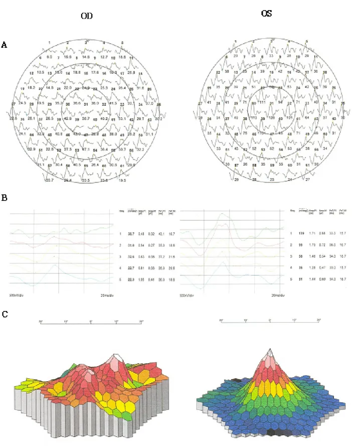
보다 작은 상대적 시야결손을 보였고, 빛간섭단층 촬영(STRATUS OCT model 3000, Zeiss Instruments INC. San Leandro, CA)상 우 안 황반 부위에 장액성 망막박리와 종양이 위치하 는 맥락막 부위에 높은 반사도의 불규칙한 외부표 면을 가진 두꺼운 판과 같은 형태가 관찰되었다 (Fig. 3A-D). 다초점 망막전위도검사상 First order kernel responses의 Trace arrays에 서 환측은 건측에 비해 중심부 뿐만 아니라 주변부 에 걸쳐 파형이 감소된 소견을 보였는데 특히 중심부가 더 현저하게 감소되었고, Ring averages는 ring 1 부터 ring 6까지 건측에 비해 진폭(amplitude)의 감소가 관찰되었으며, 3D-Topography상 건측에 서 보이는 상승부(peak)가 관찰되지 않고 중심부의 반응이 현저하게 감소된 소견을 보였다(Fig. 4A-C).
고 찰
맥락막골종은 성숙한 골로 구성된 양성 종양으로 1978년 Gass et al3이 처음으로 보고하였으며, 전형 적으로 10∼20대 건강한 여성에서 발생하지만 남성이 나 10세 이하의 어린이, 30세 이상의 성인에 대한 보고 도 있다.3-8 75% 정도에서 단안성으로 발생하지만 드 물게 양안성으로 발생하기도 하며 건안에 새로운 병변 이 발생하기도 한다.9,10 증상은 무증상에서부터 시력저 하, 변시증, 시야 결손 등 다양하게 나타나며 갑자기 시 력이 저하된 경우에는 황반부의 망막하 신생혈관에 의 한 것이 많고, 형광안저촬영상 초기부터 형광물질의 누 출을 보이며 중심와밖에 위치할 경우 Argon-green, Krypton-red 레이저를 이용한 광응고를, 중심와를 침범한 경우 광역학치료를 시도할 수 있다.11-14
Figure 1. (A) Fundus photograph of the right eye shows large, well-demarcated, round, yellow-white lesion involving the peripapillary area and macula. (B) Early fluorescein angiogram of the right eye demonstrates hyperfluorescence due to window defect corresponding to choroidal lesion. (C) Late fluorescein angiogram of the right eye demonstrates hyperfluorescence without leakage.
― 대 한 안 과 학 회 지 제 49 권 제 1 호 2008년 ―
주로 시신경주위와 시신경연접부위에 발생하지만 황반부까지 침범할 수 있으며, 드물게는 시신경연접 부위의 침범없이 황반부에만 발생하였다는 보고도 있 다.2,4,5
병변은 망막색소상피의 얇아진 정도, 탈색소 정도에 따라 황적색 또는 황백색을 띠며 심할수록 황백색을 띠 게 되고, 경계가 분명한 부채꼴 혹은 지도모양으로 난 원형이나 둥근 모양을 보인다. 병변 부위의 망막혈관과 시신경유두는 특징적으로 종양의 영향을 받지 않으며, 유리체나 전안부에 이상소견을 보이지 않는다.
형광안저촬영상 망막색소상피의 변성으로 인해 초기 부터 과형광이 나타나고 말기까지 지속되며, 시야검사 상 병변의 크기보다 작은 상대적 시야결손이 나타난 다.8
A-scan에서 맥락막골종의 전면에 해당하는 부위에 고도의 spike가 나타나고, B-scan에서 약간 융기된
high reflectivity의 맥락막 종양이 나타나며 낮은 주 사감도로 주위 조직의 echo가 사라지게 하더라도 종양 부위에 고도의 반향은 지속되고 종양 바로 뒤에 보이는 acoustic shadowing은 가성 시신경(pseudo-optic nerve) 형태를 보인다.1,15 전산화단층촬영상 후극부 맥락막 부위에 정상적인 골음영과 동일한 고음영의 석 회화 병변이 특징적으로 관찰되어 맥락막골종을 확진하 는데 중요하다. 자기공명촬영에서 본 증례에서와 같이 특이소견이 관찰되지 않는 경우도 있으나, T1WI상 과 밀도, T2WI상 상대적인 저밀도의 소견을 보이며, T1 weighted scan상 증강 소견을 보일 수 있다.16
빛간섭단층촬영은 10 µm 이하의 단면으로 망막의 미세구조를 확인할 수 있는 비접촉, 비침습적인 고해상 도 단층 검사로서, 주로 황반부 병변 혹은 맥락막망 막 질환의 검사에 사용되며 맥락막신생혈관, 망막하액, 망막부종, 얇아진 망막 등을 선명하게 볼 수 있다.17 Figure 2. (A) A-scan ultrasonography demonstrates the high-intensity echo spike corresponding to the anterior surface of choroidal osteoma. (B) B-scan ultrasonography demonstrates the slightly elevated, highly reflective choroidal mass and posterior acoustic shadowing. (C), (D) Computed tomography shows dense choroidal lesion having the density of normal bone.
Figure 3. (A) The visual field of the right eye shows a defect relatively small compared to lesion size. (B) The visual field of the left eye. (C) Optical coherence tomography of the right eye corresponding to the scanning line shown in the fundus photograph shows serous retinal detachment in the macula and thick, irregular plate-like structure with high reflectivity in choroidal mass lesion. (D) Optical coherence tomography of the left eye.
― 임 혜 진 외 : 맥 락 막 골 종 의 O C T와 M F-ER G ―
Figure 4. Multifocal electroretinography. (A) Trace arrays of the right eye show suppression of central and peripheral signal especially in the central lesion compared with unaffected left eye. (B) Ring averages of the right eye show reduced amplitude in all locations in comparison with unaffected left eye. (C) 3D-Topography of the response density in the right eye shows marked suppression of the central signal and no foveal peak in comparison with unaffected left eye.
― 대 한 안 과 학 회 지 제 49 권 제 1 호 2008년 ―
― 임 혜 진 외 : 맥 락 막 골 종 의 O C T와 M F-ER G ―
Fukasawa and Iijima17는 1명의 맥락막 골종 환 자에서 단계에 따른 빛간섭단층촬영의 소견을 보고하였 는데, 안저검사상 the creamy-white lesion은 망막 색소상피가 위축되어 병변의 구조가 잘 관찰되며 높은 반사도를 보이는 해면구조가 관찰되고, the less- intense white lesion은 성숙된 골화를 보이는 진행 된 병변으로 망막색소상피 위치에서 더 높은 반사도를 보여 깊은 곳에 위치하는 구조를 관찰하기 어려우며, the orange lesion은 망막색소상피가 거의 정상적이 어서 빛 투과가 한정되어 망막색소상피 아래로 낮은 반 사도를 보이며 맥락막 구조가 잘 관찰되지 않는다고 하 였다. Ide et al18은 맥락막 골종이 있는 3명의 환자에 서 빛간섭단층촬영을 시행하고 흔히 발견되는 2가지 소 견을 보고하였는데, 종양 뒤에 높은 반사도를 보이는 다수의 흔적(track)들이 관찰되며, 종양이 위치하는 맥락막 부위에 두껍고 불규칙한 판과 같은 형태의 높은 반사도를 보인다고 하였다. 본 증례에서 관찰된 맥락막 부위에 높은 반사도의 불규칙한 외부표면을 가진 두꺼 운 판과 같은 형태는 Ide et al18이 발표한 내용과 일 치하는 소견으로, Fukasawa and Iijima17이 발표한 논문에서 the less-intense white lesion에 해당한다.
망막전위도는 외부에서 가한 섬광 자극에 따른 망막 활동전위를 기록한 것으로 망막기능장애를 객관적으로 평가할 수 있는 검사수단이며 다초점 망막전위도검사는 망막전체의 국소반응들을 동시에 평가할 수 있다. 본 증례에서 First order kernel responses의 Trace arrays, Ring averages, 3D-Topography를 분석 하였으며, Trace arrays에서 환측은 건측에 비해 중 심부 뿐만 아니라 주변부에 걸쳐 파형이 감소된 소견을 보였고 특히 중심부가 더 현저하게 감소되었으며, Ring averages는 ring 1부터 ring 6까지 건측에 비해 진폭(amplitude)의 감소가 관찰되었고, 3D-Topography 상 건측에서 보이는 상승부(peak)가 관찰되지 않고 중 심부의 반응이 현저하게 감소된 소견을 보여 맥락막골 종으로 인한 망막기능장애를 확인할 수 있었다.
맥락막골종의 많은 경우가 수개월 혹은 수년에 걸쳐 크기가 증가하지만 맥락막골종의 성장을 막거나 치료할 수 있는 방법은 최근까지 알려진 것이 없어 주기적인 관찰로 망막하신생혈관이 발생하면 조기에 광응고술이 나 광역학치료를 시도하는 것이 도움이 된다.1,19
빛간섭단층촬영과 다초점 망막전위도검사는 낮은 특 이성으로 인해 그 자체만으로 다른 유사 질환을 감별하 는데 한계점이 있다. 빛간섭단층촬영의 경우 망막하병 변의 반사도가 망막색소상피와 맥락막모세혈관 등 인접 구조물의 이차적인 변화에 영향을 받을 수 있고, 다초 점 망막전위도검사의 경우 미약한 망막기능 저하가 있
는 질환에서 반응의 민감도에 따라 정상의 반응과 구별 이 어려울 수 있다. 하지만 다른 영상기법으로 감지할 수 없는 망막과 맥락막 병변의 미세 변화를 빛간섭단층 촬영을 이용하여 객관적으로 측정할 수 있고, 다초점 망막전위도검사를 통해 짧은 시간에 망막의 부분적인 기능을 평가할 수 있어 보조적인 진단 수단으로 유용 하게 이용될 수 있다. 결론적으로 맥락막골종은 빛간섭 단층촬영상 불규칙한 외측 표면을 가진 두꺼운 판 모양 의 높은 반사도를 보였고, 다초점 망막전위도검사를 통 해 맥락막골종으로 인한 망막의 기능 저하를 예측할 수 있었다.
참고문헌
1) Shields CL, Shields JA, Augsburger JJ. Choroidal osteoma.
Surv Ophthalmol 1988;33:17-27.
2) Gass JD. New observations concerning choroidal osteomas. Int Ophthalmol 1979;2:71-84.
3) Gass JD, Guerry RK, Jack RL, Harris G. Choroidal osteoma.
Arch Ophthalmol 1978;96:428-35.
4) Battaglia PM, Da PS, Toto L, et al. Photodynamic therapy for choroidal neovascularization associated with choroidal osteoma.
Retina 2001;2:660-1.
5) Kida Y, Shibuya Y, Oguni M, et al. Choroidal osteoma in infant. Am J Ophthalmol 1997;124:119-20.
6) Mizota A, Tanabe R, Adachi-Usami E. Rapid enlargement of choroidal osteoma in a 3-year-old girl. Arch Ophthalmol 1998;116:1128-9.
7) Noble KG. Bilateral choroidal osteoma in three siblings. Am J Ophthalmol 1990;109:656-60.
8) Kayazawa F, Shimamoto S. Choroidal osteoma. Two cases in Japanese women. Ann Opthalmol 1981;13:1053-6.
9) Joffe L, Shields JA, Fitzgerald JR. Osseous choristoma of the choroid. Arch Ophthalmol 1978;96:1809-12.
10) Sin DH, Oum BS. Choroidal osteoma: Two cases report. J Korean Ophthalmol Soc 1993;34:264-71.
11) Grand MG, Burgess DB, Singerman LJ, Ramsey J. Choroidal osteoma. Treatment of associated subretinal neovascular membranes. Retina 1984;4:84-9.
12) Hoffman ME, Sorr EM. Photocoagulation of subretinal neovascularization associated choroidal osteoma. Arch Ophthalmol 1987;105:998-9.
13) Morrison DL, Magargal LE, Ehrlich DR, et al. Review of choroidal osteoma: Successful krypton red laser photocoagulation of an associated subretinal neovascular membrane involving the fovea. Ophthalmol Surg 1987;18:299-303.
14) Baum MD, Pilkerton AR, Berler DK, Kramer KK. Choroidal osteoma. Ann Ophthalmol 1979;11:1849-51.
15) Shelds JA, Shields CL. Osseous tumors of the uvea:
Intraocular tumors: a text and atlas, 1st ed. Vol. 1.
Philadephia: WB Saunders, 1992:261-72.
― 대 한 안 과 학 회 지 제 49 권 제 1 호 2008년 ―
16) DePotter P, Shields JA, Shields CL, Rao VM. Magnetic resonance imaging in choroidal osteoma. Retina 1992;11:221-3.
17) Fukasawa A, Iijima H. Optical coherence tomography of choroidal osteoma. Am J Ophthalmol 2002;133:419-21.
18) Ide T, Ohguro N, Hayashi A, et al. Optical coherence tomography patterns of choroidal osteoma. Am J Ophthalmol 2000;130:131-4.
19) Yoon KC, Jeong JH, Seo MS. Choroidal osteoma. J Korean Ophthalmol Soc 1997;38:167-71.
― 임 혜 진 외 : 맥 락 막 골 종 의 O C T와 M F-ER G ―
=ABSTRACT=
Optical Coherence Tomography and Multifocal Electroretinography in a Patient with Choroidal Osteoma
Hye Jin Lim, M.D.1, Min Chul Bae, M.D.1, Joon Woo Park, M.D.2, Young Wook Cho, M.D.1, Dae Jin Park, M.D.1
Department of Ophthalmology, Daegu Fatima Hospital1, Daegu, Korea Department of Ophthalmology, Changwon Fatima Hospital2, Changwon, korea
Purpose: We present a case of choroidal osteoma and report a pattern of optical coherence tomography and multifocal electroretinography with a literature review.
Case summary: Using B-scan and CT, we performed OCT and MF-ERG in a patient diagnosed with choroidal osteoma. OCT revealed serous retinal detachment in the macula and a thick, irregular plate-like structure with high reflectivity in the choroidal mass lesion. MF-ERG demonstrated that Trace arrays show suppression of central and peripheral signals, especially in central lesions. Ring averages show reduced amplitudes in all locations, and 3D-topography of the response density shows marked suppression of the central signal and no foveal peak in comparison with the unaffected left eye.
Conclusions: In patients with choroidal osteoma, OCT can reveal thick plate-like high reflectivity with an irregular outer surface, and MF-ERG can predict retinal dysfunction.
J Korean Ophthalmol Soc 49(1):169-176, 2008
Key Words: Choroidal osteoma, Multifocal electroretinography, Optical coherence tomography
Address reprint requests to Dae Jin Park, M.D.
Department of Ophthalmology, Daegu Fatima Hospital
#576-31 Sinam-dong, Dong-gu, Daegu 701-600, Korea
Tel: 82-53-940-7140, Fax: 82-53-940-7149, E-mail: [email protected]